Quantcast
Channel: Anaesthesia, Pain & Intensive Care
Viewing all 555 articles
Browse latest View live

Postural cues for scapular retraction and depression promote costoclavicular space compression and thoracic outlet syndrome

$
0
0

Kjetil Larsen, CES

Corrective Exercise Specialist, Training & Rehabilitation, Oslo (Norway)
Correspondence: Kjetil Larsen, CES, Corrective Exercise Specialist, Training & Rehabilitation, Oslo (Norway); Tel.: +47 975 45 192; E-mail: Kjetil@trainingandrehabilitation.com

ABSTRACT

A commonly used postural corrective measure is to pull the shoulders back and down. This corrective measure is most likely based upon the idea that postural acromial protraction is a frequent tendency in neck and shoulder patients, as is excessive clavicular elevation during shoulder movement. However, this corrective measure is based upon logical fallacies, firstly because it will cause scapular depression and downward rotation, which has been associated with scapular dyskinesis (SD), shoulder impingement syndrome (SIS) and neck pain. Secondly, biomechanically it will set the patient in the Halstead’s costoclavicular compression (“military brace”) test position, which may result in plexopathy and thoracic outlet syndrome (TOS). The corrective measure thus opposes what it is intended to do, as it may exacerbate neck and shoulder problems rather than ameliorating them. Based on the anatomy and evidence, as well as personal clinical experience with 115 TOS patients, it is my impression that the cue in question is harmful and that its use should be discontinued. Conversely, the patient should be cued to raise his or her scapulae until the superior scapular angles are levelled with the T2 vertebra, and learn to stay there, as this will upwardly rotate the scapulae as well as decompress the costoclavicular space.
Keywords: Thoracic outlet syndrome; Costoclavicular space syndrome; Scapular posture; Shoulder impingement syndrome; Scapular dyskinesis
Citation: Larsen K. Postural cues for scapular retraction and depression promote costoclavicular space compression and thoracic outlet syndrome. Anaesth Pain & Intensive Care 2018;22(2):256-267
Received – 28 June 2017, Reviewed – 29 June 2018, Corrected & Accepted 30 June 2018

INTRODUCTION

The notion that proper scapular posture involves pulling the shoulders “back and down” is widely accepted and practiced by several current musculoskeletal practices, as well as by exercise trainers.1-6 However, pulling the clavicle back and down may compress the costoclavicular space and cause thoracic outlet syndrome: In fact, the Halstead’s “military posture” stress test, a provocative test for costoclavicular space (CCS) compression, involves these exact clavicular movements.7-14 Intentional scapular depression also promotes downward rotation and anterior tilt, which has been associated with neck pain, scapular dyskinesis (SD), shoulder impingement syndrome (SIS),1535 and neck pain.6,35-39 The aim of this article is to look at the origin of the “back and down” postural corrective measure, demonstrate its harmful implications, and to provide alternative criteria for assessment and correction.

The notion itself most likely originates from earlier studies, mainly those addressing SD and SIS where it was shown that patients with these afflictions tend to have an anteriorly situated clavicle/acromion in posture,31,34,40 implying scapular anterior tilt, downward rotation and protraction. Earlier studies have also shown that patients with SIS have a tendency of clavicular elevation as well as scapular downwards rotation and anterior tilt on the affected side during glenohumeral articulation.24-29,34-35 It has also been documented that patients with neck pain have a postural tendency of anteriorly positioned clavicles with downward scapular rotation.6,35-39 Some of these authors recommended postural correctives, which involved scapular retraction.40-43

Since the literature suggests that anterior drooping in posture, as well as anterior tilt and downward rotation during shoulder flexion and abduction, one could recommend some degree of scapular retraction as a corrective measure. However, stemming from the powerlifting and fitness communities,1-5  depression of the scapula was also included in this corrective strategy. As mentioned, scapular retraction and depression may promote CCS compression as well as SD with concomitant SIS. Therefore, it would seem that this corrective intervention is not based upon evidence nor sound biomechanics.

SCAPULAR RESTING POSTURE

Researchers have estimated that optimal longitudinal resting position of the scapula is when the superior scapular angle is levelled with the T2 spinous process, 0-5˚ of upwards rotation and approximately 20˚ of clavicular retraction and 20-25˚ of upwards clavicular inclination.6,10,11,14,16,34,44-48

By pulling the scapulae back and down, the only criteria that will be met as regards optimal position, will be retraction. It will also cause depression, anterior tilt (from squeezing the shoulders together,11 and downward rotation, which completely opposes the corrective’s original purpose i.e. to increase upwards rotation and posterior tilt, as well as retraction. Either way, this may cause continuous postural lengthening and inhibition of the trapezius and levator scapulae, often resulting in cervical stiffness,49 cervical myofascial tenderness and pain,6,15,37-39 scapular dyskinesis and shoulder impingement syndrome,6,24-29,34,35,40 and rotator cuff injuries.30,33

Although quite troublesome, shoulder and neck pain, etc. are still considered of lesser significance compared to costoclavicular compression syndrome, which normally involves compression of the brachial plexus, subclavian artery & subclavian vein between the clavicle and 1st rib, but may also occur against the 2nd rib.50  Retraction and depression of the scapulae may lead to osseous compression of the costoclavicular space,7-14  as it quite literally puts the patient’s clavicle in continuous Halstead’s costoclavicular compression test (“military brace”) position, often resulting in thoracic outlet syndrome (TOS).

21-OA-Fig1

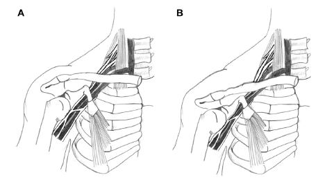
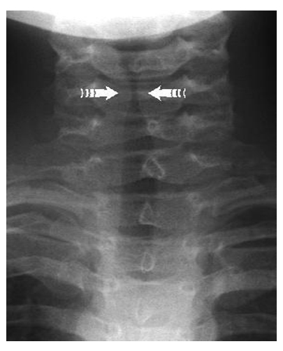
Figure 1: Costoclavicular space compression (Image source: Watson et al., 2010)

 It is well documented that TOS patients tend to have depressed scapulae, along with anterior tilt,10-14,44-46,51-60 which further depresses the clavicle. Watson states that scapular depression along with dyskinesis is commonly seen in TOS patients, and that this encourages costoclavicular compression.11 Elevating the scapulae will decompress the CCS,58 which is also an orthopedic test called the Cyriax test.8 This is one major reason why TOS surgery involves resection of the 1st rib; to free the neurovascular bundle within the CCS. It is therefore of paramount importance to ensure that patients do not overly depress their scapulae, to avoid oblivious CCS compression. Figure 1 illustrates how depression and retraction of the clavicle may compress the neurovascular bundle. Contrarily, lifting the clavicle will decompress the neurovascular bundle.

COSTOCLAVICULAR SPACE COMPRESSION

Compression of the neurovascular bundle (NVB) of the thoracic outlet may lead to plexopathy, muscular atrophy,61-65  carpal tunnel syndrome (double crush),65-68  chest pain, ulnar neuropathy,65-72 dorsal scapular neuralgia73,74  arm swelling and cyanosis.75,76  Other symptoms are Raynaud’s syndrome,75,76  positional ischemia or venous insufficiency,69,72,77-80  which may contribute to intracranial hypertension83  and migraines in addition it may present as  digital sensory loss 72), hand sweating and coldness.9899,107108  secondary dysautonomia such as atrial fibrillation and vasoconstriction.81,98103  deep vein thrombosis (DVT) development,79,88-93  weakness of the extremities, chest pain and pseudoangina,69-71,88-90 subclavian artery injuries with subsequent embolus,73,96  which  may lead to retrograde thromboembolism with sequelar stroke,91-93,97.98  and more.

Because of the vast spectrum of symptoms that may appear in the sequela of TOS development, TOS may reveal itself with any of the above-mentioned symptoms. It is well known that these symptoms are not unique,79,99 and it is hard to diagnose and treat the condition.78,100-104 This is one of the main reasons why (occult) TOS may be misdiagnosed as carpal tunnel syndrome, ulnar neuralgia, idiopathic chest pain, etc., in its beginning and intermediate stages. Neurographies (MR neurography imaging (MRN), electromyography (EMG), electroneurography (NCV), somatosensory evoked potentials (SEP) are insufficiently sensitive for detection of TOS, and may only be positive in very advanced stage.72,105-112  Rousseff et al.111 states that EMG / NCV is useless for identification of TOS, as 18 out of 20 patients with very obvious TOS symptoms had normal electrodiagnostic results. A systematic review conducted by Kwee et al.112 concludes that MRN is not sensitive nor specific enough for detection of brachial plexus neuropathies nor other peripheral neuropathies. This further complicates the likelihood of these patients (i.e. a victim of the “chronic Halstead’s maneuver”) to be properly diagnosed and treated.

It is a misfortune for patients to be iatrogenically set into the “back and down” scapular posture, as they may not be diagnosed until many years later due to the diffuse presentation of TOS.

 CLINICAL PRACTICE & APPROPRIATE CORRECTIVES

In this section I will provide some criteria for identification of TOS as well as scapular resting dysfunction. Neurogenic TOS is the utmost common variant, which makes up approximately 95% of total TOS incidences.78,79,113 Because the inferior trunk lies more susceptibly (anteriorly) placed in the CCS, symptoms of C8-T1 (ulnar) neuropathy may appear first. However, the superior trunk (C7) and middle trunk (C5-6) may be affected, especially in more progressed cases of TOS.65,72 Supraclavicular tenderness (Morley’s test) and weakness of the 5th finger are sensitive, and relatively specific tests for thoracic outlet syndrome.69-71,78-79,99,100,113-116 Weakness of the triceps (C7 myotome) is also common. There may also be positional ischemia upon shoulder elevation, demonstrated by a white hand sign,69-71,116-119 indicating severe compression of the subclavian artery.59,88,116,120,121 One test alone, e.g. Adson’s or Roos’ test, may not be specific enough to diagnose TOS, especially because only 5% of TOS incidences are considered to be of vasculogenic dominance.

Compression of the CCS may occur related to posture or intermittently during certain activities. For example, a patient may have a seemingly normal scapular resting height, but still have a tendency of pulling their scapulae back and down during exercise, or have scapular dyskinesis, which may lead to intermittent compression of the neurovascular bundle. It is important to examine the patient’s scapular position as well as loaded and unloaded  movement pattern in different scenarios. In addition Halstead’s CCS test may be used during the examination. A detailed explanation of identification and correction of scapular dyskinesis is outside the scope of this article.

The longitudinal scapular height can be measured by palpating the C7 (vertebra prominens) spinous process, then counting down to the T2 level, and comparing it to the level of the superior scapular angle. Up-/downwards rotation can be measured vertically by evaluating the angle of the medial scapular border, or horizontally, the angle of the scapular spine. The scapula is in downward rotation if the spine is pointing caudally, or if the superior angle is lateral to the inferior angle (sagittal axes). Anterior-/posterior tilting can be evaluated by measuring how far anterior the acromion is in relation to the inferior scapular angle, in the sagittal plane (coronal axis). An inclinometer, which in modern times is available for download on any smartphone, can also be used reliably to measure scapular angulation.11,23-26,40,122-128

The Cyriax release test is another orthopedic test which relieves the CCS by elevating the clavicle.8 Thus, postural CCS compression may be ameliorated by raising the shoulders slightly,58 and staying there .This will also upwardly rotate the scapula,55 which is important for SD and SIS treatment. To correct scapular slouching, ask the patient to lift their acromion until the clavicle is elevated and superior scapular angle is approximately levelled with the T2 vertebra, and the scapula is in mild upwards rotation. The patient must be educated with regards to the etiology of costoclavicular space compression syndrome, so that they become sufficiently motivated to maintain their newly acquired posture. Further, he or she must learn to maintain adequate scapular height during glenohumeral articulation.

Figure 2 shows a 25-year-old patient with chronic brachial, periscapular, chest and neck pain. She had been vigorously pulling her scapula back and down to “relax” her shoulder girdle, inevitably worsening the situation. Selmonosky’s DT was positive, as was Halstead’s CCS maneuver. Both scapulae were situated at the T4 vertebral level; very depressed (Figure 2, left). The left scapula was slightly more depressed than the right one and more caudally rotated as well. There was also bilateral scapular dyskinesia present during movement and loading of the arms. After identifying the scapular depression, the patient was cued to lift her acromion while slightly elevating the scapulae towards the back of the head, as to promote scapular elevation and upwards rotation with slight retraction, until the scapular angle was parallel with the T2 spinous process. She was told to stay there (Figure 3), and we also practiced moving and loading the arms while maintaining proper scapular height and angulation. The reason for cueing the patient specifically to lift the acromion rather than just the scapula, is that this promotes upwards rotation due to upper trapezius engagement. Some patients may unknowingly lift their scapulae up and forward by engaging the levator scapulae muscle, with sequelar downward scapular rotation. The patient experienced almost immediate remission of her symptoms that were related to loading and articulation of the arms after learning to hold her shoulders up. Also note the trapezius hypertrophy, although the muscle is clearly not over-engaging in lifting the scapulae in the “before” image. This can be misleading, which is why scapular height must be measured rather than ‘eyeballed’.

21-OA-Fig2

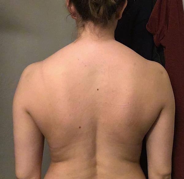
Figure 2: TOS patient with severely depressed scapulae, before correction

21-OA-Fig3

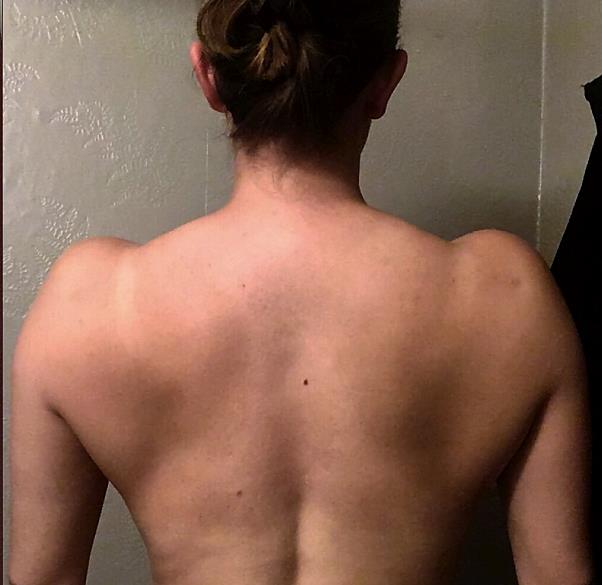
Figure 3: TOS patient after correction

DISCUSSION

It has been suggested by several authours hthat the upper trapezius is overactive in patients with SIS and SD, based on EMG test results during glenohumeral articulation.17,44,47,129-131 Yet, despite this, a conspicuous pattern of scapular downward rotation, anterior tilting and protraction is demonstrated in the very same patients. Because the UT promotes upwards rotation and posterior tilting, as well as retraction,15,35,132 the notion that the UT is truly over-engaging in scapular movement does not seem likely. Contrary to this, the levator scapuli muscle will cause scapular elevation, protraction, downward rotation and anterior tilting,132-133 and is, therefore, a culprit more compatible with the evidence, than that of the UT. Moreover, regarding the authenticity of the documented excess UT EMG results, some studies show that hypertrophied muscles with high EMG output on the symptomatic side (in this case, the symptomatic sacroiliac side), was later proven to be significantly weak,134-136  and that the excessive EMG output signal normalized after increasing the respective muscle’s strength. This may suggest that it is not as black and white as the studies which demonstrate UT overactivity suggest; it may still be weak. A weak UT attempting to stabilize and move the scapulae properly, yet fails to do so, may explain why we see increased EMG signals in the UT yet movements which contradict its true involvement, i.e. movements of downward rotation, protraction, depression, which would not be reasonably present if there was legitimate over-engagement of the UT in scapular movement.

Other researchers suggest that in order to maintain proper scapular position during glenohumeral articulation, all of the stabilizers must engage, especially the UT, SA, middle trapezius and lower trapezius.11,60  It has also been stated that a common error is to over-engage the rhomboid by squeezing the shoulder blades together, as this will promote anterior scapular tilt with concomitant depression of the scapula by the latissimus dorsi muscle.11

In accordance with Watson and Mckinnon’s papers,11,60  I have experienced that by setting a patient into the norms that were provided by Sahrmann and others,6,10,11,14,16,34,44,46- one will quickly realize that most patients with neck and shoulder problems tend to have depressed scapulae, and that they need to engage their upper trapezius, not suppress it. A main muscle that retracts and upwardly rotates the scapula, is also the upper trapezius muscle. Most of the negative biomechanical associations (scapular downward rotation, anterior tilt, protraction, depression) with SD, SIS, and TOS, are functionally countered by the upper trapezius, which promote elevation, retraction, posterior tilting and upward rotation. Based on this, and the evidence considered, it would seem quite contraindicated to pull the scapula back and down. It has been demonstrated that scapular elevation has an immediate beneficial effect on cervical pain137 as well as range of motion.138 Further, it has been documented that subjects with lower scapular resting position have a tendency of higher pain thresholds in the upper trapezius.5253 Whiplash associated disorders (WAD) patients have also been demonstrated to have low-riding clavicles.15,37 And, it has been demonstrated that patients with slouched scapulothoracic postures have decreased shoulder abduction ROM and posterior scapular tilting as well as decreased muscle force in glenohumeral abduction above 90˚.49  Finally, it has even been documented that patients with upper extremity deep vein thrombosis have a significantly narrower costoclavicular space in resting posture than that of controls.94,95 These would all make some solid points against pulling the clavicles back and down as postural means of therapeutic intervention.

Between January 1st 2017 and June 20th 2018, I evaluated 115 TOS patients for the co-presence of scapular depression, at my clinic in Oslo (Norway). They were diagnosed based on the criteria provided by Selmonosky’s diagnostic triad (DT) which involves supraclavicular tenderness (Morley’s sign) as well as relative weakness of the fifth finger, with or without a white hand sign.69,70,71,116  I classified scapular depression as having the superior scapular angle situated more than two finger widths below the T2 vertebra (approximately 2 cm). The survey revealed that 100% of the patients had scapular depression on the symptomatic side, cf. Sahrmann and colleagues’ norms. Most of these had been told to pull their scapulae back and down to correct their posture by their musculoskeletal therapist, and became considerably worse after following these cues, as a result. Five of these even had TOS surgery to decompress the CCS, but were still told to pull their shoulders back and down by their therapist, although TOS surgery clearly aims to increase the costoclavicular interval. The latter patients had severely depressed scapulae on the afflicted side, resulting in compression of brachial plexus between the clavicle and the 2nd rib. It is my impression, although clearly well-intended, that being cued to pull “back and down” obliviously and iatrogenically exacerbated the situation for these patients.

Because of the consensus that scapula has a tendency to protract, rotate forward (anterior tilt) and down (downward rotation) in patients with SD, SIS and neck pain, there may be some warrant in increasing scapular retraction alone. However, pulling back and down will cause the scapula to retract, depress and downwardly rotate, and is not compatible with any the criteria provided by the evidence, which states to increase retraction, upward rotation and posterior tilt. Increasing depression, downward rotation and anterior tilting may not only promote scapular dyskinesis and shoulder impingement syndrome, but also encroachment of the costoclavicular space and sequelar TOS.

The notion that proper scapular posture is obtained by pulling the shoulders back and down, is most likely based on only a few EMG studies, which show high upper trapezius output, as well as “pirate tales” from fitness and powerlifting communities. However, because patients with SIS and SD also demonstrate downward rotation, protraction, and depression, and, because the UT prevents these, it is unlikely to be truly over active nor over-engaged in moving the scapulae in a pathological manner. Retraction and depression of the scapulae may cause compression of the CCS as it mimics the Halstead’s CCS compression test. CCS compression implies osseous compression of the neurovascular bundle, which may lead to many diffuse and seemingly unrelated secondary problems, whose etiology may prove difficult to identify. Scapular depression has also been uniformly identified in TOS patients. There are also reports that scapular depression has been associated with increased myofascial pain in the cervical musculature, restricted range of motion, WAD, upper extremity DVT development, and more.

Therefore, often well-intended yet misunderstood cueing of pulling the shoulders “back and down”, may set the patient on a dark journey with many diffuse symptoms and few answers. Because unreliable diagnostic value of neurographic examinations, and because relatively few practitioners are versed in recognizing the signs of TOS, especially in its beginning-intermediate stages, there is likelihood that the patient will continue to pull their shoulders back and down and not suspect this as the etiology of their newfound problems. This can lead to longstanding problems for the patient and it may take a long time before his or her symptoms are finally identified as related to costoclavicular space compression.

LIMITATIONS

The survey part of this manuscript is based on patients who, on their own initiative have visited my unsubsidized private clinical practice. They may or may not fully represent the general patient population within the NHS.

CONCLUSION

In conclusion, it is of utmost importance to evaluate the patient’s scapular resting position based on the evidence, rather than generically cueing him or her to pull their scapulae back and down. If the patient presently has low-riding scapulae, and is cued further into depression, an iatrogenic sequela of maladies may develop as result. Pulling the shoulders “back and down” is a logical fallacy, which does not result in what it is thought to do, is not compatible with the evidence, the anatomy, biomechanics nor with common sense. Although the cue itself, and I reiterate, is clearly originating from well-meaning therapists, it is my impression that the SIS and SD studies have been gravely misinterpreted, and that the “back and down” cue should be abolished once and for all.

Conflict of interest

Nil

REFERENCES

  1. Osar, Evan. Forward shoulder posture and scapular retraction exercises. Accessed 2018 June 24. Available from https://www.otpbooks.com/evan-osar-forward-shoulder-posture/
  2. Scapular stabilisation exercises. Accessed 2018 June 24. Available from https://physioworks.com.au/treatments-1/scapular-stabilisation-exercises.
  3. Senger, Megan. Correct Cues for Scapular Motion. Accessed 2018 June 24. Available from https://www.acefitness.org/certifiednewsarticle/2384/correct-cues-for-scapular-motion/
  4. Bolton, Andy. How to break bench records. Accessed 2018 June 24. Available from https://www.t-nation.com/training/how-to-break-bench-records.
  5. Austin, Dan. Powerlifting. Human Kinetics Publishers; 2012.
  6. Osar, E. Corrective exercise solution to common hip and shoulder dysfunction. Lotus Publishers; 2012.
  7. Starkey C, Brown SD. Orthopedic & Athletic Injury Examination Handbook. 3rd edition. F.A. Davis Company; 2015.
  8. Magee DJ. Orthopedic physical assessment. 6th Elsevier; 2013.
  9. Mohindra M, Jain JK. Fundamentals of Orthopedics. 2nd edition. Jaypee Brothers Medical Publishers; 2017
  10. Hoppenfield S. Physical examination of the spine and extremities. 1st edition. Thomas H, Illustrator. Hutton R, Collaborator. Prentice Hall; 1976.
  11. Watson LA, Pizzari T, Balster S. Thoracic outlet syndrome Part 2: conservative management of thoracic outlet. Man Ther. 2010 Aug;15(4):305-14. [PubMed] doi: 10.1016/j.math.2010.03.002
  12. Kai Y, Oyama M, Kurose S, Inadome T, Oketani Y, Masuda Y. Neurogenic thoracic outlet syndrome in whiplash injury. J Spinal Disord. 2001 Dec;14(6):487-93. [PubMed]
  13. Skandalakis JE, Mirilas P. Benign anatomical mistakes: the thoracic outlet syndrome. Am Surg. 2001 Oct;67(10):1007-10. [PubMed]
  14. Telford ED, Mottershead S. Pressure at the cervico-brachial junction; an operative and anatomical study. J Bone Joint Surg Br. 1948 May;30B(2):249-65. [PubMed]
  15. Ludewig PM, Cook TM, Nawoczenski DA. Three-dimensional scapular orientation and muscle activity at selected positions of humeral elevation. J Orthop Sports Phys Ther. 1996 Aug;24(2):57-65. [PubMed] [Free full text]
  16. McClure PW, Michener LA, Sennett BJ, Karduna AR. Direct 3-dimensional measurement of scapular kinematics during dynamic movements in vivo. J Shoulder Elbow Surg. 2001 May-Jun;10(3):269-77. [PubMed]
  17. Ludewig PM, Cook TM. Alterations in shoulder kinematics and associated muscle activity in people with symptoms of shoulder impingement. Phys Ther. 2000 Mar;80(3):276-91. [PubMed]
  18. Endo K, Ikata T, Katoh S, Takeda Y. Radiographic assessment of scapular rotational tilt in chronic shoulder impingement syndrome. J Orthop Sci. 2001;6(1):3-10. [PubMed]
  19. Hébert LJ, Moffet H, McFadyen BJ, Dionne CE. Scapular behavior in shoulder impingement syndrome. Arch Phys Med Rehabil. 2002 Jan;83(1):60-9. [PubMed]
  20. Lin JJ, Lim HK, Yang JL. Effect of shoulder tightness on glenohumeral translation, scapular kinematics, and scapulohumeral rhythm in subjects with stiff shoulders. J Orthop Res. 2006 May;24(5):1044-51. [PubMed]
  21. Lawrence RL, Braman JP, Laprade RF, Ludewig PM. Comparison of 3-dimensional shoulder complex kinematics in individuals with and without shoulder pain, part 1: sternoclavicular, acromioclavicular, and scapulothoracic joints. J Orthop Sports Phys Ther. 2014 Sep;44(9):636-45, A1-8. doi: 10.2519/jospt.2014.5339. [PubMed] [Free full text]
  22. Warner JJ, Micheli LJ, Arslanian LE, Kennedy J, Kennedy R. Scapulothoracic motion in normal shoulders and shoulders with glenohumeral instability and impingement syndrome. A study using Moiré topographic analysis. Clin Orthop Relat Res. 1992 Dec;(285):191-9. [PubMed]
  23. Watson L, Balster SM, Finch C, Dalziel R. Measurement of scapula upward rotation: a reliable clinical procedure. Br J Sports Med. 2005 Sep;39(9):599-603. [PubMed] [Free full text]
  24. Struyf F, Nijs J, Baeyens JP, Mottram S, Meeusen R. Scapular positioning and movement in unimpaired shoulders, shoulder impingement syndrome, and glenohumeral instability. Scand J Med Sci Sports. 2011 Jun;21(3):352-8. doi: 10.1111/j.1600-0838.2010.01274.x. [PubMed]
  25. Struyf F, Nijs J, De Graeve J, Mottram S, Meeusen R. Scapular positioning in overhead athletes with and without shoulder pain: a case-control study. Scand J Med Sci Sports. 2011 Dec;21(6):809-18. doi: 10.1111/j.1600-0838.2010.01115.x. [PubMed]
  26. Kibler WB, McMullen J, Uhl T. Shoulder rehabilitation strategies, guidelines, and practice. Orthop Clin North Am. 2001 Jul;32(3):527-38. [PubMed]
  27. Lukasiewicz AC, McClure P, Michener L, Pratt N, Sennett B. Comparison of 3-dimensional scapular position and orientation between subjects with and without shoulder impingement. J Orthop Sports Phys Ther. 1999 Oct;29(10):574-83; discussion 584-6. [PubMed] [Free full text]
  28. Green RA, Taylor NF, Watson L, Ardern C. Altered scapula position in elite young cricketers with shoulder problems. J Sci Med Sport. 2013 Jan;16(1):22-7. doi: 10.1016/j.jsams.2012.05.017. [PubMed]
  29. Ludewig PM, Braman JP. Shoulder Impingement: biomechanical Considerations in Rehabilitation. Man Ther. 2011;16(1):33-39. doi:10.1016/j.math.2010.08.004. [PubMed] [Free full text]
  30. Greenfield B, Catlin PA, Coats PW, Green E, McDonald JJ, North C. Posture in patients with shoulder overuse injuries and healthy individuals. J Orthop Sports Phys Ther. 1995 May;21(5):287-95. [PubMed] [Free full text]
  31. Roche SJ, Funk L, Sciascia A, Kibler WB. Scapular dyskinesis: the surgeon’s perspective. Shoulder Elbow. 2015 Oct;7(4):289-97. doi: 10.1177/1758573215595949. [PubMed] [Free full text]
  32. Smith J, Dietrich CT, Kotajarvi BR, Kaufman KR. The effect of scapular protraction on isometric shoulder rotation strength in normal subjects. J Shoulder Elbow Surg. 2006 May-Jun;15(3):339-43. [PubMed]
  33. Borstad JD. Resting position variables at the shoulder: evidence to support a posture-impairment association. Phys Ther. 2006;86:549–57. [PubMed]
  34. Ludewig PM, Reynolds JF. The Association of Scapular Kinematics and Glenohumeral Joint Pathologies. J Orthop Sports Phys Ther. 2009 Feb; 39(2): 90–104. doi: 10.2519/jospt.2009.2808 [PubMed] [Free full text]
  35. Ludewig PM, Cook TM. The effect of head position on scapular orientation and muscle activity during shoulder elevation. J Occup Rehabil. 1996 Sep;6(3):147-58. doi: 10.1007/BF02110752. [PubMed]
  36. Helgadottir H, Kristjansson E, Mottram S, Karduna AR, Jonsson H Jr. Altered scapular orientation during arm elevation in patients with insidious onset neck pain and whiplash-associated disorder. J Orthop Sports Phys Ther. 2010;40:784–91. doi: 10.2519/jospt.2010.3405. [PubMed] [Free full text]
  37. Helgadottir H, Kristjansson E, Mottram S, Karduna A, Jonsson H Jr. Altered alignment of the shoulder girdle and cervical spine in patients with insidious onset neck pain and whiplash-associated disorder. J Appl Biomech. 2011;27:181–91. [PubMed]
  38. Martínez-Merinero P, Lluch E, Gallezo-Izquierdo T,  Pecos-Martín D, Plaza-Manzano G, Nuñez-Nagy S, et al. The influence of a depressed scapular alignment on upper limb neural tissue mechanosensitivity and local pressure pain sensitivity. Musculoskelet Sci Pract. 2017 Jun;29:60-65. doi: 10.1016/j.msksp.2017.03.001. [PubMed]
  39. Azevedo DC, de Lima Pires T, de Souza Andrade F, McDonnell MK. Influence of scapular position on the pressure pain threshold of the upper trapezius muscle region. Eur J Pain. 2008 Feb;12(2):226-32. [PubMed]
  40. Kibler WB. Scapular involvement in impingement: signs and symptoms. Instr Course Lect. 2006;55:35-43 [PubMed]
  41. Struyf F1, Nijs J, Mottram S, Roussel NA, Cools AM, Meeusen R. Clinical assessment of the scapula: a review of the literature. Br J Sports Med.2014 Jun;48(11):883-90. [PubMed] doi:10.1136/bjsports-2012-091059
  42. Kaur K, Das PG, Lenka PK, Anwer S. Immediate effect of posture correction of trapezius activity in computer users having neck pain – an electromyographic analysis. The Internet Journal of Allied Health Sciences and Practice. 2013 Oct 01;11(4), Article 10 [Free full text]
  43. Tate A, McClure P, Kareha S, Irwin D. Effect of the scapula reposition test on shoulder impingement symptoms and elevation strength in overhead athletes. J Orthop Sports Phys Ther. 2008;38:4–11. [PubMed] [Free full text]
  44. Kendall FP, McCreary EK, Provance PG. Muscles: Testing and Function with Posture and Pain. Williams and Wilkins. 4thBaltimore.1993;pp 286–293
  45. Swift TR, Nichols FT. The droopy shoulder syndrome. Neurology. 1984 Feb;34(2):212-5 [PubMed]
  46. Todd TW. The Descent of the Shoulder after Birth. Anat. Anz. Bd. Aufsatze. 1912:41(14):385-97
  47. Sahrmann, S.A. Diagnosis and Treatment of Movement Impairment Syndrome. St. Louis:Mosby;2002. 30-31p.
  48. Sahrmann S. Movement System Impairment Syndromes of the Extremities, Cervical and Thoracic Spines. Elsevier Health Sciences; 2010.
  49. Kebaetse M, McClure P, Pratt NA. Thoracic position effect on shoulder range of motion, strength, and three-dimensional scapular kinematics. Arch Phys Med Rehabil. 1999 Aug;80(8):945-50. [PubMed] [Free full text]
  50. Wijeratna MD, Troupis JM, Bell SN. The use of four-dimensional computed tomography to diagnose costoclavicular impingement causing thoracic outlet syndrome. Shoulder Elbow. 2014;6(4):273–75 [PubMed] [Free full text] DOI: 10.1177/1758573214533781
  51. Walsh MT. Therapist management of thoracic outlet syndrome. J Hand Ther. 1994 Apr-Jun;7(2):131-44. [PubMed]
  52. Kenny RA, Traynor GB, Withington D, Keegan DJ. Thoracic outlet syndrome: a useful exercise treatment option. Am J of Surgery.1993;165(2):282-84. [Link]
  53. Press JM, Young JL. Vague upper-extremity symptoms? Phys Sportsmed. 1994 Jul;22(7):57-64 [PubMed] doi: 10.1080/00913847.1994.11947668.
  54. Pascarelli EF, Hsu YP. Understanding work-related upper extremity disorders: clinical findings in 485 computer users, musicians, and others. J Occup Rehabil. 2001;11(1):1e21. [PubMed]
  55. Ranney D. Thoracic outlet: an anatomical redefinition that makes clinical sense. Clin Anat. 1996;9(1):50-2. [PubMed]
  56. Sucher BM. Thoracic outlet syndromeea myofascial variant: part 2. Treatment. J Am Osteopath Associ.1990;90(9):810-2, 817-823. [PubMed] [Free full text]
  57. Aligne C, Barral X. Rehabilitation of patients with thoracic outlet syndrome. Ann Vasc Surg. 1992;6(4):381e9. [PubMed]
  58. Kitamura T, Takagi K, Yamaga M, Morisawa K. Brachial plexus stretching injuries: microcirculation of the brachial plexus. J Shoulder Elbow Surg. 1995;4(2):118-23. [PubMed]
  59. Thompson, R.W. Neurogenic TOS. Accessed 2016 March 15. Available at http://tos.wustl.edu/What-is-TOS/Types-of-TOS/Neurogenic-TOS.
  60. Mackinnon SE, Novak CB. Thoracic outlet syndrome. Curr Probl Surg. 2002;39(11):1070-145. [PubMed]
  61. Kieffer E. Actualités de chirurgie vasculaire. Les syndromes de la traversée thoraco-brachiale. Paris: A.E.R.C.V. éditions; 1989. [Free full text]
  62. Merle M. Les syndromes de la traversée cervico-thoraco-brachiale. Monographie des Annales de Chirurgie de la Main. 1995;7:29–47. [Free full text]
  63. Alexandre J, Le Dû C, Corcia P, Laulan J. Formes déficitaires du syndrome de la traversée thoraco-brachiale. A propos de 16 cas. 41e congrès du GEM; Paris, décembre. 2005.
  64. Allieu Y, Benichou M, Touchais S, Desbonnet P, Lussiez B. Les formes neurologiques du syndrome du hile du membre supérieur: le rôle du scalène moyen. Ann Chir Main. 1991;10:308–312
  65. Laulan J, Fouquet B, Rodaix C, Jauffret P, Roquelaure Y, Descatha A. Thoracic outlet syndrome: definition, aetiological factors, diagnosis, management and occupational impact. J Occup Rehabi. 2011;21(3):366-73. doi:10.1007/s10926-010-9278-9. [PubMed] [Free full text]
  66. Dahlin LB, Lundborg G. The neurone and its response to peripheral nerve compression. J Hand Surg. 1990;15(1):5–10. [PubMed]
  67. Upton AR, McComas AJ. The double crush in nerve entrapment syndromes. Lancet. 1973;2:359–62. [PubMed]
  68. Narakas AO. The role of thoracic outlet syndrome in the double crush syndrome. Ann Chir Main Memb Super. 1990;9:331–40. [PubMed]
  69. Selmonosky CA, Byrd R, Blood C, Blanc JS. Useful triad for diagnosing the cause of chest pain. South Med J. 1981; 74:947-49. [PubMed]
  70. Selmonosky CA. The white hand sign. A new single maneuver useful in the diagnosis of thoracic outlet syndrome. Southern Med Journal. 2002; 85:557. [Abstract]
  71. Selmonosky CA, Poblete Silva R. The diagnosis of thoracic outlet syndrome. Myths and Facts. Chilean J of Surg. 2008 June; 60(3):255-261.
  72. Sanders RJ, Hammond SL, Rao NM. Thoracic outlet syndrome: a review. Neurologist. 2008 Nov;14(6):365-73. [PubMed] doi: 10.1097/NRL.0b013e318176b98d.
  73. Durham JR, Yao JS, Pearce WH, Nuber GM, McCarthy WJ 3rd. Arterial injuries in the thoracic outlet syndrome. J Vasc Surg. 1995 Jan;21(1):57-69; discussion 70. [PubMed]
  74. Chen D, Gu Y, Lao J, Chen L. Dorsal scapular nerve compression. Atypical thoracic outlet syndrome. Chin Med J. 1995;108:582-5 [PubMed]
  75. Akgun K, Aktas I, Terzi Y. Winged scapula caused by a dorsal scapular nerve lesion: a case report. Arch Phys Med Rehabil. 2008;89:2017-20. [PubMed] doi: 10.1016/j.apmr.2008.03.015.
  76. Aralasmak A, Karaali K, Cevikol C, Uysal H, Senol U. MR Imaging Findings in Brachial Plexopathy with Thoracic Outlet Syndrome. American Journal of Neuroradiology. 2010 March;31(3):410-417. [Free full text]
  77. Riddell DH, Smith BM. Thoracic and vascular aspects of thoracic outlet syndrome. Clin Orthop. 1986;207:31–6. [Abstract]
  78. Foley JM, Finlayson H, Travlos A. A Review of thoracic outlet syndrome and the possible role of botulinum toxin in the treatment of this syndrome. Toxins. 2012;4(11):1223-35. [PubMed] [Free full text] doi:10.3390/toxins4111223
  79. Hooper TL, Denton J, McGalliard MK, Brismée JM, Sizer PS Jr. Thoracic outlet syndrome: a controversial clinical condition. Part 1: anatomy, and clinical examination/diagnosis. J Man Manip Ther. 2010 Jun;18(2):74-83. [PubMed] [Free full text] doi: 10.1179/106698110X12640740712734
  80. Sanders RJ, Hammond SL, Rao NM. Diagnosis of thoracic outlet syndrome. J Vasc Surg. 2007 Sep;46(3):601-4. [PubMed]
  81. Archie M, Rigberg D. Vascular TOS—Creating a Protocol and Sticking to It. Diagnostics. 2017;7(2):34. [PubMed] doi:10.3390/diagnostics7020034.
  82. Pemberton, HS. Sign of submerged goitre. Lancet. 1948;248:509. doi:10.1016/s0140-6736(46)91790-4
  83. Sina F, Razmeh S, Habibzadeh N, Zavari A, Nabovvati M. Migraine headache in patients with idiopathic intracranial hypertension. Neurol Int. 2017;9(3):7280. doi:10.4081/or.2017.7280. [PubMed] [Free full text]
  84. De Simone R, Ranieri A, Montella S, et al. Intracranial pressure in unresponsive chronic migraine. J Neurol. 2014;261: 1365-73. [PubMed] [Free full text]
  85. Ninan T. Mathew, K. Ravishankar, Luis C. Sanin. Coexistence of migraine and idiopathic intracranial hypertension without papilledema. Neurology. 1996;46(5):1226-30. [PubMed]
  86. Yri HM, Rönnbäck C, Wegener M, Hamann S, Jensen RH. The course of headache in idiopathic intracranial hypertension: a 12-month prospective follow-up study. Eur J Neurol. 2014;21:1458–64. doi:10.1111/ene.12512 [PubMed]
  87. Ozdemir O, Ozcakar L. Thoracic outlet syndrome: Another cause for unilateral palmar hyperhidrosis. Clin Rheumatol. 2007;26(8):1375-76. [PubMed]
  88. Suderland S, ed. Nerve Injury. 2nd ed. NY:Edinburg NY Churchill Livingston; 1970 [1981 Printing].
  89. Urschel Jr HC, Razzuk MA. Upper plexus thoracic outlet syndrome: optimal therapy. Ann Thorac Surg. 1997;63(4):935-39. [Free full text]
  90. Urschel HC Jr, Kourtis H, Jr. Thoracic outlet syndrome: a 50 year experience at Baylor University. Proc (Bayl Union Med Cent).2007;20(2):125-35. [PubMed] [Free full text]
  91. Urschel HC, Razzuk MA, Hyland JW, Matson JL, Solis RA, Wood RE, et al. Thoracic outlet syndrome masquerading as coronary artery disease (pseudoangina). Ann Thorac Surg.1973;16(3):239-48 [Abstract]
  92. Vemuri C, McLaughlin LN, Abuirqeba AA, Thompson RW. Clinical presentation and management of arterial thoracic outlet syndrome. J Vasc Surg. 2017;65(5):1429-39 [Free full text]
  93. Shreeve MW, La Rose JR. Chiropractic care of a patient with thoracic outlet syndrome and arrhythmia. J Chiropr Med.2011;10(2):130-34 [PubMed] [Free full text] doi: 1016/j.jcm.2010.09.002
  94. Yamagami T, Handa H, Higashi K, Kaji R. Brachial plexus injury with cough attack: case report. Neurosurgery. 1994;34(6):1084-6; discussion 1086. [PubMed]
  95. Arnhjort T, Nordberg J, Delle M, Borgis CJ, Rosfors S, Larfars G. The importance of the costoclavicular space in upper limb primary deep vein thrombosis, a study with magnetic resonance imaging (MRI) technique enhanced by a blood pool agent. Eur J Intern Med. 2014;25(6):545-49. [PubMed] [Free full text]
  96. Fiorentini C, Mattioli S, Graziosi F, Bonfiglioli R, Armstrong TJ, Violante FS. Occupational relevance of subclavian vein thrombosis in association with thoracic outlet syndrome. Scand J Work Environ Health. 2005;31:160–163. [PubMed]
  97. Olin T.(1961) Arterial Complications in Thoracic Outlet Compression Syndrome, Acta Radiologica. 1961;56:97-112 [Free full text] , DOI: 10.3109/00016926109176643
  98. Lee TS, Hines GL. Cerebral embolic stroke and arm ischemia in a teenager with arterial thoracic outlet syndrome: a case report. Vasc Endovascular Surg. 2007;41(3):254-57 [PubMed]
  99. Palmer OP, Weaver FA. Bilateral cervical ribs causing cerebellar stroke and arterial thoracic outlet syndrome: a case report and review of the literature. Ann Vasc Surg.2015;29(4)840.e1-4 [PubMed] doi: 10.1016/j.avsg.2014.12.008.
  100. Boezaart AP, Haller A, Laduzenski S, Koyyalamudi VB, Ihnatsenka B, Wright T. Neurogenic thoracic outlet syndrome: A case report and review of the literature. Int J Shoulder Surg. 2010;4(2):27-35. doi:10.4103/0973-6042.70817. [PubMed] [Free full text]
  101. Atasoy E. Thoracic outlet compression sydnrome. Orthop Clin North Am. 1996;27(2):265-303. [PubMed]
  102. Povlsen B, Hansson T, Povlsen SD. Treatment for thoracic outlet syndrome. Cochrane Database Syst Rev. 2014 Nov 26;(11):CD007218. doi: 10.1002/14651858.CD007218.pub3. [PubMed]
  103. Redman L, Robbs J. Neurogenic thoracic outlet syndrome: Are anatomical anomalies significant? S Afr J Surg. 2015 Oct 8;53(1):22-5. [PubMed] [Free full text]
  104. Köknel TG. Thoracic outlet syndrome. Agri. 2005 Apr;17(2):5-9. [PubMed] [Free full text]
  105. Robey JH, Boyle KL. Bilateral Functional Thoracic Outlet Syndrome in a Collegiate Football Player. N Am J Sports Phys Ther. 2009 Nov; 4(4): 170–81. [PubMed] [Free full text]
  106. Tolson TD. “EMG” for thoracic outlet syndrome. Hand Clin. 2004;20: 37– 42. [PubMed]
  107. Passero S, Paradiso C, Giannini F, Cioni R, Burgalassi L, Battistini. Diagnosis of thoracic outlet syndrome. Relative value of electrophysiological studies. Acta Neurol Scand. 1994;90:179–85. [PubMed]
  108. Tsao BE, Ferrante MA, Wilbourn AJ, Shields RW. Electrodiagnostic features of true neurogenic thoracic outlet syndrome. Muscle Nerve. 2014;49(5):724-27 [PubMed] doi: 10.1002/mus.24066.
  109. Veilleux M, Stevens JC, Campbell JK. Somatosensory evoked poten- tials: lack of value for diagnosis of thoracic outlet syndrome. Muscle Nerve. 1988;11:571–75. [PubMed]
  110. Aminoff MJ, Olney RK, Parry GJ, Raskin NH. Relative utility of different electrophysiologic techniques in the evaluation of brachial plexopathies. Neurology. 1988;38:546–50. [PubMed]
  111. Komanetsky RM, Novak CB, Mackinnon SE, Russo MH, Padberg AM, Louis S. Somatosensory evoked potentials fail to diagnose thoracic outlet syndrome. J Hand Surg Am. 1996 Jul;21(4):662-6. [PubMed]
  112. Rousseff R, Tzvetanov P, Valkov I. Utility (or futility?) of electrodiagnosis in thoracic outlet syndrome. Electromyogr Clin Neurophysiol. 2005 Apr-May;45(3):131-33. [PubMed]
  113. Kwee RM, Chhabra A, Wang KC, Marker DR, Carrino JA. Accuracy of MRI in diagnosing peripheral nerve disease: a systematic review of the literature. Am J Roentgenol.2014;203:1303-09 [PubMed] [Free full text]
  114. Sanders RJ, Annest SJ. Pectoralis Minor Syndrome: Subclavicular Brachial Plexus Compression. Diagnostics. 2017;7(3):46. [PubMed] [Free full text] doi:10.3390/diagnostics7030046
  115. Brantigan CO, Roos DB. Diagnosing thoracic outlet syndrome. Hand Clin. 2004;20:27–36. [PubMed] [Free full text]
  116. Liu JE, Tahmoush AJ, Roos DB, Schwartzman RJ. Shoulder-arm pain from cervical bands and scalene muscle anomalies. J Neurol Sci. 1995;128:175–80 [PubMed]
  117. Selmonosky, C.A. Diagnosis of Thoracic Outlet Syndrome. Accessed on 2015 May 15. Available at http://www.tos-syndrome.com.
  118. Fried, SM. Nazarian LN. Dynamic neuromusculoskeletal ultrasound documentation of brachial plexus/thoracic outlet compression during elevated arm stress testing. Hand (NY) 2013;8:358-365, [PubMed] [Free full text] doi: 10.1007/s11552-013-9523-8
  119. Braun RM. Rechnic M. Shah KN. Pulse oximetry measurements in the evaluation of patients with possible thoracic outlet syndrome. J Hand Surg Am. 2012;37:2564-69. [PubMed] doi: 10.1016/j.jhsa.2012.09.020
  120. Thompson RW. Bryan’s Story. Accessed on 2016 March 3. Available at http://tos.wustl.edu/Patient-Features/Bryans-Story.
  121. Illig KA, Doyle AJ. A comprehensive review of Paget-Schroetter syndrome. J Vasc Surg. 2010;51:1538-47

[PubMed] [Free full text] Doi:10.1016/j.jvs.2009.12.022

  1. Guzzo JL, Chang K, Demos J, Black JH, Freischlag JA. Preoperative thrombolysis and venoplasty affords no benefit in patency following first rib resection and scalenectomy for subacute and chronic subclavian vein thrombosis. J Vasc Surg. 2010;52(3):658-62 [PubMed] doi: 10.1016/j.jvs.2010.04.050
  2. Laudner KG, Stanek JM, Meister K. Differences in scapular upward rotation between baseball pitchers and position players. Am J Sports Med. 2007 Dec;35(12):2091-5 [PubMed] doi: org/10.1177/0363546507305098
  3. Johnson MP, McClure PW, Karduna AR. New method to assess scapular upward rotation in subjects with shoulder pathology. J Orthop Sports Phys Ther. 2001 Feb;31(2):81-9. [PubMed] [Free full text]
  4. Borsa PA, Timmons MK, Sauers EL. Scapular-positioning patterns during humeral elevation in unimpaired shoulders. J Athl Train. 2003;38(1):12-17. [Free full text]
  5. Su KP, Johnson MP, Gracely EJ, Karduna AR. Scapular rotation in swimmers with and without impingement syndrome: practice effects. Med Sci Sports Exerc. 2004 Jul;36(7):1117-23. [PubMed]
  6. Scibek JS, Carcia CR. Validation of a new method for assessing scapular anterior‐posterior tilt. Int J Sports Phys Ther. 2014 Oct;9(5):644–56. [PubMed] [Free full text]
  7. Shobush DC, Simoneau GG, Dietz KE, Levene JA, Grossman RE, Smith WB . The Lennie test for measuring scapular position in healthy young adult females: a reliability and validity study. J Orthop Sports Phys Ther. 1996;23(1):39-50. [PubMed] [Free full text]
  8. O’Shea A, Kelly R, Williams S, McKenna L. Reliability and Validity of the Measurement of Scapular Position Using the Protractor Method. Phys ther. 2016;96(4):502 [PubMed] doi: 10.2522/ptj.20150144.
  9. Ekstrom RA, Donatelli RA, Soderberg GL. Surface electromyographic analysis of exercises for the trapezius and serratus anterior muscles. J Orthop Sports Phys Ther. 2003 May;33(5):247-58. [PubMed] [Free full text]
  10. Cools AM, Witvrouw EE, Declercq GA, Vanderstraeten GG, Cambier DC. Evaluation of isokinetic force production and associated muscle activity in the scapular rotators during a protraction–retraction movement in overhead athletes with impingement symptoms Br J Sports Med. 2004;38:64-68 [Free full text]
  11. Cools AM, Declercq GA, Cambier DC, Mahieu NM, Witvrouw EE. Trapezius activity and intramuscular balance during isokinetic exercise in overhead athletes with impingement symptoms. Scand J Med Sci Sports. 2007 Feb;17(1):25-33 [PubMed]
  12. Paine R, Voight ML. The role of the scapula. Int J Sports Phys Ther. 2013;8(5):617–29. [PubMed] [Free full text]
  13. Reinold MM, Escamilla R, Wilk KE. Current concepts in the scientific and clinical rationale behind exercises for glenohumeral and scapulothoracic musculature. J Orthop Sports Phys Ther. 2009;39:105–117 [PubMed] [Free full text] doi: 10.2519/jospt.2009.2835.
  14. Vleeming A, Schuenke MD, Masi AT, Carreiro JE, Danneels L, Willard FH. The sacroiliac joint: an overview of its anatomy, function and potential clinical implications. J Anat. 2012;221(6):537-67. [PubMed] [Free full text] doi:10.1111/j.1469-7580.2012.01564.x.
  15. Mooney V, Pozos R, Vleeming A, Gulick J, Swenski D. Exercise treatment for sacroiliac pain. Orthopedics. 2001 Jan;24(1):29-32. [PubMed]
  16. Massoud Arab A, Reza Nourbakhsh M, Mohammadifar A. The relationship between hamstring length and gluteal muscle strength in individuals with sacroiliac joint dysfunction. J Man Manip Ther. 2011 Feb;19(1):5-10 [PubMed] [Free full text]doi:10.1179/106698110X12804993426848.
  17. Van Dillen LR, McDonnell MK, Susco TM, Sahrmann SA. The immediate effect of passive scapular elevation on symptoms with active neck rotation in patients with neck pain. Clin J Pain. 2007 Oct;23(8):641-7. [PubMed]
  18. Andrade GT, Azevedo DC, De Assis Lorentz I, Galo Neto RS, Sadala Do Pinho V, Ferraz Goncalves RT, et al. Influence of scapular position on cervical rotation range of motion. J Orthop Sports Phys Ther. 2008 Nov;38(11):668-73. [PubMed] [Free full text]21-OA-Fig1

Evaluation of a low-cost videolaryngoscope – a randomized controlled pilot study

$
0
0

Dear Sir,

With regard to your article “Evaluation of a low-cost videolaryngoscope – a randomized controlled pilot study”1 Please note that the initial work on inexpensive videolaryngoscopy technique using an USB endoscope camera was demonstrated publicly in India as early as in 2012 and published online as a case report.2 Hence the statement in the article of it being reported only later in 2016 is not true.

There have been several authors who have published similar reports after 2012, regrettably claiming it as a new technique. A blog, published later on, after the first case report in 2013, describes the development of this technique.3

Regards,
John George Karippacheril.
E-mail: johngeorgedon@gmail.com

 References:

1. Vadhanan P, Balakrishnan K, TripatyDK. Evaluation of a low-cost videolaryngoscope – a randomized controlled pilot study. Anaesth Pain & Intensive Care 2017;21(4):406-412 [Free full text]
2. Karippacheril JG, Umesh G, Nanda S. Assessment and confirmation of tracheal intubation when capnography fails: a novel use for an USB camera. J Clin Monit Comput. 2013 Oct;27(5):531-3. doi: 10.1007/s10877-013-9458-1 [PubMed]
3. Ketamine. Evolution of inexpensive videolaryngoscopy: from concept to practice. August 20, 2015. Available online at https://prehospitalmed.com/2015/08/20/evolution-of-inexpensive-videolaryngoscopy-from-concept-to-practice/

Evaluation of a low-cost videolaryngoscope – a randomized controlled pilot study Author’s reply:

Sir

The low cost video laryngoscope has been around for several years and even a Mumbai Based manufacturer has been producing this device attached to a curved blade. We have modified the scope in two ways – by using a Miller’s Blade, and secondly by keeping the original bulb of the Millers blade also functioning to provide adequate illumination, as the USB borescopes have low powered LED bulbs which might not be enough in the presence of secretions apart from using an android based smart phone instead of a personal computer. Even though various claims for this device do exist, including Dr Wickrama Wickramasinghe – a Sri Lanka based anesthetist receiving the WFSA innovation award for the same in 2015.1 we would like to exert that we make no claims as being the inventor or the first person to report such a use for borescopes. We have just done a randomized controlled trial upon a Millers blade based low cost video laryngoscope which to the best of our knowledge is the first. This fact is highlighted in the end of the original youtube video posted by the author.2 However, omission of acknowledgement of the previously done commendable trial by Karippacheril et al  in 2013 using a Macintosh blade is regretted.

We thank Karippacheril et al. for the clarification and congratulate them for their efforts.

Sincerely
Prasanna Vadhanan
vadhanan.prasanna@gmail.com

References:
1. WFSA Announces Winners of Innovation Awards. 2015. Available at https://www.wfsahq.org/latest-news/latestnews/481-wfsa-announces-winners-of-innovation-awards
2. A low cost video laryngoscope. Prasanna v. YouTube. Published on Mar 19, 2015. Available at

https://youtu.be/NTTD9RwzarM

Observation of intravenous cannula: an utmost important issue

$
0
0

Teena Bansal*, Jatin Lal**, Bharat Singh***, Somesh***

* Associate  Professor (DA, DNB ),**Professor (MD), ***Junior Resident
Department of Anaesthesiology & Critical Care,  Pt. B.D. Sharma University of Health Sciences Rohtak-124001 ( Haryana ), India


Corresponding Author:
Dr. Teena bansal
19/6 J, Medical Campus
PGIMS, Rohtak-124001 (Haryana) India
e-mail: aggarwalteenu@rediffmail.com
Mobile: +91-9034239374


Needlestick injury is very dangerous. To provide protection against such injury, safety cannulae are very important safety measure. Injection port of these cannulae provide needleless drug injection, thus preventing injuries. These cannulae have inbuilt valve mechanism to facilitate administration of drug without any backflow.

A 28 year old female was posted for diagnostic laparoscopy for primary infertility. 20 G intravenous cannula was placed. Intraoperatively fluid and drugs were given through the same cannula. The arm board was draped intraoperatively, so neither the arm nor the cannula was visible. Surgery lasted for 2 hours. At the end of the surgery, the drapes were removed. While giving reversal agent, suddenly a pop was heard and the drug could be injected but after removing the syringe, there was leakage of fluid through the injection port. Even after covering the injection port, the leak continued and when Ringer Lactate was stopped, blood started flowing back through the port. New intravenous cannula was placed and the previous one was removed.

On inspecting the cannula, the valve of the cannula was found to be displaced. One way valve of the cannula is an elastomeric sleeve. During the administration of drug via syringe, there is deflection of elastomeric sleeve allowing passage of drug through the port and after injecting drug, the sleeve returns to its original position preventing leakage or backflow.1 Our patient was under drapes intraoperatively and during that period, there was no problem in administration of drug. The problem occurred after removal of  drapes. Had it been occurred intraoperatively, though we could have been able to administer drug with pop but the leakage of fluid and blood would have remained unnoticed. We wish to highlight that intravenous cannula should be observed intraoperatively particularly if there is resistance to administration of drug and then a pop is heard and the drug can be injected. It becomes an utmost important issue if the arm board is draped to prevent blood loss and air embolism as ingress of air can cause air embolism also.

25-OA-Fig3

REFERENCES

  1. Behura A, Ahuja M. A leaky intravenous cannula. Anaesthesia 2006;61:411. [PubMed] [Free full text] doi: 10.1111/j.1365-2044.2006.04603.x


A comparison between intravenous metoprolol and labetalol in prevention of cardiovascular stress response to laryngoscopy and intubation

$
0
0

Nishee R Swami, MBBS, DNB1 MBBS, DNB1, Vaishalee K Badhe, MBBS, DNB1, Vaishali V. Deshpande, MD, DA2, Vaijayanti K. Badhe, MD, DA3, Shidhaye Ramchandra Vinayak, MD, DA3

1Assistant Professor, 3Professor?
Department of Anesthesiology and Critical Care, Pravara Institute of Medical Sciences, Loni 413736 (India)
2Professor / Head, Department of Anesthesiology, Seth Nandlal Dhoot Hospital, Aurangabad (India)

Correspondence: Dr. Vaishalee K. Badhe, MBBS, DNB, Assistent Professor, Department of Anesthesiology and Critical Care, Pravara Institute of Medical Sciences, Loni 413736 (India); E-mail: drvaishalee@rediffmail.com

ABSTRACT

Introduction: A prospective, randomized, double blind, clinical study was designed to compare intravenous metoprolol 30 µg/kg versus intravenous labetalol 0.2 mg/kg single dose given 5 min prior to intubation in for prevention of cardiovascular stress response to laryngoscopy and intubation in patients undergoing spine surgeries under general anesthesia.
Methodology: Sixty ASA grade I patients of either sex , comprising age group of 25-50 years , undergoing elective spine surgeries under general anesthesia were randomly distributed in two equal groups. Inj metoprolol hydrochloride 30 µg/kg in Group M and inj labetalol 0.2 mg/kg in Group L respectively were given intravenously 5 min prior to induction. Heart rate, systolic, diastolic and MBP recorded at different time intervals before and after intubation.
Results: Significant rise noted in heart rate, systolic, diastolic and MBP immediately after intubation in both groups though less in Group L that remained up to 2 min; returned to baseline between 2 to 5 min and became significantly lower than baseline at 5 min and onwards.
Conclusion: Labetalol is superior to metoprolol in attenuating the cardiovascular stress response to laryngoscopy and intubation.
Keywords: Labetalol, Metoprolol, Presser response, Laryngoscopy, Tracheal intubation
Citation: Swami NR, Badhe VK, Deshpande VV, Badhe VK, Shidhaye RV. A comparison between intravenous metoprolol and labetalol in prevention of cardiovascular stress response to laryngoscopy and intubation. Anaesth Pain & Intensive Care 2018;22(2):180-186
Received: 17 May 2018 Reviewed: 25 Jun 2018 Corrected: 26 Jun 2018 Accepted: 30 Jun 2018

INTRODUCTION

The frequent occurrence of cardiovascular responses to laryngoscopy and tracheal intubation has attracted the attention of anesthesiologists for several years. Marked circulatory effects of laryngoscopy and tracheal intubation like reflex tachycardia (rise up to 20%) and hypertension (rise up to 40-50%) is encountered during intubation1. A number of responses to intubation occur, including hypertension, tachycardia, arrhythmia, raised intracranial and intraocular pressure. The cardiovascular responses may have serious consequences including myocardial ischemia, dysrrthymias, pulmonary edema, sudden left ventricular failure, cerebrovascular hemorrhage and at times even cardiac arrest.2 These changes are tolerated quite well by healthy patients, because of their transient nature and there are no grave sequelae. However patients suffering from coronary artery disease, hypertension, valvular heart disease, stroke, penetrating eye lesion, intracranial lesions are not able to withstand them. In principle this response can be modified by using methods which act locally, centrally or peripherally. Cardiovascular response to laryngoscopy and intubation is a reflex phenomenon with afferent stimuli carried over glossopharyngeal and vagal pathways which activate the suprategmental and hypothalamic sympathetic centers to cause peripheral sympathoadrenal response with release of adrenaline and nor adrenaline. The elevation of blood pressure is associated with norepinephrine release whereas changes in heart rate are epinephrine related.3 Norepinephrine levels may increase on laryngoscopy and intubation from (60-310 pg/ml) and continue to rise for 4 to 8 min, Epinephrine levels may raise 4 times from 70 to 280 pg/ml 4. Above data indicates role of sympathoadrenergic receptor blockers (α and β blockers) in attenuating the cardiovascular stress response to intubation by attenuating the effects of catecholamines with the act of laryngoscopy and intubation. Beta blockers (e.g. etoprolol, esmolol.5,6 labetalol7,8 or landiolol9) with bradycardia, antihypertensive, antiarrythmic and anti-ischemic properties have found a role in preventing cardiovascular responses to intubation. Metoprolol is selective β1 blocker and labetalol is selective α1 and non-selective β blocker. Several studies on metoprolol10-13 and labetalol7,8,14-16 showed their effectiveness in attenuating the cardiovascular stress response to intubation. Additionally metoprolol and Labetalol found role in providing controlled hypotension to provide bloodless field as required in spine surgeries .Though researchers have compared esmolol and different other drugs with metoprolol and labetalol10,11,14, 16 we couldn’t find studies comparing metoprolol and labetalol in prevention of cardiovascular stress response to laryngoscopy and intubation. With this background we designed this prospective, randomized, double blind, comparative clinical study to assess the degree of effectiveness of IV metoprolol 30 µg/kg versus IV labetalol 0.2 mg/kg single dose given 5 min prior to intubation in prevention of cardiovascular stress response to laryngoscopy and intubation in patients undergoing spine surgeries under general anesthesia.

 METHODOLOGY

After approval from the hospital ethical committee, a prospective randomized double blind clinical study was conducted on 60 ASA grade I patients (30 in each group) of either sex , comprising age group of 25-50 y, undergoing elective spine surgeries under general anesthesia .Excluding criteria were: systemic disorder like diabetes mellitus, hypertension, heart disease, having bradycardia or heart block, respiratory disease like asthma, COPD, having history of any drug allergy and patients not willing to participate. After valid informed written consent selected patients were randomly allocated to two groups.

Group M: Inj metoprolol hydrochloride 30 µg/kg intravenously 5 min prior to induction
Group L: Inj labetalol 0.2 mg/kg intravenously 5 min prior to induction

Method of Randomization: Method of randomization was blocked randomization. Randomization was carried out based on blocking. A total of 15 blocks of size 4 with treatment allocation of 1:1 for Group M and Group L were created with the help of computer software. Coded envelopes (fifteen) were used and each envelope was used for four patients leading to random assignment of one subject to one group.

Sample size calculation was based on previous studies.14 Sample size was estimated to be 46 (23 in each group) to get the difference of 10 % in mean arterial pressure (MAP) measured at intubation in Group L and Group M using a two sample t test, assuming a two sided type I error of 5% (α = 0.05) and power at 80 (β = 0.20)

All patients received tablet diazepam 10 mg orally on the night prior to surgery. After conforming nil by mouth status and written informed consent every patient was taken to operation table. Intravenous (IV) line was secured with 20G Angiocath™ cannula, ringer lactate drip was started and midazolam 0.02 mg/kg and pentazocine 0.3 mg/kg was injected IV as premedication.

Multipara monitor (Vista 120™, Drager, Germany) measuring pulse rate, ECG, noninvasive blood pressure (NIBP), capnography (EtCO2) and oxygen saturation (SO2) was applied. Heart rate and blood pressure [systolic blood pressure (SBP), diastolic blood pressure (DBP) and MAP] were measured at base line. (10 min prior to induction). Inj metoprolol 30 µg/kg and inj labetalol 0.2 mg/kg were given intravenously to respective group patients 5 min prior to induction by an anesthetist who was blinded for the study. Preoxygenation was done with 100% oxygen for 5 min .Induction of anesthesia was done with inj thiopentone 5 mg/kg. Intubation was facilitated with inj suxamethonium hydrochloride 2 mg/kg. Laryngoscopy and endotracheal intubation was done by the same anesthetist trained in the technique for 2 y. Anesthesia was maintained with nitrous oxide and oxygen (50:50%) + isoflurane (0.6%). Muscle relaxation was achieved using inj atracurium (0.5 mg/kg) with subsequent boluses of (0.1 mg/kg) as per requirement. Mechanical ventilation was done targeting EtCO2 32-36 mmHg. Neuromuscular block was reversed at the end of surgery with inj. neostigmine 0.05 – 0.06 mg/kg and inj. glycopyrrolate 0.008-0.01 mg/kg. Heart rate, SBP, DBP, MAP (non-invasive) were measured at 10 min and 5 min prior to induction, immediately after induction, during intubation (0 min), at 01, 02, 3, 5,10, 30 and 60 min post intubation by the anesthetist who were blinded to the drug given. No surgical stimulation was allowed in first 10 min. Cases where intubation was difficult and that took more than 20 sec and required more than one attempt were excluded from the study. Intra operative oozing and clarity of surgical field was assessed by oral questionnaire to surgeon. Upon completion of surgery patients were extubated after reversal of neuromuscular block and were observed for 8hrs in the postoperative period for side effects like bradycardia, hypotension, nausea, vomiting, difficult respiration. Appropriate treatment for side effects was planned. For severe bradycardia atropine 7 µg/kg was given. For persistent bradycardia isoprenaline infusion 2-25 µg/min was kept ready. Hypotension was treated by fluid challenge. Vasopressors like Dopamine 5-15 µg/kg /min was given if required. Increase in airway resistance if any was treated with terbutaline inhalation

Statistical analysis:

All the observations for the above mentioned parameters were collected in a master chart. Demography parameters were analyzed by student’s unpaired t test. Categorical data was analyzed by Chi square test. For finding the statistical significance between the two groups unpaired t test was applied to ascertain the pattern and magnitude of differences. Paired t test was applied for intra group comparison. A p value of < 0.05 was considered as significant and P value of < 0.01 was considered as highly significant.

RESULTS

The patients in both the groups were comparable with respect to age, sex, height, weight, duration of surgery and time required for intubation. (Table 1). They were also comparable with respect to baseline heart rate, SBP, DBP and MAP measured at 10 min before induction (Table 2-A).

Table 2-A shows the hemodynamic parameters at different time intervals in both groups and Table 2-B shows the percent change in the hemodynamic parameters at different time intervals in both groups. There was no significant difference in heart rate after premedication and induction in both groups. The maximum change in the heart rate was seen immediately after intubation in both groups. The rise in heart rate remained up to 2 min ; returned to baseline between 2 to 5 min and became significantly lower than baseline at 5 min and onwards . However the difference between the two groups at all the time intervals was not significant. No group showed arrhythmias following intubation. Fall in SBP clinically not significant to require treatment was observed in both groups as a result of induction of anesthesia. It was more in Group L. There was a significant rise in SBP in both the groups at intubation, 1 min and 2 min post intubation but the rise was less in Group L. This rise in SBP at intubation remained up to 2 min post intubation and returned to baseline between 2 to 5 min post intubation and became significantly lower than baseline thereafter. Both groups were comparable regarding systolic BP at 2 min post intubation and thereafter though initial rise at intubation was less in Group L. Similarly there was decrease in DBP and MAP at induction in both groups, more being in Group L. There was rise in DBP and MAP in both groups at intubation, 1 min and 2 min post intubation. The rise in DBP and MAP was more in Group M as compared to Group L till 2 min post intubation. At 5 min and onwards DBP and MAP were significantly less than the baseline values in both groups. DBP and MAP were lower in Group L as compared to Group M at 10, 30 and 60 min post intubation. Significant hypertension or hypotension (> 30% change from baseline) was not found in any group at intubation. One patient in Group M developed bradycardia up to 48 beats/min during reversal of residual muscle relaxation with neostigmine and glycopyrrolate, however, blood pressure was normal. This change in heart rate was more than 30% of baseline and it responded to atropine 7 µg/kg. In Group L one patient developed transient hypotension up to 80/50 mmHg, which responded by putting off isoflurane and giving Ringer lactate 200ml. One patient in Group L developed transient hypotension in recovery room on changing to sitting position up to 84/50 mmHg which got improved by giving supine position and giving ringer lactate .No other specific treatment was required .Two patients in Group M and three patients in Group L had nausea and vomiting post operatively in recovery room. Question was asked to operating surgeon regarding oozing and clarity of operating field. Operating field was clear and bloodless in 80 % of patients in Group M as against in 83.33% patients in Group L.

DISCUSSION

The cardiovascular response to the act of tracheal intubation is a reflex phenomenon with the afferent stimuli carried over both glossopharyngeal and vagal pathways. Such stimuli activate suprategmental and hypothalamic sympathetic centers to cause a peripheral sympathoadrenal response with release of adrenaline and noradrenaline.19 Metoprolol and labetalol do not decrease release of catecholamines but attenuate responses of elevated catecholamines following laryngoscopy and intubation. Different researchers have studied different doses of metoprolol ranging from 0.5 mg to 4 mg10-13 and different doses of labatalol8,14-17 ranging from 0.15 mg/kg to 2 mg/kg for prevention of cardiovascular stress response to Laryngoscopy and intubation. We used optimum doses of metoprolol 30 micrograms /kg and labetalol 0.2 mg/kg so as to get desired effect and avoid undesirable side effects, 5 min prior to induction considering peak action of both drugs being at 5 min after administration. To avoid confounding effect on cardiovascular response to intubation by drugs used for induction, we induced all patients with inj thiopentone sodium 5 mg/kg like previous investigators12,14,15 and inj Suxamethonium hydrochloride 2 mg/kg to facilitate intubation. Propofol was avoided as it attenuates the presser response to intubation 17. As duration of laryngoscopy and intubation also has confounding effect all patients having prediction for difficult intubation were excluded. Laryngoscopy was done by trained anesthetist with two years of experience. Same type of laryngoscope blade, Macintosh blade size 3 was used for laryngoscopy considering the observations of Haidry MA et al.18 that the use of different type of laryngoscope blades can attenuate the presser response to intubation. King BD et al.1 observed the onset of the presser response within 5 to 15 sec of elevating epiglottis during laryngoscopy and returning at the end of 5 min. Bruder et al. 2observed that the response lasted for 5 to 10 min .Hence we monitored the parameters till 10 min after intubation. Readings at 30 and 60 min were taken to observe hemodynamic status. In our study both the groups showed significant increase in heart rate after intubation which remained up to 2 min, reached to baseline between 2 and 5 min and became significantly lower than baseline at 5 min and onwards. Though the percentage change in heart rate immediately following intubation was found to be less in Group M (7.44%) than in Group L (9.79%) this intergroup difference was not statistically significant. Comparable changes in heart rate with metoprolol and Labetalol were found by previous investigators. Kumar et al.12 found increase in heart rate by 10.26% and Pratheeba N et al.11 found increase in heart rate by 16.59 % immediately following intubation in metoprolol pretreated group. While Shende SY et al.10 who used higher dose of metoprolol (80 micrograms /kg) found only 3.37 % rise in heart rate. Ekta Ratnani et al.8 found 4.9 % rise and Lakshmi BS et al.16 found 13.94 % rise in pulse rate immediately following intubation in labetalol pretreated group similar to our findings. King BD teal 19511stated a marked cardiovascular response to laryngoscopy and intubation with increase in pulse rate up to 20 % without any preventive medication. B. Sowbhagya Lakshmi et al.16found even upto39 % increase in pulse rate without any preventive medication. Thus metoprolol and Labetalol both significantly attenuated the increase in the pulse rate during intubation .In metoprolol pretreated group at 1 min post intubation we found 5.38 % increase in pulse rate like Gurudatta KN et al.13 who found 5.08 % and Shende SY et al.10who found 6.52 % .In labetalol pretreated group at 1 min post intubation we found 7.35 % increase in pulse rate while EktaRatnani et al.8 found 2.6 % rise and Lakshmi BS et al.16 found 14.66 % rise in pulse rate. In our study, none of the patients had tachycardia or arrhythmias during intubation. One patient in Group M developed sinus bradycardia during reversal of residual nondepolarizers with neostigmin and glycopyrrolate which responded to atropine 7 µg/kg IV. Kumar et al. 12 similarly observed sinus bradycardia in one patient in Group M during reversal of nondepolarizers. None of the patient in Group L developed bradycardia. No other episodes of arrhythmias with few exceptions of transitory sinus tachycardia were observed in our study .Readings at 30 and 60 min post intubation in Group M (-11.80% and -12.13%) and Labetalol (-9.09% and -9.49%) group showed heart rate being significantly lower than baseline indicating controlled hemodynamic status. SBP decreased significantly than baseline immediately after induction in both groups (-8.33% vs. -12.84%). Decrease in SBP was significantly more in Group L than Group M during induction (p = 0.0227).However during intubation there was an increase of 15.62% and 8.01% in SBP in Group M and Group L respectively .Thus the rise in Group M was more than that of the Group L, which was statistically significant . King BD et al.1stated a marked rise in blood pressure up to 40-50% without any preventive medication. Lakshmi BS et al.16 also found up to 32.71 % increase in pulse rate without any preventive medication. Thus both metoprolol and labetalol significantly attenuated the increase in SBP during intubation. But labetalol is significantly more effective in attenuating the presser response to intubation than metoprolol. The readings at 01, 02, 05, 10 min post intubation in both metoprolol and Group L showed that the significant increase in SBP than baseline at intubation remained up to 2 min reached to baseline in 2-5 min and became significantly lower than baseline at 5 and 10 min onwards. Comparable results were obtained in metoprolol study by other researchers.8,10,11,13,16 Readings at 30 and 60 min post intubation were significantly lower than baseline in Group M (-10.05% and -10.22%) and Group L (-12.50% and -12.43%) indicating controlled hemodynamic status. DBP and MAP showed similar behavior like SBP. Metoprolol and labetalol significantly attenuated the increase in DBP and MAP during intubation. But labetalol is significantly more effective in attenuating the presser response to intubation than metoprolol. Comparable results were obtained by other investigators in the study of metoprolol10, 11, 13 and labetalol.8, 16 Rate pressure product was also calculated at intubation and 1 min post intubation in metoprolol and labetalol and it was 11746.4 ± 2128.15 vs. 11345.1 ± 2047.19 and 11176 ± 2006 vs. 10877 ± 1995.4 respectively. Rate pressure product in Group L was lower than Group M at intubation, 1 min post intubation and onwards. An intra-op study of anesthetized patients by Barash PG20 found development of ischemic electrocardiographic changes in patients with rate pressure product greater than 12000. In both metoprolol and Group Ls of our study rate pressure product at intubation and 1 min post intubation remained below 12000.

CONCLUSION

We conclude that both metoprolol 30 µg/kg and labetalol 0.2 mg/kg given 5 min prior to induction significantly attenuate the cardiovascular stress response to intubation (heart rate and blood pressure). Though metoprolol is more effective in attenuating heart rate response to intubation than labetalol statistically there is no significant difference.

Labetalol is superior to metoprolol in attenuating the blood pressure (systolic, diastolic and mean arterial blood pressure) response to intubation.

Both metoprolol and labetalol are effective in maintaining controlled hypotension and minimizing the blood loss and improving the surgical view. Side effects of both drugs are few; bradycardia being observed with metoprolol and hypotension with labetalol which are easily treatable.

Conflict of interest: None declared by the authors
Authors’ contribution: The authors declare that all individuals named as authors qualify for authorship in that each has: made substantial contributions to conception and design or data acquisition or data analysis and interpretation; drafted the article or revised it critically for important intellectual content; and approved the final version to be published, and as such, all persons listed as authors have participated sufficiently in the work to take public responsibility for the content of the manuscript.

REFERENCES

  1. King BD, Harris LC Jr, Griefenstein FE, Elder JD Jr, Dripps RD. Reflex circulatory responses to direct laryngoscopy and tracheal intubation performed during anesthesia. Anesthesiology. 1951;12(5)56-66. [PubMed]
  2. Bruder N, Ortega D, Granthil C. Consequences and prevention method of hemodynamic changes during laryngoscopy and intratracheal intubation. Ann Fr Anesth Reanim 1992; 11(1):57-71. [PubMed]
  3. Takki S. Tammisto T, Nikki P, Jattella A. Effect of Laryngoscopy and Intubation on plasma catecholamine levels during intravenous induction of anesthesia. Br J Anaesth. 1972; 44(12):1323-3328. [PubMed] [Free full text]
  4. Corck RC, Weiss JL, Homerroff SR, Bentley J. Fentanyl preloading for rapid sequence of anesthesia. Anaesth. Analg. 1984;63(1):60-64. [PubMed]
  5. Miller Dr, Martineau RJ, Wynands JE, Hill J. Bolus administration of Esmolol for controlling hemodynamic response to tracheal intubation: The Canadian Multi Center Trial. Can J Anaesth. 1991;38(7):849-58. [PubMed]
  6. Singh H, Vichitvejpaisal P, Guines AY, White PF. Comparative effects of Lidocaine ,Esmolol and Nitroglycerin in modifying the hemodynamic response to laryngoscopy and intubation. J Clin Anesth. 1995;7(1):5-8. [PubMed]
  7. Ramanathan J, Sibai BM, Mabie WC, Chauhan D, Ruiz AG. The use of Labetalol for attenuation of the hypertensive response to endotracheal intubation in preeclampsia. Am J Obstet Gynecol. 1988;159(3):650-4. [PubMed]
  8. Ratnani E, Sanjeev OP, Singh A, Tripathi M, Chourasia.HK. Comparative Study of Intravenous Esmolol, Labetalol and Lignocaine in Low Doses for Attenuation of Sympathomimetic Responses to Laryngoscopy and Endotracheal Intubation. Anesth Essays Res. 2017;11(3):745–750. doi: 10.4103/aer.AER_9_17 [PubMed] [Free full text]
  9. YamazakiA, Kinoshita H, Shimogai M, Fujii K, Nakahata K, Iraami H, et al. Landiolol attenuates tachycardia in response to endotracheal intubation without affecting blood pressure. Can J Anaesth. 2005;52(3):254-4.[PubMed]
  10. Shende SY, Gorgile RN, Sanyogita V, Naik, Marathe Comparison of Effect of IV Esmolol and I.V. Metoprolol For Attenuation of Pressor Response to Laryngoscopy And Intubation During Elective General Surgical Procedures Under General Anaesthesia. IOSR J Dent Med Sci. 2017;16 (1):1-6. [Free full text]
  11. Pratheeba N, Remadevi R, Bhat R, Kumar SB. Attenuation of hemodynamic response to laryngoscopy and endotracheal intubation – comparison of fentanyl, esmolol and metoprolol in normotensive individuals. Ind J Clin Anaesth. 2017;4(1):88-93. [Free full text]
  12. Kumar, Tikle. Attenuation of circulatory responses to Laryngoscopy and tracheal intubation with Metoprolol. Ind J Anaesth. 1995;43:385.
  13. Gurudatta KN, Kumara AB. Attenuation of Cardiovascular Responses to Laryngoscopy and Intubation by Intravenous Metoprolol. Journal of Evolution of Medical and Dental Sciences. 2014;3(20):5390-5398. [Free full text]
  14. Singh SP, Quadir A, Malhotra P. Comparison of esmolol and labetalol, in low doses, for attenuation of sympathomimetic response to laryngoscopy and intubation. Saudi J Anaesth 2010;4(3):163–168. doi: 10.4103/1658-354X.71573.[PubMed] [Free full text]
  15. Kumar R , Gandhi R, Mallick I, Wadhwa R, Adlakha N, Bose M. Attenuation of hemodynamic response to laryngoscopy and endotracheal intubation with two different doses of labetalol in hypertensive patients. Egypt J Anaesth. 2016;32:339–344. [PubMed] [Free full text]
  16. Lakshmi BS, Sree SM, Prasad PK, Rao V. To Evaluate effect of IV Esmolol (1 mg/kg) Compared to I. V. Labetalol (0.5 mg/kg) in Attenuating Pressor response during Laryngoscopy & Intubation in General Anesthesia. Journal of Evolution of Medical and Dental Sciences. 2014;3(35):9371-9378.[PubMed] [Free full text]
  17. Harris CE,Murray AM, Anderson JM, Grounds RM, Morgan M. Effects of thiopentone, etomidate and propofol on the haemodynamic response to tracheal intubation. 1988;43 Suppl:32-6. [PubMed]
  18. Haidry MA, Khan FA. Comparison of hemodynamic response to tracheal intubation with Macintosh and McCoy laryngoscopes. J Anaesthesiol Clin Pharmacol. 2013;29(2):196-9. doi: 10.4103/0970-9185.111710.[PubMed] [Free full text]
  19. Burstein CL, Woloshin G, Newman W. Electrographic studies during endotracheal intubation: effect during anaesthesia and intravenous Procaine. Anaesthesiology.1950;11:299-14.
  20. Barash PG,Kopriva CJ. The rate-pressure product in clinical anesthesia: boon or bane? Anesth Analg. 1980;59(4):229-31. [PubMed]

 Table 1: Demographic characteristics

Parameter Group M

Mean ± SD

Group L

Mean ± SD

P value
Age (y) 33 ± 5.40 33.5 ± 5.32 > 0.05


Weight (Kg) 54.26 ± 3.46 54.4 ± 3.60
Height (cm) 159.83 ± 3.16 159.63 ± 2.89
Sex (M/F) 15/15 15/15
Duration of surgery (min) 95.66 ± 9.25 96.16 ± 8.57
Time required for intubation (sec) 11.76 ± 1.95 12 ± 1.87

p-value significant at < 0.05


Table 2-A: Hemodynamic parameters

Time HR

(beats / min)


SBP

(mmHg)

DBP

(mmHg)

MBP

(mmHg)


GROUP M

Mean ± SD


Group L

Mean ± SD


Group M

Mean ± SD


Group L

Mean ± SD


Group M

Mean ± SD


Group L

Mean ± SD


Group M

Mean ± SD


Group L

Mean ± SD

10 Min before induction

(Base Line)

82.46 ± 13.08 83.3 ± 12.96 $ 114.53 ± 8.88 114.73 ± 8.49$ 73.73 ± 5.29 73.7 ± 5.27$ 87.4 ± 6.27 87.43 ± 6.08$
5 min before induction

( After premedication)

80.93 ± 13.85* 81.76 ± 12.83*$ 112.9 ± 9.50* 113.1 ± 8.39$* 72.43 ± 7.01* 72.13 ± 5.8$* 85.53 ± 7.25** 86.06 ± 5.77$**
 

Immediately after

Induction

83.66 ± 13.332* 84.9 ± 12.43*$ 105 ± 8.2

***

100 ± 6$$

***

67 ± 5

***

64 ± 4$$

***

79.43 ± 5.70

***

76.16 ± 4.83$$

***

During intubation

(0 min)

88.6 ± 14.00*** 91.46 ± 14.18 $ ***


132.43 ± 9.03

***

123.93 ± 8.97$$$

***

85.03 ± 5.10

***

79.26 ± 5.40$$$

***

100.76 ± 6.03

***

94.16 ± 6.22$$$

***

1 min post-intubation 86.9 ± 13.68*** 89.43 ± 14.06 $ *** 128.5 ± 9.0

***

121.5 ± 8.93$$$

***

82.2 ± 6.00

***

77.8 ± 5.05$$$

***

97.53 ± 6.54

***

92.37 ± 6.05$$$

***

2 min post intubation 83.9 ± 12.97*** 85.43 ± 12.89 $ *** 121.2 ± 8.83

***

117.9 ± 8.87$

***

77.93 ± 4.55

***

75.3 ± 5.52$

***

92.33 ± 5.52

***

89.6 ± 6.35$

***

5 min post intubation 79.53 ± 13.04

***

81.9 ± 13.44 $

***

112.5 ± 8.78

***

112.1 ± 8.17$

***

72 ± 5.23

***

71 ± 5.06$

***

85.53 ± 6.18

***

84.77 ± 5.82$

***

10 min post intubation 77.36 ± 12.89*** 80.13 ± 12.09$

***

104.6 ± 6.25

***

101.77 ± 7.62$

***

63.06 ± 3.67

***

61.4 ± 4.36$$

***

77.6 ± 3.97

***

74.9 ± 5.29$$

***

30 min post intubation 72.73 ± 11.57

***

75.73 ± 11.71$

***

103.03 ± 5.67

***

100.4 ± 6.13$

***

63.3 ± 3.81

***

61.2 ± 4.40

***$$

76.5 ± 3.73

***

74.26 ± 4.75$$

***

60 min post intubation 72.46 ± 11.01*** 75.4 ± 11.63 $ *** 102.03 ± 5.15

***

100.47 ± 6.01$

***

63.13 ± 3.81

***

61.6 ± 4.24$

***

76.7 ± 3.59

***

74.23 ± 4.57$$

***


*p-value > 0.05 ** p-value significant at 0.05 ***p-value significant at 0.01

When intra groupheart ratevariation at various time inter-valsvas compared by using student paired t test with two tailed distribution

$p-value > 0.05 $$ p-value significant at 0.05$$$p-value significant at 0.01

When intergroup heart rate variation at various time inter-valsalvas compared by using student unpaired t test with two tailed distribution

Table II B :HEMODYNAMIC PARAMETERS(Percent Change with respect to Base line)


TIME


HEART RATE (BEATS PER MIN)


SBPMm of Hg DBP Mm of Hg MBPMm of Hg
GROUP M

(Percent Change of Base line)

GROUP L

(Percent Change of Base line)

GROUP M

(Percent Change of Base line)

GROUP L

(Percent Change of Base line)

GROUP M

(Percent Change of Base line)

GROUP L

(Percent Change of Base line)

GROUP M

(Percent Change of Base line)

GROUP L

(Percent Change of Base line)

10 Min before induction

( Base Line)


82.46 ± 13.08


83.3 ± 12.96


114.53 ± 8.88


114.73 ± 8.49


73.73 ± 5.29


73.7 ± 5.27

87.4 ± 6.27  

87.43 ± 6.08

5 min before induction

( After premedication)

-1.86% -1.85% -1.434% -1.431%


-1.77 % -2.14% -2.14% -1.61%
 

Immediately after

Induction

1.45% 1.92% -8.33% -12.84%


-9.13% -13.17% -9.12% -12.9%
During intubation

(0min)

7.44% 9.79% 15.62% 8.01% 15.32% 7.54% 15.28% 7.69%
1 min post intubation 5.38% 7.35% 12.19% 5.90%


11.48% 5.56% 11.59% 5.65%
2 min post intubation 1.74% 2.55% 5.82% 2.76%


5.69% 2.17% 5.64% 2.48%
5 min post intubation -3.56% -1.69% 1.78% -11.30%


-2.35% -3.67% -2.14 -3.05%
10 min post intubation -6.19% -3.81% – 8.6% -11.29 % -13.12% -16.69% -11.22% -14.34%
30 min post intubation -11.80 -9.09% -10.05% -12.50% -14.15% -16.97% -12.48% -15.07%
60 min post intubation -12.13% -9.49% -10.22% -12.43% -14.38% -17.16% -12.25% -15.10%

Anesthesia using target-controlled infusion of propofol during elective pediatric surgery: Kataria versus Paedfusor pharmacokinetic model

$
0
0

Wan Mohd Nazaruddin Wan Hassan, MD, MMed (Anesth)1, Azelia Mansor, MBBS, MMed (Anesth)2, Rhendra Hardy Mohamad Zaini, MBBS, MMed (Anesth) 1

1
Senior Lecturer; 2Anesthesiologist
Department of Anesthesiology & Intensive Care, School of Medical Sciences, Universiti Sains Malaysia, Kota Bharu, Kelantan, (Malaysia)

Correspondence: Dr.Wan Mohd Nazaruddin Wan Hassan, Department of Anesthesiology and Intensive Care, School of Medical Sciences, Universiti Sains Malaysia, 16150 Kota Bharu, Kelantan, (Malaysia); Tel: +60199630385; Fax: +6097653370; E-mail: drnaza_anaest@yahoo.co.uk

ABSTRACT

Background: Kataria and Paedfusor are two validated target-controlled infusion (TCI) pharmacokinetic (PK) models in pediatric population. The aim of this study was to compare the effectiveness of these two different PK models of TCI in pediatric patients during elective surgery.
Methodology: 38 patients of ASA I and II, aged 3-12 year-old, who underwent elective surgery under general anesthesia, were randomized into two groups; Group Kataria (Group K) (n = 19) and Group Paedfusor (Group P) (n = 19). All patients initially received 1 µg/kg loading dose of intravenous (IV) remifentanil over 1 minute 15 seconds and followed with infusion at 0.1-1 µg/kg/min. Group K was subsequently started with Kataria model at target plasma concentration (Cpt) of 6 µg/ml, whereas Group P was started with Paedfusor model also at Cpt of 6 µg/ml. Success rate of induction and induction time were recorded. Anesthesia for both groups was maintained at Cpt of 3-6 µg/ml. After completion of surgery, remifentanil infusion and TCI propofol were stopped. Recovery time and plasma concentration (Cp) of propofol at recovery were recorded.
Results: All patients in both groups were successfully induced at Cpt of 6 µg/ml and induction time was also comparable. Cp at recovery was significantly lower in Group K than Group P; [1.5 ± 0.1 vs. 1.6 ± 0.1; p = 0.01]. However, there was no significant difference in time of recovery.
Conclusions: Kataria and Paedfusor PK models were comparably effective for induction of anesthesia and recovery of pediatric patients. However, Cp at recovery was lower in Kataria than Paedfusor model.
Keywords: Kataria; Paedfusor; Paedfusor PK model; Propofol; Remifentanil; Target-controlled infusion; Pharmacokinetic
Citation: Wan Hassan WMN, Mansor A, Zaini RHM. Anesthesia using target-controlled infusion of propofol during elective pediatric surgery: Kataria versus Paedfusor pharmacokinetic model. Anaesth Pain & Intensive Care 2018;22(2)207-211′
Received – 21 Nov 2017, Reviewed – 8 Jun 2018, Corrected –21 Jun 2018, Accepted 21 Jun 2018

INTRODUCTION

Total intravenous anesthesia (TIVA) is a method of anesthesia using solely the combination of intravenous anesthetic drugs which is becoming more popular technique in pediatric anesthesia. It can be administered either in manually controlled or target-controlled infusion (TCI) techniques.1 TCI is an advanced method of TIVA using a special infusion pump which is incorporated with software, consisted of an algorithm that based on pharmacokinetics (PK) profile of the specific drugs and age appropriate parameters. The PK concepts related to TIVA/TCI in children are different from adults. Children tend to have a large central compartment volume and rapid clearance of IV drugs. Therefore, they require relatively higher dose of intravenous (IV) agent per unit body weight and higher maintenance infusion rates than the weight corrected dose for adults.2 TCI for pediatric is currently available with the availability of infusion pump that is incorporated with validated PK models for pediatrics. The only currently available and validated PK models are Kataria and Paedfusor for propofol.3

The Paedfusor system was developed in the early 1990s as a variant of the Diprifusor, which is an adult TCI software and its performance was found to be within the accepted limits.4 The lower age limit for the use of  Paedfusor is 1 year and the lower weight limit is 5 kg. Another validated TCI model for pediatric is Kataria model which was developed from the study over 600 plasma propofol samples from 53 children at various stages of induction, maintenance and recovery from anesthesia. The lower age limit for the use of Kataria model is 3 years and the lower weight limit is 15 kg.5 The two models of TCI differ in PK profile. The derived keo (the constant for rate of drug removal from the effect site) values for the Paedfusor model is 0.91/min (t1/2 keo 0.8 min) and for Kataria models is 0.41/min (t1/2 keo 1.7min) for children aged between 3 to 11 years.6 Other differences are initial volume of distribution (VD) and clearance. VD for Paedfusor is 9.2 l and Kataria is 7.6 l, whereas clearance in Paedfusor is 0.58 l/min and Kataria is 0.74 l/min.7

There were limited studies comparing the use of different PK models of propofol in pediatric patients and the question was raised in term of the difference in clinical effects between these two PK models. We hypothesized that Paedfusor PK model might provide better anesthetic effects than Kataria PK model in pediatric patients. Therefore, our aim was to compare the success rate of induction, induction time recovery time and plasma concentration at recovery of these two models for elective pediatric surgery.

METHODOLOGY

This study was a prospective, double-blinded, randomized controlled trial, conducted in Hospital Universiti Sains Malaysia, which is the teaching university hospital. After approval from the university ethics committee and written inform consent from all parents of the patients, 38 patients undergoing elective surgery under general anesthesia, with age between 3 to 12 years and American Society of Anesthesiologists (ASA) class I-II, were randomized into two groups; Group Kataria (Group K) (n = 19) and Group Paedfusor (Group P) (n = 19). Those patients with history of allergy to study drugs, co-morbidities related to the heart and history of inborn error metabolism of lipid were excluded from the study. Patients were withdrawn from the study if they were not cooperative during IV line insertion and developed either severe hypotension or bradycardia after starting infusion of study drugs, which required optimization with rescue drugs, for instance IV atropine or IV ephedrine. The randomization was based on computer-generated randomization.

This study was a double blinded study where the patient and the second medical officer who assessed the patient in the operation theatre did not know which model of TCI propofol was actually used. Both groups received TIVA/TCI from standard Alaris™ PK TIVA/TCI pump, United Kingdom, for TCI propofol and manual infusion of remifentanil. The set up and conduct of TCI pump was performed based on randomization by the anesthesiology registrar in charge in that particular OT. The conduct of anesthesia was performed by a second medical officer and data collection was done by the first investigator.

All selected patients were applied eutectic mixture of local anesthetic cream on both hands during pre-operative visit and IV cannula was inserted after an hour in the ward. No premedication was prescribed in the morning of the surgery. In OT, all patients were monitored for non-invasive blood pressure, pulse oximeter, electrocardiogram, capnography and bispectral index (BIS) monitoring. After pre-oxygenation for 3 minutes, all patients received a slow bolus of 1 µg/kg remifentanil infusion for 1 minute 15 seconds as initial analgesia. During induction, Group K was induced with the Kataria model of TCI propofol at target plasma concentration (Cpt) of 6 µg/ml, whereas Group P was induced with a Paedfusor model also at Cpt of 6 µg/ml. Success rate of induction and induction time was recorded. After successful induction, the laryngeal mask airway (LMA) was inserted and the patients were breathing spontaneously throughout the surgery. During maintenance of anesthesia, both groups were maintained at Cpt of 3-8 µg/ml based on BIS index of 40-60 with combination of remifentanil infusion at 0.1-1.0 µg/kg/min. Supplement analgesia was provided appropriately with suppository paracetamol 20 mg/kg and/or suppository diclofenac sodium 1 mg/kg. The regional block was given to patients if no contraindication. After completion of surgical closure, TCI propofol and remifentanil infusion were discontinued and patients were extubated when they were fully recovered. Plasma concentration (Cp) at recovery and the recovery time during emergence were recorded. The success rate of induction was defined as successful loss of consciousness and verbal response at starting Cpt. The induction time was defined as the time taken from starting of infusion of propofol to loss of consciousness. Cp at recovery was defined as the concentration of propofol at the plasma level, which was displayed on the TCI pump monitor at extubation. The recovery time during emergence was defined as the time taken from discontinuation of propofol to extubation.

The sample size calculation was based on expected significant difference in the time to peak effect of 0.4, standard deviation of 0.35, power of 0.8 and α = 0.05.  The calculated sample size was 17 per group using Power and Sample size software, version 3.0.10. After considered 10 % of potential drop out, the total samples were 38 patients.

All measurement data were analyzed for normal distribution and homogeneity variance. Categorical data were analyzed with either khi-square or Fisher exact test, whereas numerical data were analyzed with either independent t-tests or Mann Whitney test. The statistical analysis was performed by SPSS version 22 software and p < 0.05 was considered as a significant difference.

RESULTS

There was no significant difference in terms of age, height, weight, genders, types of surgery and ASA health status between the two study groups (Table 1).

Table 1: Demographic
characteristics in both groups

Parameters Group Kataria

(n = 19)

Group Paedfusor

(n = 19)

p-value
Age (years) 6.2 ± 2.7 6.3 ± 2.9 0.61
Height (cm) 105.0 ± 21.4 108.7 ± 20.4 0.90
Weight (kg) 22.8 ± 11.5 23.5 ± 11.5 0.92
ASA:
I 18 (94.7) 17(89.4) 0.32
II 1 (5.3) 2 (10.6)
Sex:
Males 18 (94.7) 19 (100) 0.32
Females 1 (5.3) 0 (0)
Type of Surgery:
General Surgery 15 (78.9) 17(89.4) 0.32
Orthopedic 4 (21.1) 2 (10.6)

Data were expressed as Mean ± SD or n (%)

All patients in both groups were successfully induced at Cpt of 6 µg/ml and induction time was also comparable [Group K, 0.5 ± 0.1 vs. Group P, 0.5 ± 0.1 µg/ml; p = 0.89]. Cp at recovery was significantly lower in Group K than Group P; [1.5± 0.1 vs. 1.6 ± 0.1 µg/ml; p = 0.01]. However, there was no significant difference in time of recovery [Group K, 14.6 ± 2.3 vs. Group P, 15.1 ± 2.5 µg/ml; p = 0.51] (Table 2).

Table 2: Success rate of induction, induction time, plasma concentration at recovery and time of recovery in both groups

Parameters Group Kataria

(n = 19)

Group Paedfusor

(n = 19)

p-value
Success rate of induction 19 (100) 19 (100)
Induction time (min) 0.5 ± 0.1 0.5 ± 0.1 0.89
Plasma concentration at recovery (µg/ml) 1.5± 0.1 1.6 ± 0.1 0.01*
Time of recovery (min) 14.6 ± 2.3 15.1 ± 2.5 0.51

Data were expressed as Mean ± SD or n (%).*p < 0.05 was significant

 DISCUSSION

The use of TIVA for pediatric anesthesia is not quite popular before the availability of TCI pump with validated models for pediatric population. A survey concerning the use of propofol infusions among 388 pediatric anesthetists in United Kingdom showed that 26% of anesthetists used propofol infusions with at least a monthly frequency and only 2% regularly used BIS monitoring.8 The availability of TCI pump with both validated models for pediatric, Kataria and Paedfusor models has facilitated the practice of TIVA and has increased the safety of its practice. The comparison between Kataria and Peadfusor models of TCI propofol in our study showed that both models were comparable in success rate of induction, induction time and recovery time. The significant difference was only in Cp at recovery where Cp for Kataria model was lower than Cp for Paedfusor model.

In our study, Cpt of propofol 6 µg/ml with remifentanil infusion was used for induction. The plasma concentration 6 µg/ml had been chosen based on the Malaysian protocol on pediatric TCI.9 To the best of our knowledge, there was no study that investigated the comparison of anesthesia effects between these two different TCI PK models for pediatric. There were few available studies looking more into the predictive performance of various PK models of TCI including pediatric models. Varveris DA, et al. conducted a study to evaluate the ease of use and efficacy of the Paedfusor for children down to the age of 6 months and weighing 5 kg which involved thirty ASA I children. Target plasma and calculated effector site propofol readings were recorded on insertion of the LMA, insertion of regional block, surgical incision and on removal of LMA. The results showed that Cpt level of 8 µg/ml induced sleep universally within 1 minute. Mean calculated effector site concentration was 4.29 µg/ml for insertion of the LMA and 2.78 µg/ml for LMA removal.10 Our result in Paedfusor group also showed 100% success rate at Cpt 6 µg/ml which was even lower than 8 µg/ml as in Varveris DA et al. study. The induction time for Paedfusor group in our study was 0.5 ± 0.1 min, which was faster than described in above study. If based on Kataria PK model, Fuentes et al. has conducted a study to determine effect-site concentration (Ce) targets associated with induction success rates of 50% (Ce50) and 95% (Ce95) among children 3-11 years of age. The results identified useful propofol targets to be used with the Kataria effect-site model were 3.8 µg/ml (95% CI: 3.1-4.4 µg/ml) for Ce50 and 6.1 µg/ml (95% CI: 4.6-7.6 µg/ml) for Ce95.11 The result was almost similar in our study where success rate of induction in Kataria group at Cpt 6 µg/ml was 100%. Although Kataria and Paedfusor differ in PK algorithm dataset, but from our study all subjects were successfully induced with TCI propofol at Cpt of 6 µg/ml and no significant difference in induction time between Kataria and Paedfusor. Furthermore, remifentanil might have reduced Cpt of propofol that was required for induction and also shortened the induction time. On top of remifentanil, we also provided appropriate supplement of analgesia either suppository medication or regional block for both groups which might also contribute to reduction of anesthesia and analgesia requirement intraoperatively.

This study was also aimed to look for any differences between Kataria and Paedfusor PK models on the emergence. Our study failed to demonstrate the difference in emergence time between these two groups of TCI models. The mean emergence time following termination of propofol infusion was 14.6 ± 2.3 min in Kataria group and 15.1 ± 2.5 min in Paedfusor group respectively. There were no Ce-awake displayed on TCI pump for pediatrics; therefore we could only compare the Cp of the patient during recovery. Cp at recovery was significantly lower in Group Kataria than Group Paedfusor; [1.5± 0.1 vs. 1.6 ± 0.1; µg/ml; p = 0.01]. McCormack JG et al. conducted a study on the predictability of recovery from anesthesia using Paedfusor as TCI model, using ke0 of 0.26 min-1. The result from 90 patients between aged 3 months to < 10 years, showed that a wide variation in emergence time was observed, with a mean ± standard deviation (SD) of 16.9 ± 7 min, and a trend to more rapid emergence in older subjects. Emergence time was the time of first purposeful spontaneous movement occurred at a mean ± SD predicted Ce of 2.0 ± 0.5 µg/ml and state entropy of 79 ± 11.12

There were few studies comparing between these two PK models. Munoz et al. conducted a study to estimate the value of ke0 for propofol in children using the time to peak effect (tpeak) method and two pharmacokinetic models of propofol for children, Kataria and Paedfusor models. The median ke0 in children was 0.41 min-1 with the model of Kataria and 0.91 min-1 with the Paedfusor model (p < 0.01). The corresponding t1/2 ke0 values, in minutes, were 1.7 and 0.8, respectively. This study showed that the values of ke0 of propofol calculated for children depend on the pharmacokinetic model used and only can be used with the appropriate set of pharmacokinetic parameters to target effect site in this population.13

Cortinez et al. studied the dose-response relationship by comparing the predicted effect-site concentration (Ce) and the level of hypnosis measured by a monitor of depth of anesthesia based on auditory evoked potential in the adult based on Schnider and for children based on the models of Kataria and of the Paedfusor system. The Ce associated with auditory evoked potentials at 50% of the maximum effect (Ce50) estimated by Kataria was, 2.06 [0.24] µg/ml and Paedfusor was 3.56 [0.22] µg/ml.14 In term of cost, there should not be much difference between the two pharmacokinetic models because both models are available in current TCI/TIVA infusion pump. As in our study, we used Alaris™ PK TIVA/TCI pump, United Kingdom, whereby both models are available and either one can be selected. To the best of our knowledge, there is no study comparing the amount of propofol consumption between the two pediatric pharmacokinetic models to compare the cost of drugs consumed by different techniques. In term of user interface, Paedfusor model has advantage in term of usage for lower age limit at minimum age of one year old and lower body weight limit at minimum of 5 kg. The age limit for Paedfusor model is between 1-16 years and body weight limit is between 5-60 kg. Whereas for Kataria model, the age limit is between 3-16 years and body weight limit is between 30-60 kg.9

CONCLUSION

Kataria and Paedfusor PK models are comparably effective for target controlled infusion of propofol for induction of anesthesia and in recovery of pediatric patients. However, Kataria model shows a lower Cp at recovery than Paedfusor model.

 Conflict of Interest: None
Acknowledgement: We would like to thank USAINS Tech Services Sdn Bhd for providing USAINS Research Grant Universiti Sains Malaysia for this research.
Author’s contribution:
WMNWH
– Concept of study, statistical analysis, manuscript writing, editing
AM – Data collection, statistical analysis, manuscript writing
RHMZ – Manuscript writing, editing

REFERENCES                                                                                      

  1. Strauss JM, Giest J. [Total intravenous anesthesia. On the way to standard practice in pediatrics]. Anaesthesist. 2003;52:763-77. [PubMed]
  2. Mani V, Morton NS. Overview of total intravenous anesthesia in children. Paediatr Anaesth. 2010;20:211-22. [PubMed] doi: 10.1111/j.1460-9592.2009.03112.x.
  3. Constant I, Rigouzzo A, Louvet N. [Update on propofol TCI in children]. Ann Fr Anesth Reanim. 2013;32:e37-42. [PubMed] doi: 10.1016/j.annfar.2012.10.023.
  4. Absalom A, Amutike D, Lal A, White M, Kenny GN. Accuracy of the ‘Paedfusor’ in children undergoing cardiac surgery or catheterization. Br J Anaesth. 2003;91:507-13. [PubMed]
  5. McCormack JG. Total intravenous anaesthesia in children. Curr Anaesth Criti Care. 2008;19:309-314. [Free Full Text]
  6. Munoz HR, Cortinez LI, Ibacache ME, Leon PJ. Effect site concentrations of propofol producing hypnosis in children and adults: comparison using the bispectral index. Acta Anaesthesiol Scand. 2006;50:882-87. [PubMed]
  7. Constant I, Rigouzzo A. Which model for propofol TCI in children. Pediatr Anesth. 2010;20:233-239. [PubMed] doi: 10.1111/j.1460-9592.2010.03269.x.
  8. Hill M, Peat W, Courtman S. A national survey of propofol infusion use by paediatric anaesthetists in Great Britain and Ireland. Paediatr Anaesth. 2008;18:488-93. [PubMed] doi: 10.1111/j.1460-9592.2008.02459.x.
  9. College of Anaesthesiologist, Academy of Medicine of Malaysia & Malaysian Society of Anaesthesiologists Total intravenous anaesthesia for paediatrics. A practical guidebook. 1st edition. 2016. p1-26. [Free Full Text]
  10. Varveris DA, Morton NS. Target controlled infusion of propofol for induction and maintenance of anaesthesia using the paedfusor: an open pilot study. Paediatr Anaesth. 2002;12:589-93. [PubMed]
  11. Fuentes R, Cortinez I, Ibacache M, Concha M, Munoz H. Propofol concentration to induce general anesthesia in children aged 3-11 years with the Kataria effect-site model. Paediatr Anaesth. 2015;25:554-559. [PubMed] doi: 10.1111/pan.12657.
  12. McCormack J, Mehta D, Peiris K, Dumont G, Fung P, Lim J, et al. The effect of a target controlled infusion of propofol on predictability of recovery from anesthesia in children. Paediatr Anaesth. 2010;20:56-62. [PubMed] doi: 10.1111/j.1460-9592.2009.03196.x.
  13. Munoz HR, Cortinez LI, Ibacache ME, Altermatt FR. Estimation of the plasma effect site equilibration rate constant (ke0) of propofol in children using the time to peak effect: comparison with adults. Anesthesiology. 2004;101:1269-74. [PubMed] [Free Full Text]
  14. Cortinez LI, Munoz HR, Lopez R. [Pharmacodynamics of propofol in children and adults: comparison based on the auditory evoked potentials index]. Rev Esp Anestesiol Reanim. 2006;53:289-96. [PubMed]

50 Studies Every Intensivist Should Know

$
0
0

book review
Paperback
Published: 23 February 2018
360 Pages
6-1/8 x 9-1/4 inches
ISBN: 9780190467654
Also Available As: Ebook
Also Available In: Oxford Medicine Online
Publisher: Oxford University Press

An attractive title quite resembling “50 shades of Grey” one of the most famous novels of all times and also a hit motion picture of Hollywood. Whether or not the authors of 50 Studies series had this resemblance in their mind, we have no idea. They’ve done an excellent job writing this great recollection of landmark research that is the first step towards the staircase of evidence based intensive care medicine in a precise, concise and accurate way. Although many books are written on the research trials of critical care in the past but the design of this book is too different and unique in its own way.

The book was published in New York; USA by Oxford university press in 2018.

It’s editor Dr Edward A. Bittner is not only the program director of fellowship in critical care medicine at MGH, but also he has contributed as author, co-author and editor in a number of critical care books. The most worth mentioning is Critical Care Handbook of Massachusetts general hospital which is among the bestselling books of Intensive Care Medicine and is read by almost every doctor or nurse around the globe who works in ICU.

When we read the table of contents, the first thing comes to mind is that many important studies are not included. But the author gives the answer on the very next page that second edition is on way with title, “Another 50 Studies Every Intensivist Should Know”.

Every topic starts with a research question followed by year of study, sponsors, location, inclusion and exclusion criteria, number of patients enrolled and finally the results. What makes it different from other books is the criticism, limitations and shortcomings of the study. Also, trials similar to relevant study are mentioned with brief explanation and references. And at the conclusion of topic, a case scenario is given with answer to teach the clinical application of respective trial.

This book is divided into nine sections. Each section deals with a specific system.

First section deals with Neurology, sedation and analgesia. It includes hypothermia after cardiac arrest to improve neurological outcome. Sedation vacation to facilitate weaning and decrease ventilator days in ICU. Decompressive craniectomy for traumatic brain injury and importance of intracranial pressure monitoring.

Second section deals with cardiovascular system and resuscitation. It starts with critical appraisal of goal-directed therapy in severe sepsis. PACMAN trial discussing role of pulmonary artery catheter in ICU. Next comes role of IABP in MI related cardiogenic shock and left ventricular assist devices in advanced heart failure. Compression-only CPR vs traditional CPR with rescue breaths found no difference between both groups in non-cardiac causes whereas improved outcomes when compression-only CPR was adapted.

A comparison of various vasopressors and inotropes found negligible difference in overall outcome. In acute MI comparison of primary PCI with thrombolysis showed improved results in PCI group.

Third section starts with the landmark ARDSnet trial in which low tidal volume and high PEEP strategy improved morbidity and mortality when compared to traditional strategies.  Next comes the PROSEVA study implicating effects of prone position in ARDS. One good turn deserve another was concluded. Conservative fluids versus liberal fluids strategies in ARDS where major design limitation was inability to blind physicians that may have caused bias. The role of steroids in ARDS was a controversy and it still remains to be.

One of the trials that was not supposed to be included in this book is a small, underpowered study of early versus late tracheostomy in critical patients. There were a lot of deficiencies in this trial. The respiratory section ends with the role of NIV in acute COPD.

Fourth section starts with risk factors for gastrointestinal bleeding followed by various trials on nutrition. The role of nasogastric and nasoenteric feeding in acute pancreatitis. The final trial was once again a negative trial with conflicting results about glutamine and antioxidants supplements in critical patients.

Fifth section covers nephrology. First trial demonstrates lack of efficacy of renal-dose of dopamine in AKI. Next comes the role of hemodialysis versus CRRT in AKI that showed same outcome and similar adverse events in both groups.

Sixth section covers hematology. It starts with TRICC trial giving a transfusion trigger of 7.0 g/dl hemoglobin in non-cardiac and 10 g/dl in cardiac patients. The role of tranexamic acid in bleeding trauma patients showed mild benefits. The studies determining the protocols for massive transfusion favored a ratio of 1:1:1, that means 6 pints of platelets, FFPs and RCC each in severe trauma.

Seventh section covers infections. It starts with a trial on VAP showing no clinical advantage of prolonging antibiotics to 15 days compared with 8 days. Bronchoalveolar lavage in routine wasn’t found to be superior to endotracheal suction in reducing VAP. Procalcitonin levels in reduction of use of antibiotics was uncertain.

Last section deals with endocrinology. NICE-SUGAR; a large pragmatic clinical trial found that intensive blood glucose control increases the risk of mortality. CORTICUS trial couldn’t find significant benefits of steroids in septic shock.

From junior postgraduate residents to senior consultants, this book is a must read. After finishing this volume, the readers would be satisfied about their knowledge of evidence-based critical care medicine.
Dr Muhammad Haroon
MBBS, MCCM, MRCP, FCCM
Consultant Intensive Care Medicine
Maxhealth Hospital, G-8 Markaz, Islamabad (Pakistan)
haroonoptimist@gmail.com 


Professor Khursheed Begum

$
0
0

Obituary 1 Obituary2
It was August 1983 when I joined as house surgeon later to be the junior most medical officer of the Anaesthesia Department of Services Hospital Lahore then affiliated with Allama Iqbal Medical College Lahore. The department was headed by Prof. Khursheed Begum. I had joined the specialty of Anaesthesia, and today after working for almost thirty five years in the specialty, when I look back the strength, devotion, learning, mutual bondage and respect we earned under her leadership has no match even to this day. Though it was not a long tenure served under her, it was very effective and impressionable one.

She was not only a professional head of department but more of a mother to us all. A very congenial atmosphere existed in the department in her days. We worked selflessly and were willing to undertake critical cases always despite limited resources compared to armamentarium of today. She was very protective to all staff members and stood by us through thick and thin. Punctuality was foremost, not just for her own staff but for surgeons as well. We seldom waited for the surgeons as is the practice nowadays; they had to be in the theatres on time. She ruled the theatres out of sheer respect.

She had a command and passion for regional anesthesia, performed blocks boldly and smoothly which even today cannot be performed by many. It was due to her guidance that we had had thorough training, and confidence that enabled us to work under all odds in various institutions later in life.

There used to be closed door administrative scolding sessions whenever needed, but never to be revealed outside her office to non-anesthetic fraternity. It gave us remarkable pride and fondness. What an outspoken, bold, straight forward soul! Very fair and just lady, not influenced by any position or pressure, who upheld dignity of even the junior most staff. Above all, she was approachable to seniors and juniors alike.

Once a respected ENT professor asked his staff which department was the strongest in Services Hospital Lahore; that no professor of any specialty dared touch even a house officer of that Department? The unanimous roar was “Anesthesia Department”- a victorious bliss for us even today.

May Allah (ST) reward Professor Khursheed Begum the highest ranks in Jannat-ul-Firdaus. Ameen! You will always be missed Ma’am.

Dr Leena Aziz

Consultant Anesthesiologist,
Surgimed Hospital, 1-Zafar Ali Road,
Gulberg V, Lahore (Pakistan)

I joined anesthesia department at Nishter Hospital on 1st February 1970 as SHO. At that time Miss Khursheed Begum was senior anesthetist and head of the department. She taught me the basics of anesthesia and patient care according to the provisions at that time. She was very considerate, kind and helpful but a strict supervisor. Just after one month, on 1st January1970, she sent me to Mayo Hospital Lahore for my training for DA. When I came back from UK to Nishter Hospital, Dr Khursheed Begum had been promoted as professor and head of the department at that time. She welcomed me as assistant professor and guided me to adjust myself in the department. She was transferred to King Edward Medical College Lahore in December 1979. She was equally liked by the surgeons as well as the junior staff. Her services to her patients and the department in Nishter Hospital Multan will always be well appreciated. I will not hesitate to say that she was an ‘Iron Lady’ as far as discipline was concerned, a willing worker, kind to juniors and an uncontroversial personality.

May Allah bless her soul in heavens; Ameen!

Professor Mehdi Hasan Mumtaz

Professor of Anesthesiology & Intensive Care
Multan (Pakistan)

TRENDS & TECHNOLOGY

$
0
0

http://www.apicareonline.com/wordpress/wp-content/uploads/2018/08/june/30=T&T-287.pdf


Comparison of hemodynamic stability and recovery profile with sevoflurane as inhalational agent versus propofol as total intravenous anesthesia during laparoscopic surgeries

$
0
0

Jigna Shah1, Niraj Varma2

1
Assistant Professor; 2 DNB fellow
Department of Anesthesiology, GMERS Medical College, 225, Sola Rd, Shenbhai Nagar, Sola, Ahmedabad, Gujarat 380081, (India)

Correspondence: Dr Jigna Shah, 2-Friends Avenue, Sindhubhavanmarg, Thaltej, Ahmedabad,. Gujarat 380059. (India); E-mail:drjignars@yahoo.co.in

 ABSTRACT

Background and Aim: An ideal day care anesthetic agent should have rapid smooth induction, hemodynamic stability and provide rapid recovery with minimal intra-operative and post-operative side effects. Both propofol and sevoflurane meet these criteria. The present study investigated the hemodynamic stability and recovery profile while maintaining anesthesia with sevoflurane as inhalational agent versus propofol as total intravenous anesthesia during laparoscopic surgeries.
Methodology: This was a prospective study conducted for one year at our hospital. Using convenient sampling technique, a total of 50 adult patients of American Society of Anesthesiologists (ASA) physical status I or II, aged between 18-60 years, of either sex, who were scheduled for elective day care surgeries of less than 2-hour duration under general anesthesia were selected for the study after informed consent. All the patients were randomly allocated into one of the two groups using computer generated random number table. Group-S received induction with propofol and maintenance with sevoflurane, while Group-P was induced and maintained with propofol only. Hemodynamic and recovery profile were then compared.
Data were expressed as percentages and proportions or mean and standard deviation. The differences between two groups were analysed using unpaired t-test while categorical variables were analysed using chi-square test. All the statistical tests were performed in Epi Info 3.5.1 software by CDC, USA.6  p < 0.05 was considered as statistically significant while p < 0.01 was considered as statistically highly significant.
Results: The baseline demographic analysis showed that the two groups did not differ significantly in age, weight, sex, ASA grade and operative times. During the course of surgery, heart rate was significantly low in Group-P at 45 to 60 min than in Group-S. Systolic and diastolic blood pressure were significantly low during maintenance of anesthesia with propofol as compared to sevoflurane. Group-S showed significantly shorter time for spontaneous eye opening and recalling names and recognizing surroundings. Post-operative nausea and vomiting was significantly low in Group-P.
Conclusion: The present study concludes that patients in both groups were hemodynamically stable. Sevoflurane has the added advantage of providing rapid emergence and recovery of cognitive function. Hence it can be considered as a useful alternative to propofol for maintenance of anesthesia.
Keywords: Anesthesia; Day care surgery; Laparoscopic surgery; Propofol; Sevoflurane
Citation: Shah J, Varma N. Comparison of hemodynamic stability and recovery profile with sevoflurane as inhalational agent versus propofol as total intravenous anesthesia during laparoscopic surgeries. Anaesth Pain & Intensive Care 2018;22(2):212-218
Received – 7 Jun 2017, Reviewed – 13, 18 Jun, 18 Dec 2018, Corrected – 11, 17 Jul 2018, Accepted 18 Jul 2018

INTRODUCTION

Daycare surgery is a planned surgery wherein the patients, requiring early recovery and discharge, are admitted for short stay for surgery on a non-resident basis.1 It is one of the most common surgical procedures performed worldwide and widely used nowadays for laparoscopic appendectomy, lap cholecystectomy, lap hernioplasty, other urology surgeries and gynaecological surgeries like diagnostic laparoscopy for infertility, hysteroscopy, embryo transfer etc. Current practices for establishing an anesthetic state consists of initial administration of an intravenous sedative-hypnotic as an induction agent followed by inhalational agents for maintenance of anesthesia. However, one common problem encountered during such practice is the phase of transition from the induction to maintenance. This has promoted the rediscovery of single agent anesthesia, which avoids problems associated with transition phase.

An ideal day-care anesthetic agent should have rapid smooth induction and provide rapid recovery with minimal intra-operative and post-operative side effects.2 These are the characteristics desirable for early hospital discharge. It is nearly improbable that a single anesthetic agent completely satisfies all these requirements, however pharmacological developments over the past decades have brought us considerably closer.

By virtue of its kinetic properties, propofol has become the preferred intravenous (IV) anesthetic agent for day-care surgeries.3 Propofol allows for rapid induction of anesthesia, adequate maintenance and rapid recovery with minimal post-operative nausea vomiting (PONV). Sevoflurane, a newer volatile halogenated inhalational anesthetic agent with relatively low blood solubility also provides both rapid induction and recovery time.4 The non-pungent odor of the drug makes it agreeable for most patients especially during an inhalational induction of anesthesia. Sevoflurane has been successfully used as an alternative to propofol in various daycare procedures.5

As the recovery characteristics of propofol are comparable with many newer inhalational agents, we conducted a study to determine if sevoflurane offered advantages in terms of hemodynamic stability, recovery profile and emergence times as compared to conventional intravenous propofol induced anesthesia.

 METHODOLOGY

This study was conducted in department of Anesthesiology within the premises of Sterling Hospital, Ahmedabad from January 2011 till December 2011. This was a prospective study designed to compare the hemodynamic and recovery profile of patients administered with propofol versus sevoflurane for general anesthesia. Appropriate ethical clearance was obtained from Hospital Ethics Committee. Each patient was included in the study only after informed consent.

Using convenient sampling technique, a total of 50 adult patients of American Society of Anesthesiologists (ASA) physical status I or II, aged between 18-60 y, of either sex, who were scheduled for elective day care laparoscopic surgeries of less than 2-h duration under general anesthesia were selected for the study after informed consent. Patients who provided consent to be included in the study or patients with correlated cardiovascular, pulmonary, renal disease or history of hypersensitivity to halogenated anesthetic agents were excluded from the study. All the patients were randomly allocated into one of the two groups using computer generated random number table. Hence each group contained a total of 25 patients.

Pre anesthetic checkup was performed the day before and on the day of surgery. Basic routine investigations like hemoglobin, renal function tests, serum electrolytes, random blood sugar, electrocardiogram (ECG) and chest x-ray PA view were done and recorded. In the operating room, all standard monitors like non-invasive blood pressure (NIBP), pulse-oximetry (SpO2), electrocardiogram (ECG) and capnography (EtCO2) were attached and vital parameters of the patient recorded. All the patients in both groups were pre-medicated with inj glycopyrrolate 4 µg/kg iv, inj fentanyl 1 µg/kg iv and inj lidocaine 1.5 mg/kg iv. In both groups, after pre-oxygenation with 100% O2 for three min, anesthesia was induced using inj propofol 2 mg/kg iv. This was followed by endotracheal intubation facilitated using inj succinylcholine 2 mg/kg iv. Intubation was confirmed with EtCO2 and inj vecuronium 0.05 mg/kg iv was given after return of respiration. In both groups patients were put on Circle absorber system (ventilator) with IPPV mode with tidal volume 7 ml/kg, respiratory rate 16/min with PEEP 5 cmH2O. In Both groups intra operative non-opioid analgesia was given in the form of inj diclofenac and inj paracetamol to all the patients.

In Group-S, anesthesia was maintained using sevoflurane (1-2%) dial concentration, nitrous oxide (50%) and oxygen (50%) with intermittent injection of vecuronium. In Group-P, anesthesia was maintained with propofol (100-120 µg/kg/min), nitrous oxide (50%) and oxygen (50%) with injection of vecuronium intermittently.

At the end of the surgery, in both groups sevoflurane and propofol were discontinued, especially when deflation of pneumoperitoneum and closure started and onset of spontaneous respiration also. Neuromuscular blockade was reversed with inj neostigmine 50 µg/kg iv and inj glycopyrrolate 8 µg/kg iv. Extubation of trachea was done when patients were adequately recovered from the effects of neuromuscular blockade with regular breathing pattern and were able to respond to verbal commands. Time of extubation and the times at which patients were able to state their name were recorded.

The heart rate, non-invasive blood pressure, oxygen saturation (SpO2) and end tidal CO2 (EtCO2) were recorded pre-operatively, every minute from induction and intubation for 5 min, at 15 min intervals during surgery and after extubation at 5, 10, 15 and 30 min. Emergence was assessed at 15 sec intervals after discontinuation of the volatile anesthetic. Times since discontinuation of anesthetic agent were recorded. The time at which the patients opened their eyes and responded to verbal command were recorded. Anesthesia time and operative time were also recorded. Postoperative follow up for complications like nausea, vomiting and general discomfort was done for 24 h.

Data analysis: Qualitative data were expressed as percentages and proportions. Quantitative data were expressed as mean and standard deviation. The differences between two groups with respect to continuous variables were analysed using unpaired t-test while categorical variables were analysed using chi-square test. All the statistical tests were performed in Epi Info 3.5.1 software by CDC, USA.6  p < 0.05 was considered as statistically significant while p < 0.01 was considered as statistically highly significant.

 RESULTS

A total of 50 patients aged 18-60 years belonging to ASA grade I-II were included in the study in two equal random groups. The baseline demographic analysis showed that the two groups did not differ significantly in age, weight and sex. Both the groups were comparable with respect to ASA grade and operative time (the difference was non-significant) (Table 1).

Table 1: Baseline characteristics of the patients

Characteristic Group-S

N=25

Group-P

N=25

p value Inference*
Age (Mean ± SD) (y) 41.1 ± 12.2 38.1 ± 12.7 0.39 NS
Weight (Mean ± SD) (kg) 52.9 ± 13.9 58.4 ± 14.1 0.55 NS
Sex Male 9 12 0.38 NS
Female 16 13
ASA grade Grade I 10 10 1.0 NS
Grade II 20 20
Operative time (min) 72.0 ± 15.4 75.6 ± 16.0 0.49 NS

*NS = non-significant

There was no significant difference in heart rate between the two groups during intra-operative intervals, except at 45 and 60 min. Post-operatively, the heart rate was significantly higher in Group-S at 5, 10 and 15 min intervals (Table 2).

Table 2: Mean heart rate (beats per min) with standard deviation at various intervals

Time (in min) Group-S

N=25

Group-P

N=25

P value Inference*
Pre-op baseline 73.2 ± 2.9 75.2 ± 3.5 0.07 NS
Intubation 78.6 ± 4.8 80.5 ± 4.1 0.13 NS
Post-intubation 5 m 76.1 ± 1.3 75.3 ± 4.0 0.11 NS
Insufflation 10 m 83.8 ± 4.9 81.9 ± 2.5 0.09 NS
15 m 73.4 ± 3.9 72.3 ± 3.7 0.12 NS
30 m 69.4 ± 2.5 69.5 ± 3.4 0.18 NS
45 m 72.0 ± 3.5 67.0 ± 3.3 0.02 S
60 m 72.2 ± 5.4 65.8 ± 3.8 0.01 S
75 m 69.7 ± 4.3 67.0 ± 3.7 0.15 NS
90 m 68.8 ± 3.8 67.1 ± 2.7 0.11 NS
105 m 67.8 ± 3.5 68.0 ± 2.4 0.21 NS
120 m/END 71.6 ± 3.2 68.7 ± 3.5 0.20 NS
Post-op 5 m 89.6 ± 7.9 82.8 ± 5.4 0.01 S
Post-op 10 m 87.2 ± 6.3 83.4 ± 5.0 0.01 S
Post-op 15 m 86.0 ± 5.3 82.8 ± 5.4 0.02 S
Post-op 30 m 84.24 ± 5.79 80.72 ± 5.47 0.16 NS

*S = significant, NS = non-significant


Table 3: Mean systolic and diastolic non-invasive blood pressure (mmHg) at various intervals

Time (in min) Systolic blood pressure p Diastolic blood pressure p


Group-S

N=25

Group-P

N=25

Group-S

N=25

Group-P

N=25

Pre-op baseline 125.6 ± 8.4 123.2 ± 8.5 0.11 72.0 ± 5.4 71.3 ± 7.0 0.12
Intubation 137.6 ± 12.1 146.3 ± 15.4 0.06 78.2 ± 7.2 77.0 ± 8.1 0.22
Post-intubation 5 m 107.8 ± 7.7 106.4 ± 13.6 0.12 68.9 ± 5.2 60.4 ± 4.9 0.06
Insufflation 10 m 142.3 ± 8.0 138.1 ± 8.0 0.06 80.3 ± 5.1 73.6 ± 6.5 0.01
15 m 134.6 ± 5.8 126.8 ± 7.7 0.01 78.9 ± 4.6 71.0 ± 4.6 0.01
30 m 135.0 ± 14.0 121.1 ± 6.9 0.02 78.3 ± 4.2 71.6 ± 3.9 0.02
45 m 133.4 ± 14.1 113.4 ± 9.3 0.01 76.8 ± 4.4 68.5 ± 4.4 0.00
60 m 130.0 ± 5.4 109.3 ± 7.2 0.03 75.3 ± 4.3 67.5 ± 4.4 0.01
75 m 130.5 ± 4.9 116.2 ± 8.9 0.01 78.5 ± 4.9 64.8 ± 3.6 0.00
90 m 132.0 ± 3.5 115.0 ± 6.8 0.01 78.3 ± 2.6 65.6 ± 4.2 0.00
105 m 133.6 ± 4.3 113.0 ± 6.7 0.00 78.4 ± 4.7 65.3 ± 1.6 0.01
120 m/END 131.0 ± 4.2 116.0 ± 5.3 0.01 85.0 ± 1.4 64.0 ± 4.0 0.00
Post-op 5 m 143.4 ± 6.5 150.2 ± 6.6 0.23 87.0 ± 5.0 87.2 ± 5.9 1.10
Post-op 10 m 134.3 ± 4.3 134.0 ± 6.3 1.01 81.1 ± 5.8 78.2 ± 6.9 0.11
Post-op 15 m 130.5 ± 3.9 129.8 ± 6.8 0.25 78.8 ± 4.8 77.7 ± 4.6 0.19
Post-op 30 m 127.1 ± 4.3 121.9 ± 13.3 0.06 77.2 ± 4.2 78.3 ± 5.9 0.16


The systolic blood pressure was significantly low in Group-P from 15 min after insufflations till the end of surgery. Similarly, the diastolic blood pressure was significantly low in Group-P from 10 min of insufflation till the end of surgery. There was no significant difference in systolic and diastolic blood pressure between the two groups during induction as well as post-operatively (Table 3).

The end tidal CO2 levels between the two groups did not differ significantly during induction and intubation. However, the EtCO2 was significantly higher in Group-S from 10 min of insufflations till 60 min (Table 4).

Table 4: Mean end tidal CO2 (EtCO2) (mmHg) levels at various intervals

Time (in min) Group-S

N=25

Group-P

N=25

p value Inference*
Intubation 32.7 ± 1.3 33.3 ± 1.8 0.18 NS
Post-intubation 5 m 28.9 ± 1.0 30.3 ± 1.4 0.27 NS
Insufflation 10 m 37.7 ± 1.7 33.4 ± 2.0 0.01 S
15 m 35.4 ± 1.6 32.9 ± 1.5 0.03 S
30 m 34.2 ± 1.6 32.1 ± 1.5 0.02 S
45 m 34.8 ± 2.0 32.0 ± 1.7 0.01 S
60 m 33.6 ± 1.6 30.5 ± 1.5 0.01 S
75 m 31.4 ± 1.1 30.8 ± 2.9 0.22 NS
90 m 31.0 ± 1.2 30.2 ± 2.8 0.13 NS
105 m 30.8 ± 1.0 30.0 ± 3.3 0.06 NS
120 m/ END 30.0 ± 1.4 28.5 ± 4.9 0.02 S

*S = significant, NS = non=significant


Table 5: Recovery characteristics and post-operative complication in both groups

Profile Group-S

N=25

Group-P

N=25

P value Inference*
Recovery profile (emergence)
Open eyes (Mean ± SD) (min)

Orientation (Mean ± SD) (min)

Seat (Mean ± SD) (h)

Walk (Mean ± SD) (h)

3.4 ± 1.2

5.8 ± 1.5

3.6 ± 1.1

6.4 ± 1.4

8.0 ± 0.7

11.4 ± 0.1

3.4 ± 0.7

6.7 ± 0.8

0.00

0.00

0.15

0.09

S

S

NS

NS

Complications
PONV (0-4 hours) (N)

PONV (4-48 hours) (N)

Pain VAS (Mean ± SD)

20

16

4.5 ± 0.6

11

0

4.8 ± 0.6

0.00

0.00

0.11

S

S

NS

*S = significant, NS = non-significant

Propofol group showed significant delay in spontaneous eye opening compared to sevoflurane group. Propofol also showed significant delay in recalling name and recognizing surroundings compared to sevoflurane group (Table 5). But the time to seat upright and walk without support, shows no significant difference in both groups. Post-operative nausea and vomiting was significantly low in group II, while no significant different was found in visual analog scale of pain between the two groups (Table 5).

 DISCUSSION

Laparoscopic procedures are rapidly increasing nowadays in day care procedures because of reduced hospital stay and health cost.7 Rapid emergence and post-operative recovery as well as hemodynamic stability are important requisites of modern day anesthesia.8Generally both propofol and sevoflurane meet these criteria. Propofol is preferred intravenous agent in day care surgeries as it has smooth induction and rapid recovery with some antiemetic properties.9 Sevoflurane is nowadays widely used in anesthesia because of its relative lack of airway irritation and myocardial depressant effect.10 Sevoflurane has a low blood gas partition coefficient of 0.69 which contributes to rapid induction and emergence than with other volatile agents.11

The present study investigated the hemodynamic and recovery profile of propofol versus sevoflurane in day care surgeries. In our study, the mean age was 41.1 years and mean weight 52.9 kgs. In a similar study conducted by Sahu DK et al.1 the mean age was 40.9 years and mean weight 57.1 kg. Shah A et al.12 reported in their study that mean age of the ASA grade I-II patients was 35.5 years while mean weight was 52.8 kg. The mean age and weight in a study conducted by Singh SK et al.3 was 38.7 years and 56.6 kg respectively. Thus there was no wide variation in mean age and weight across different studies.

Reduction in pulse rate was noticed in both the groups post induction as patients were induced with propofol. During the course of surgery, heart rate was significantly low in Group-P at 45 to 60 min than in Group-S. This could be due to maintenance of anesthesia in Group-P with propofol. Juckenhöfel S, et al.13and Yao XH14 et al. observed a significant decrease in mean heart rate during maintenance of anesthesia with propofol but not with sevoflurane.

In present study, systolic and diastolic blood pressures were significantly low during maintenance of anesthesia with propofol as compared to sevoflurane. Similar findings were reported by several studies conducted by Orhon ZNet al.15, Joo HS et al.16, Shah A et al.12, where blood pressure significantly decreased intra-operatively with propofol group, although patients remained hemodynamically stable. Samantaray A et al.17observed that the intra operative haemodynamic parameters like heart rate and blood pressure were within acceptable range in both the groups during his study on spine surgery. Frink et al.,18 found that compared to baseline values, sevoflurane anesthesia decreased systolic and diastolic arterial blood pressures 3-5 min before surgical incision.

The patients in our study belonging to sevoflurane group showed significantly shorter time for spontaneous eye opening and recalling names and recognizing surroundings, as compared to propofol group. Similar findings were reported by Wandel C et al.19 Our findings also concurred with studies done by Yao XH et al.14 and Singh SK3which reported emergence and recovery were significantly faster in sevoflurane group than propofol group. Contrary to our findings, Gupta et al.20 reported no significant difference in eye opening time between the sevoflurane and propofol group whereas Larsen et al.21 reported better recovery characteristics in propofol group.

Post-operative nausea vomiting still affects the level of content of patients after anesthesia. Out study reported significantly low PONV in propofol group. This could be due the intrinsic anti-emetic properties of propofol. Many studies22-26 reported similar findings.

CONCLUSION

The present study concludes that patients in both group were hemodynamically stable, though there were slight variations in heart rate, blood pressure and EtCO2 in both groups. Sevoflurane has the added advantage of providing rapid emergence and recovery of cognitive function. Hence it can be considered as a useful alternative to propofol for maintenance of anesthesia. However, it is advisable to administer anti-emetic prophylaxis when sevoflurane is to be used to maintain anesthesia.

Source of funding: None
Conflict of interest: None
Authors’ contribution:
JS:
Concept, study design, manuscript writing
NV: Data collection and analysis, study conduction

REFERENCES

  1. Sahu DK, Kaul V, Parampill R. Comparison of isoflurane and sevoflurane in anaesthesia for day care surgeries using classical laryngeal mask airway. Indian J Anaesth. 2011;55(4):364-66. [PubMed] [Free full text] doi: 10.4103/0019-5049.84857.
  2. Ghatge S, Lee J, Smith I. Sevoflurane: An ideal agent for day case anesthesia? Acta Anaesthesiol Scand. 2003;47:917–31. [PubMed] [Free full text]
  3. Singh SK, Kumar A, Mahajan R, Katyal S, Mann S. Comparison of recovery profile for propofol and sevoflurane anaesthesia in cases of open cholecystectomy. Anesthesia, Essays and Researches. 2013;7(3):386-89.
  4. Bharti N, Chari P, Kumar P. Effect of sevoflurane versus propofol-based anaesthesia on the hemodynamic response and recovery characteristics in patients undergoing micro laryngeal surgery. Saudi J Anaesth. 2012;6(4):380-82. [PubMed] [Free full text] doi: 10.4103/1658-354X.105876.
  5. Philip BK, Lombard LL, Roaf ER, Drager LR, Calalang I, Philip JH. Comparison of vital capacity induction with sevoflurane to intravenous induction with propofol for adult ambulatory anaesthesia. Anesth Analg 1999;89:623–7. [PubMed] [Free full text]
  6. Centres for Disease Control and Prevention. Epi Info version 3.5.1, 2008. Available from: www.cdc.gov/epiinfo. (Accessed on 15/04.2010)
  7. Erk G, Erdogan G, Sahin F, Taspinar V, Dikmen B. Anesthesia for laparoscopic surgery a comparative evaluation-desflurane/sevoflurane vs. propofol. Middle East J Anesthesiol 2007;19(3):553-62. [PubMed]
  8. Raeder J, Gupta A, Pedersen FM. Recovery characteristics of sevoflurane or propofol based anaesthesia for day care surgery. Acta Anaesthesiol Scand 1997;41(8):988-94. [PubMed] [Free full text]
  9.  Reves JG, Glass P, Lubarsky DA, McEvoy MD. Intravenous anesthesia. In: Miller RD, editor. Anesthesia. 7th ed. 2010. New York: Churchill Livingstone. 719-58.
  10. Doi M, Ikeda K. Airway irritation produced by volatile anaesthetics during brief inhalation: comparison of halothane, enflurane, isoflurane and sevoflurane. Can J Anesth.1993;40:122-6. [PubMed] [Free full text]
  11. Lu CC ,Tsai CS, Ho ST, Chen WY, Wong CS, Wang JJ, et al. Pharmacokinetics of sevoflurane uptake into the brain and body. Anaesthesia.2003;58:951-956. [PubMed] [Free full text]
  12. Shah A, Adaroja RN. Comparison of haemodynamic changes with propofol and sevoflurane anaesthesia during laparoscopic surgery. Emergence 2011;4(5):6-8. [Free full text]
  13. Juckenhöfel S, Feisel C, Schmitt HJ, Biedler A. TIVA with propofol-remifentanil or balanced anesthesia with sevoflurane-fentanyl in laparoscopic operations. Hemodynamics, awakening and adverse effects. Anaesthesist 1999;48:807-12. [PubMed] doi:  10.1007/s001010050789
  14. Yao XH, Zhou P, Xiao ZK, Wang B, Chen CY, Qing ZH, et al. Comparison of target controlled propofol infusion and sevoflurane inhalational anaesthesia in laparoscopic cholecystectomy. Nan Fang Yi Ke Da XueBao 2007;27(8):1280-84. [PubMed]
  15. Orhon ZN, Devrim S, Celik M, Dogan Y, Yildirim A, Basok EK. Comparison of recovery profiles of propofol and sevoflurane anesthesia with bispectral index monitoring in percutaneous nephrolithotomy. Korean J Anesthesiol 2013;64:223-8. [PubMed] [Free full text]  doi: 10.4097/kjae.2013.64.3.223.
  16. Joo HS, Perks WJ. Sevoflurane versus propofol for anesthetic induction: a meta-analysis. Anesth Analg 2000; 91:213-9. [PubMed] [Free full text]
  17. Samantaray A, Rao MH. Comparative effects of propofol infusion versus sevoflurane for maintenance of anesthesia for spine surgery. Internet J Anesthesiol 2007;11(2).23-25.
  18. Frink EJ Jr, Malan TP, Atlas M, Dominguez LM, DiNardo JA, Brown BR. Clinical comparison of sevoflurane and isoflurane in healthy patients. Anesth Analg 1992;74:241–5. [PubMed] [Free full text]
  19. Wandel C, Neff S, Böhrer H, Browne A, Motsch J, Martin E. Recovery characteristics following anaesthesia with sevoflurane or propofol in adults undergoing out-patient surgery. Eur J Clin Pharmacol 1995;48:185-8. [PubMed]
  20. Gupta A, Stierer T, Zuckerman R, Sakima N, Parker SD, Fleisher LA. Comparison of recovery profile after ambulatory anesthesia with propofol, isoflurane and desflurane: A systematic review. Anesth Analg 2004;98:632-41. [PubMed] [Free full text] doi: 10.1213/ANE.0000000000000860.
  21. Larsen B, Seitz A, Larsen R. Recovery of cognitive function after remifentanil-propofol anesthesia: A comparison with desflurane and sevoflurane anesthesia. Anesth Analg 2000;90:168-74. [PubMed] [Free full text]
  22. Chen HP, Hsu YH, Hua KC, Lin CC, Lo YF, Yu HP. Comparison of sevoflurane versus propofol under auditory evoked potential monitoring in female patients undergoing breast surgery. Biomed J 2013;36:125–31. [PubMed] doi: 10.4103/2319-4170.113228.
  23. Pollard BJ, Elliott RA, Moore EW. Anaesthetic agents in adult day care surgery. Eur J Anaesthesiol 2003;20:1–9. [PubMed]
  24. Vari A, Gazzanelli S, Cavallaro G, De Toma G, Tarquini S, Guerra C, et al. Post­operative nausea and vomiting (PONV) after thyroid surgery: A prospective, randomized study comparing totally intravenous versus inhalational anesthetics. Am Surg 2010;76:325–8. [PubMed]
  25. Won YJ, Yoo JY, Chae YJ, Kim DH, Park SK, Cho HB, et al. The incidence of postoperative nausea and vomiting after thyroidectomy using three anaesthetic techniques. J Int Med Res 2011;39:1834–42. [PubMed] [Free full text]
  26. Singh Y, Singh AP, Singh DK. Comparative evaluation of cost effectiveness and recovery profile between propofol and sevoflurane in laparoscopic cholecystectomy. Anesth Essays Res 2015;9(2):155–160. [Free full text]

Acute ischemia by radial artery cannulation

$
0
0

Acute ischemia of the thumb caused by radial artery cannulation. Once diagnosed, the first treatment is the removal of the arterial line; after compression, ischemia should recede in less than a few hours; otherwise, urgent surgical consultation becomes mandatory.

a b


Evaluating the efficacy of Valsalva’s maneuver and music therapy on peripheral venous cannulation: a prospective study

$
0
0

Hakan Tapar, MD1, Tugba Karaman, MD2, Serkan Dogru, MD1, Aynur Sahin, MD1, Serkan Karaman, MD2, Mustafa Suren, MD2, Fatih Altıparmak, MD3

1Assistant Professor; 2Associate Professor; 3 Specialist
Department of Anesthesiology and Reanimation, Medical Faculty, Gaziosmanpasa University, Tokat, Turkey

Correspondence: Dr Hakan Tapar, Department of Anesthesiology and Reanimation, Medical Faculty, Gaziosmanpasa University, Tokat 60100, Turkey; Phone: 05056844496; E-mail: hakantapar@hotmail.com

ABSTRACT

Aim: Peripheral venous cannulation (PVC) is a painful but necessary procedure for patients undergoing surgery. Various distraction techniques have been used to reduce the pain. This study was designed to compare the effect of music therapy and the Valsalva maneuver (VM) on patients’ perioperative pain, anxiety, and satisfaction associated with the PVC.
Methodology: This study was performed in patients that underwent surgery from April 2017 to July 2017, at the Gaziosmanpasa University School of Medicine Hospital. One hundred and fifty patients were randomized into three groups. One listened to music (Group M), one underwent the VM (Group V), and one had no intervention (the control group, Group C) during PVC. A visual analog scale (VAS) was used to assess the pain and anxiety of the patients two minutes after venipuncture. A 5-point Likert scale was used to evaluate each patient’s satisfaction.
Results: The study found significant differences in pain score, anxiety level, and patient satisfaction between Group C and Group M (for pain, p = 0.001; for anxiety, p = 0.003; for patient satisfaction, p = 0.004). The only difference measured between groups C and V was in pain score (p = 0.034).
Conclusions: Music and the Valsalva maneuver can be useful to reduce the perception of pain. Additionally, music has a positive effect on reduces patient anxiety in a way that the VM does not.
Keywords: Venous cannulation; Pain; Valsalva maneuver; Music therapy
Citation: Tapar H, Karaman T, Dogru S, Sahin A, Karaman S, Suren M, Altıparmak F. Evaluating the efficacy of Valsalva’s maneuver and music therapy on peripheral venous cannulation: A prospective study. Anaesth Pain & Intensive Care 2018;22(2):219-223

Received – 13 Nov 2017, Reviewed – 23 Jan 2018, Corrected –12 Jun 2018, Accepted 13 Jun 2018

INTRODUCTION

Pain is a subjective experience that is influenced by environmental, socio-cultural, and personal factors and has behavioral and emotional aspects.1 Pain is also important, so much so that it was identified as the fifth vital sign by the Pain Association of America (PAA).2

Peripheral venous cannulation (PVC) should be applied for anesthesia during surgery. The PVC is mostly a painful procedure that may lead to anxiety and discomfort.3 Many pharmacological and non-pharmacological methods have been used to reduce pain and anxiety during PVC. Methods such as parental presence, verbalization, topical local anesthetics, hypnosis, and ice have been shown to reduce PVC pain.4

Some studies show that listening to music – a non-pharmacological method, reduces PVC pain and anxiety.5 The most common explanation for this involves music’s distractive and mild sedative effects.6 Music also causes an increase in the listener’s levels of s-oxytocin and reduces s-cortizol.7

Applying the Valsalva maneuver (VM) during PVC reduces the frequency and severity of patients’ pain.8 The VM increases intrathoracic pressure9, which causes a vagal response by stimulating the vagus nerve.10 Vagus nerve stimulation has an antinociceptive effect, reducing pain perception.11

The VM is a simple and non-pharmacological method used in PVC. As previously mentioned that listening music can be an effective technique to decrease the anxiety and pain intensity. However, there has been no study conducted to prove the positive effects of listening music on pain during PVC in which also compares the impacts of music with Valsalva maneuver. Therefore, this study aims to compare the effects of VM with those of listening to music on patients’ pain and anxiety during PVC.

METHODOLOGY

This prospective, randomized study was performed during venipuncture on 150 patients undergoing surgery. Study approval was obtained from the Gaziosmanpasa University Clinical Research Ethics Board (17-KAEK-009) after registering at www.clinicaltrials.gov (NCT03125317). This study was performed during PVC in patients that underwent elective surgery (Grade I or II surgical procedure) from April 2017 to July 2017. Patients with an American Society of Anesthesiologists (ASA) physical status score of I or II, aged between 18 and 65, and had given written informed consent were included in the study. Patients with a history of drug addiction, anxiety disorders, hearing problems, chronic consumption of analgesics, or peripheral neuropathy and patients with verbal communication problems were excluded. In addition, patients with failed first-attempt cannulation were excluded from the study. Patients were randomly allocated to one of three groups. None of the patients were given any medication before the procedure. Peripheral venous cannulation was performed using a 20 G venous cannula, Plusflon® (Mediplus, Haryana, India) by inserting on the right hand dorsal side of the patient by healthcare professionals. A researcher that was blind to the study evaluated and selected the participating patients.

The three randomized groups were a control group (Group C), a group that underwent the VM (Group V), and one that listened to music (Group M) (Figure 1).

Figure 1: CONSORT flow diagram


13-OA-Fig1


Group assignments were given in a sealed, opaque envelope and opened in the preoperative care room (PCR). The outcome assessors were unaware of the group allocations. In Group C, no action was performed during PVC. In Group V, patients were instructed to perform VM just before PVC: patients were asked to inhale deeply and then hold their breath after application of the tourniquet. PVC was performed during this time (VM lasted no longer than 20 s). Patients were asked to resume breathing after PVC. In Group M, patients were asked about their music preferences before PVC and listened to their selected music during the procedure (music was played for five minutes using speakers linked to an MP3 player (Sony, MP3, NWZ-B183B 4GB, China).

The primary goal of this study was to assess patients’ pain scores during PVC. The patients’ pain scores were evaluated two minutes after cannulation using a 10 cm visual analog scale (VAS). Patients’ anxiety levels before the application of the VM or music (A1) and their anxiety levels after cannulation (A2) were evaluated using VAS. Patient satisfaction was measured using the 5-point Likert scale (0: worst to 4: best) 15 min after cannulation. Researchers who were unaware of the study recorded each patient’s pain, anxiety, and satisfaction levels. The demographic data, age, sex, and ASA physical status of the patients were all noted.

A pilot study revealed that the mean VAS score was 3.00 ± 1.7. Assuming a 40% decrease in VAS score by music therapy during PVC with a two-side type 1 error of 0.05 (α = 0.05), and a power of 0.80 (β = 0.02), we calculated that a minimum of 45 patients per group would be required. One-sample Kolmogorov-Smirnov test was used to assess the normality of the distribution. Descriptive data were presented as mean (± SD) for the continuous variables, median (range) for the ordinal variables, and as numbers (frequencies) for the categorical variables. The VAS score which is the primary outcome, anxiety levels and five point Likert scale scores of the groups were compared using one-way ANOVA and posthoc analysis was completed by Tukey’s HSD test. All statistical analyses were performed using Statistical Package for the Social Sciences (SPSS) software version 20.0 (SPSS Inc., Chicago, IL, USA). P-values of p < 0.05 were considered statistically significant.


RESULTS

Statistical analysis was performed on 150 patients. There were no differences in demographic profiles (age, sex, BMI) among the different groups (p > 0.05) Table 1.

Table 1: Demographic characteristics

Variable Valsalva group

(Mean ± SD)

Music group

(Mean ± SD)

Control group

(Mean ± SD)

p
Age (years) 45.13 ± 15.05 44.30 ± 17.48 47.68 ± 15.62 0.553
Weight (kg) 74.89 ± 14.42 76.92 ± 12.53 76.94 ± 11.63 0.674
Height (cm) 164.11 ± 0.16 168.82 ± 0.07 167.28 ± 0.09 0.126
ASA I/II 36/14 32/18 34/16 0.965

Data were expressed as mean ± SD or numbers. ASA, American Society of Anesthetisiologists

The patients’ mean anxiety score (A1) was 4.66 in the control group (Group C), 4.39 in the VM group (Group V), and 4.44 in the music group (Group M). The different groups’ anxiety scores were not significantly different (p > 0.05).

The patients’ pain scores during PVC were significantly higher in Group C than in groups M and V (p = 0.001 and p = 0.034 respectively) Figure 2.

13-OA-Fig2

Figure 2. Comparison of the pain scores, Likert scores, anxiety scores between groups.

The patients’ anxiety scores after PVC (A2) were significantly higher in Group C than in Group M (p = 0.003). There were no significant differences between the A2 scores of Group C and Group V (p = 0.166) or between those of Group M and Group V (p = 0.320).

The patients’ Likert scale scores after PVC were significantly higher in Group M than in Group C (p = 0.004). There were no differences between Group C and Group V (p = 0.330), and no differences between Group M and Group V (p = 0.184) Table 2.

Table 2: The comparison of the pain, anxiety and satisfaction scores between groups

Parameter Valsalva group

(Mean ± SD)

Music group

(Mean ± SD)

Control group

(Mean ± SD)

p
Anxiety scores (A1) 4.39 ± 1.54 4.44 ± 1.78 4.66 ± 1.23 > 0.05
Pain scores 3.41 ± 0.74 3.20 ± 0.92 3.94 ± 1.30 < 0.05a,b
Anxiety scores (A2) 4.28 ± 1.22 3.84 ± 1.50 4.84 ± 1.70 0.003a
Likert scores 3.04 ± 0.55 3.24 ± 0.62 2.80 ± 0.80 0.004a

 aSignificantly difference between music group and control group, bSignificantly difference between valsalva  group and control group

 DISCUSSION

This study demonstrated that music therapy and the VM had positive effects on patient pain during PVC.

Distraction of the patient’s attention is one of the non-pharmacological techniques used in pain management. Music has been acknowledged as a safe and cheap non-pharmacologic technique. Music activates the cingulo-frontal cortex, mitigating perceived pain.12 Additionally, music increases hormonal secretions and nociceptive reflexes.13

Chang et al. conducted a study involving 76 male patients who underwent TRUS-guided prostate biopsy divided into two groups: a 38-person control group and a second group of 38 patients who listened to music. Chang et al. found that listening to music during surgery significantly reduced patients’ feelings of pain and dissatisfaction.14 A study conducted by Zengin et al. found that listening to music significantly reduces pain levels and anxiety scores compared to groups that do not listen to music during invasive interventions.15 On the other hand, in their 2014 study, Abraham et al. found no differences between the groups’ (music vs. no music) scores.16 Despite the lack of statistical evidence, most of the participants still reported feeling a positive effect, similar to the results obtained by Martindale et al. in their study of colonoscopy patients. They compared a music-listening group of 17 patients to a control group of 17 patients and found no difference between the groups’ pain and anxiety, but the patients reported a preference for listening to music.17 We found that the pain and anxiety scores (A2) in Group M were significantly lower than those of Group C. This can be explained by the distractive capabilities of music and its effects on endorphin release.18

Patient satisfaction is an important issue in health care, and it is positively impacted by music therapy.19 We used a 5-point Likert scale to evaluate patient satisfaction. Our results show significantly higher levels of patient satisfaction in Group M than in the non-music groups. Music has a slight sedative effect and provides a pleasant distraction.20 Dubois et al. investigated the effect music on satisfaction for patients of bronchoscopy, conducting a study where an intervention group was compared to a control group. Satisfaction was found to be significantly greater in the intervention group.21 Likewise, Kilic et al. also confirmed the beneficial effects of music therapy on patient satisfaction.22

The VM is a simple and effective method of reducing venous cannulation pain.11 VM stimulates the vagus nerve, producing an antinociceptive effect. Agarwal et al. found that VM decreased the VAS scores compared to those of control group patients undergoing venipuncture.8 Başaranoglu et al. showed that VM significantly decreased NRS score according to control group.11 In our study, we determined that VM decreases VAS score but does not affect patient anxiety.

Preoperative anxiety can lead to long recovery times, perioperative complications, and pain.23 In this study, preoperative music listening had a positive effect on anxiety. People with autonomic dysfunction may faint or become lightheaded when undergoing the VM, making music a preferable anxiety-reducing method.24

 LIMITATIONS

One limitation of this study was that it was not blind because of the different techniques used among the groups. Secondly, patient demographics were limited to a single hospital in one region of Turkey, and the sample size was relatively small.

 CONCLUSION

We conclude that the music significantly reduces patients’ pain and anxiety during peripheral venous cannulation, while Valsalva maneuver only provides a reduction in pain.

Conflict of interest: None declared by the authors
Authors’ contribution:
HT: study design, data analysis, data collection, and writing first draft
TK, SD, AS, SK, FA: patient recruitment, and data collection 

REFERENCES

  1. Spacek A. Modern concepts of acute and chronic pain management. Biomed Pharmacother. 2006;60:329-35. [Pubmed]
  2. Vosoghi N, Chehrzad M, Abotalebi GH, Atrkar Roshan Z. Effects of Distraction on Physiologic Indices and Pain Intensity in children aged 3-6 undergoing IV injection. J Hayat 2011;16:39-47. [Abstract]
  3. Hosseinabadi R, Biranvand S, Pournia Y, Anbari K.The effect of acupressure on pain and anxiety caused by venipuncture. J Infus Nurs. 2015;38:397-405. [Pubmed] doi: 10.1097/NAN.0000000000000065.
  4. Akdas O, Basaranoglu G, Ozdemir H, Comlekci M, Erkalp K, Saidoglu L. The effects of Valsalva maneuver on venipuncture pain in children: comparison to EMLA(®) (lidocaine-prilocaine cream). Ir J Med Sci. 2014;183:517-20. [Pubmed] doi: 10.1007/s11845-013-1037-4.
  5. Klassen JA, Liang Y, Tjosvold L, Klassen TP, Hartling L. Music for pain and anxiety in children undergoing medical procedures: A systematic review of randomized controlled trials. Ambul Pediatr 2008;8:117-28. [Pubmed] doi: 10.1016/j.ambp.2007.12.005.
  6. Balan R, Bavdekar SB, Jadhav S. Can Indian classical instrumental music reduce pain felt during venepuncture? Indian J Pediatr. 2009;76:469-73. [Pubmed] doi: 10.1007/s12098-009-0089-y.
  7. Nilsson U. The effect of music intervention in stres response to cardiac surgery in a randomized clinical trial. Heart Lung. 2009:38;201-7. [Pubmed] doi: 10.1016/j.hrtlng.2008.07.008.
  8. Agarwal A, Sinha PK, Tandon M, Dhiraaj S, Singh U. Evaluating the efficacy of the valsalva maneuver on venous cannulation pain: a prospective, randomized study. Anesth Analg. 2005;101:1230-2. [Pubmed]
  9. Henry TR. Therapeutic mechanisms of vagus nerve stimulation. Neurology. 2002;59:3-14. [Pubmed]
  10. Kirchner A, Stefan H, Bastian K, Birklein F. Vagus nerve stimulation suppresses pain but has limited effects on neurogenic inflammation in humans. Eur J Pain. 2006;10:449-55. [Pubmed]
  11. Basaranoglu G, Basaranoglu M, Erden V, Delatioglu H, Pekel AF, Saitoglu L. The effects of Valsalva manoeuvres on venepuncture pain. Eur J Anaesthesiol. 2006;23:591-3. [Pubmed]
  12. Valet M, Sprenger T, Boecker H, Willoch F, Rummeny E, Conrad B, et al.
    Distraction modulates connectivity of the cingulo-frontal cortex and the midbrain during pain – an fMRI analysis. Pain. 2004;109:399-408. [Pubmed]
  13. Kahloula M, Mhamdia S, Nakhlia MS, Sfeyhi AN, Azzaza M, Chaouch A, et al. Effects of music therapy under general anesthesia in patients undergoing abdominal surgery. Libyan J Med. 2017;12:1260886 [Pubmed] [Free Full Text] doi: 10.1080/19932820.2017.1260886.
  14. Chang YH, Oh TH, Lee JW, Park SC, Seo IY, Jeong HJ, et al. Listening to music during transrectal ultrasound-guided prostate biopsy decreases anxiety, pain and dissatisfaction in patients: a pilot randomized controlled trial. Urol Int. 2015;94:337-41. [Pubmed] doi: 10.1159/000368420.
  15. Zengin S, Kabul S, Al B, Sarcan E, Doğan M, Yildirim C. Effects of music therapy on pain and anxiety in patients undergoing port catheter placement procedure. Complement Ther Med. 2013;21:689-96. [Pubmed] doi: 10.1016/j.ctim.2013.08.017
  16. Abraham A, Drory V Listening to music during electromyography does not influence the examinee’s anxiety and pain levels. Muscle Nerve 2014;50:445-7. [Pubmed] doi: 10.1002/mus.24291.
  17. Martindale F, Antonina A, Bartlomiej P, Hannah K, Jane M. The effects of a designer music intervention on patients’ anxiety, pain, and experience of colonoscopy. Gastroenterol Nurs. 2014;37:338-42. [Pubmed] doi: 10.1097/SGA.0000000000000066
  18. Valizadeh F, Shahabi M, Mehrabi YE. A comparsion the two methods effect on divagatqn of mind: Music hay-ho rhythmic breathing tchnique. Yafteh. 2004;3:43-51.
  19. Richards T, Johnson J, Sparks A, Emerson H. The effect of music therapy on patients’ perception and manifestation of pain, anxiety, and patient satisfaction. Medsurg Nurs. 2007;16:7-14. [Pubmed]
  20. Nguyen TN, Nilsson S, Hellstrom AL, Bengtson A. Music therapy to reduce pain and anxiety in children with cancer undergoing lumbar puncture: a randomized clinical trial. J Pediatr Oncol Nurs. 2010;27:146-55. [Pubmed] doi: 10.1177/1043454209355983.
  21. Dubois JM, Bartter T, Pratter MR. Music improves patient comfort level during outpatient bronchoscopy. Chest. 1995;108:129-30. [Pubmed]
  22. Parlar Kilic S, Karadag G, Oyucu S, Kale O, Zengin S, Ozdemir E, et al. Effect of music on pain, anxiety, and patient satisfaction in patients who present to the emergency department in Turkey. Jpn J Nurs Sci. 2015;12:44-53. [Pubmed] doi: 10.1111/jjns.12047.
  23. Kain ZN, Mayes LC, Caldwell-Andrews AA, Karas DE, McClain BC. Preoperative anxiety, postoperative pain, and behavioral recovery in young children undergoing surgery. Pediatrics. 2006;118:651-8. [Pubmed] [Free Full Text]
  24. Zhang R, Crandall CG, Levine BD. Cerebral hemodynamics during the Valsalva maneuver: insights from ganglionic blockade. Stroke. 2004;35:843-7. [Pubmed]

Anesthesia Clinical Services Accreditation by Royal College of Anaesthetists UK: An example to follow

$
0
0

Zahid Sheikh

Consultant Anesthesiologist, Royal Derby Hospitals NHS Foundation Trust, Derby (UK)
Correspondence: Dr Zahid Sheikh,
Consultant Anesthesiologist
Royal Derby Hospitals NHS Foundation Trust
Uttoxeter Road
Derby DE22 3NE, (UK)
E-mail: sheikhzis@aol.com; zahid.sheikh@nhs.net

ABSTRACT

There has been an increasing awareness about the need of a system of quality assurance in the healthcare services throughout the world. Many of the advanced countries have developed meticulous guidelines and checklists to assure quality and safety, and prevent medical errors at every step of the healthcare and minimise the iatrogenic mortality and morbidity, and have introduced accreditation systems to offer incentives to the best of the institutions. A system of awarding a certificate of ‘Anesthesia Clinical Services Accreditation’ (ACSA) has been evolved by Royal College of Anaesthetists UK (RCoA) to be awarded to the suitable healthcare institutions. This editorial offers an outline of this system to introduce the need of such a system in every country with the aim of enhancing quality of the care being provided by the healthcare institutions.
Keywords:  Accreditation; Anesthesia; Medical Errors/prevention & control; Safety Management/standards; Quality Improvement
Citation: Sheikh Z. Anesthesia Clinical Services Accreditation by Royal College of Anaesthetists UK: An example to follow. Anaesth Pain & Intensive Care 2018;22(3):297-300
Received: 16 Sep 2018, Reviewed: 21 Sep 2018, Corrected & Accepted: 23 Sep 2018

Royal College of Anaesthetists UK (RCoA) has a system of awarding a certificate of Anesthesia Clinical Services Accreditation (ACSA) to the suitable healthcare institutions, who apply for this certificate. At the time of writing this editorial, there have been only 20 departments of anesthesiology in UK, which were judged eligible to obtain this prestigious award. As part of the process, the applicant department is required to demonstrate to the RCOA, that it meets all of the 155-quality standards within 5- main domains being tested, including patient safety training and teaching. The quality control teams of RCOA visit the hospital every year to monitor progress in the improvement of anesthetic services as per the standards of ACSA.

Most recently the anesthetic department of The Royal Derby Hospital in the East Midlands UK has been granted the RCoA accreditation in anesthesia services. The application for the accreditation had been submitted four years back and the accreditation process was completed according to the standards of accreditation laid down by the RCoA. The standards committee visited the hospital every year to monitor the progress in improving the standards of care and the measures taken by the anesthetic department to fulfil the mandatory requirements. The accreditation was awarded only after the whole process of the setting of the standards and demonstration of the successful implications of all domains recommended by the College were completed and the standards committee was fully satisfied.

A summary of the 51-page comprehensive document for accreditation by the Royal College and the salient points of the process to fulfil the criteria for a successful accreditation is presented here.

The process of Anaesthesia Clinical Services Accreditation (ACSA) has five main Domains:

  • The Care Pathway
  • Equipment, Facilities and Staffing
  • Patient Experience
  • Clinical Governance
  • SubspecialtiesAll domains are divided into subdomains;

    The Care Pathway includes preoperative care. Preoperative assessment clinics need to be run by appropriately trained staff, trainee anesthesiologists with input from senior anesthesiologists. Any investigations required and support provided by other medical specialities in preoptimization of the patients and for the post-op care of patients is ensured.

    Specialty-specific anesthesiologists as appropriate should be assigned elective surgical lists. All lists have named anesthesiologists and the lists are compiled 24 hrs preop.

    Post-op care of patient includes the provision of adequate pain relief and risk stratification is discussed and documented clearly in the pre-op notes. Patients and their careers are given adequate information upon which to base their decision regarding anesthesia, post-operative care, and pain relief

    All patients should have a named and documented supervisory anesthesiologist who has overall responsibility for the care of the patient. This should be visible on the anesthetic record, on the rota and on display in the department. Named senior anesthesiologists supervising trainees in all areas of anesthetic services is documented and published on a weekly working rota.

    There are policies and documentation for the handover of care of patients from one team to another throughout the perioperative pathway. A copy of policies and protocols should be provided. Handovers should be visible on the anesthetic record. A rolling audit of handover quality would be useful to demonstrate compliance with this standard.

    Current guidelines for the management of anesthetic emergencies are appropriately displayed and immediately and reliably available in sites where anesthesia and sedation is provided and include guidelines for children. Copies of policies which are required for emergencies that may occur (based on the services being provided) should be appropriately displayed and immediately and reliably available.

    There are policies for the management of acute pain and post-operative nausea and vomiting, including for those with special requirements, e.g. chronic pain, drug dependency. There is a policy for the management of morbidly obese patients. A copy of the policy should be provided.

    An appropriate early warning score is in use for all patients including emergencies, obstetric patients and children. Early warning scores should be visible on patient observation charts. Arrangements are in place for the multidisciplinary management of patients with significant comorbidities. Pediatrics early warning scores should be visible on all age-specific observation charts. Charts should be modified for the obstetric patients.

    Policies for children’s surgical services are formulated and reviewed by a multidisciplinary team- including leads from the following specialties; pediatrics, anesthesia, surgery and nursing.

    There is a documented policy for the interdisciplinary management of critically ill children including short term admission to a general ICU. There are clear criteria and standards for pediatric day surgery with regards the children attending, discharge pathway and also about the environment and staff where it is delivered. When a child undergoes anesthesia, all staff (operating department / practitioners / assistants / anesthetic nurses / recovery) have pediatric competencies and experience, including basic as well as advanced life support competency.

    Where there are elective cesarean section lists, there needs to be dedicated obstetric, anesthesia, theatre and midwifery staff.

    Arrangements are in place for the multidisciplinary management of vulnerable older patients.

    There should be policies for the 24-hour cover of emergency surgery, prioritization of emergency cases according to clinical urgency, and seniority of clinical staff according to patient risk. The local arrangements should be verbally relayed by staff members and clearly visible on duty charts.

    There is a policy to address the airway management of adults and children in the emergency department.

    Equipment, Facilities, and Staffing levels.


All anesthetic equipment is checked before use according to published guidelines and the checks are documented. These guidelines can be published by the local competent authority.

A copy of documented checks should be provided

Equipment for monitoring including capnography, ventilation and resuscitation including defibrillation must be available at all sites where patients are anesthetized or sedated and on the delivery suite.

In areas that treat children, this must include equipment specifically designed for children.

Defibrillators, bag and masks and capnography should be available, including in remote locations.

Trained anesthetic assistance / nurse should be present to help anesthesiologist where patients are being anesthetized. Staff should be proficient in using the available equipment models and frequently asked if they encounter any difficulties with equipment in any sites; specifically at all situations where a patient will be intubated, including the ward. Equipment must be available to administer oxygen to all patients undergoing procedures under sedation by an anesthesiologist. There must be the ability to monitor continuous CO2 output.

Devices for monitoring and maintaining or raising the temperature of the patient should be available throughout the perioperative pathway including control of theatre temperature.

There is either a fully equipped obstetric theatre in the delivery suite or an adjacent theatre that is always available for this purpose

After general or regional anesthesia, or sedation, all patients recover in a specially designated recovery areas equipped with appropriate monitoring facilities / emergency drugs and intubating equipment. The recovery area should have oxygen delivery system and suction. The recovery room staff must be appropriately trained in all relevant aspects of post-operative care. A written policy should be provided describing which members of staff, based on their qualifications, should be present in recovery for each of the procedures being undertaken. Until patients can maintain their airway, breathing and circulation they are cared for on a one-to-one basis by an appropriately trained member of staff, with an additional member of staff available at all times. Critically ill patients in the recovery area are cared for by appropriately trained staff and have appropriate monitoring and support

A written policy should be provided and this should be seen in the recovery area.

There is a recognized process in place for the referral of patients requiring critical care, including pediatric and obstetric patients, to an appropriate facility. A written policy should be provided for adults and children.

There are agreed criteria for discharge from recovery. After these criteria have been met, an appropriately trained member of staff accompanies patients during transfer

A written policy should be provided for adults and children.

Specialist acute pain management advice and intervention is available at all times including escalation plans. A system by which anesthesiologists can be called at any time for advice should be relayed verbally by any member of staff, including nursing staff, for adults and children. There is a dedicated acute pain nurse specialist service which also covers the needs of children.

There is a trained resuscitation team for adults, including pregnant women, children and neonates as appropriate.

There are anesthetic clinical leads with responsibility in the following areas: pre-operative

assessment, emergency anesthesia, remote sites, pediatrics, obstetrics, day surgery, acute pain management, perioperative medicine, resuscitation, ICM, anesthetic equipment, governance, simulation/human factors training, research, airway management, and safety and others as appropriate. This list is not exhaustive. PSA must work with the higher authorities to start fellowship programs of adequate duration in these disciplines at well-suited centers of excellence.

Trainees have specific training and demonstrated competence in relevant areas before working with or without distant supervision. They have unimpeded access to a nominated consultant for advice and supervision at all times.

A duty anesthesiologist is available for the obstetric unit 24 hours a day, where there is a 24-hour epidural service, the anesthesiologist is immediately available to the delivery suite.

Patient Experience: Evidence be provided for appropriate pre-op assessment times and clinics. Anesthetic notes must include the explanation of anesthesia, risk stratification and the anesthesia including the provision of post-op pain relief discussed and recorded clearly in pre-operative notes.

The patients given the choice between the general and regional anesthesia as appropriate and informed consent obtained from the patient.

Any support needed for patients with individual or special requirements including children must be mentioned in the records.

Information given to patients and/or advocates includes what to expect in the anesthetic room, operating theatre and recovery room and obstetrics department, as appropriate

Copies of written information should be provided.

Procedure specific leaflets that cover a variety of ages and levels of understanding appropriate to the patient are produced by the administration and provided in the pre-op period.


Clinical Governance: Accurate, contemporaneous, clear and complete information about operating lists is printed and displayed and any changes to lists are agreed by all relevant parties.

Written documentation should be provided and displayed


The whole theatre team engage in the use of the WHO surgical check list including team brief and debrief in all settings where anesthesia is administered.

Patient’s identification including the surgical process and surgical site marked checked and confirmed with patient and the published list.  “Stop before Block “is carried out in order to avoid wrong side block.


Where relevant there must be adequate number of doctors available to simultaneously cover commitments in obstetrics, critical care and emergency theatres.

There is a formal handover process between shifts, multidisciplinary where appropriate.


There is a system in place to allow reporting and regular presentation of: audit projects, complaints, critical incidents and other untoward incidents and near misses, with demonstrated learning and improved outcomes. The department has evidence of engagement with, and implementation of national audit projects and quality improvement programs, including obstetrics.

Continuous measurements of the outcome of elective and emergency anesthesia is undertaken.

The emergency surgery workload is continually monitored and reviewed and is used to plan future demand. Rolling audit data should be available.

Continuity of high quality of anesthesia care and safety for patients is demonstrated in the specialized surgical services supported by audits / studies with measurable outcomes.


An understanding and review of the information regarding this accreditation can be helpful towards standardisation of services and to improve quality of care of the anaesthetic services in other countries as well.


The guidelines and the requirements can be modified as appropriate to best fit for the local anaesthesia services in other countries. It is suggested that a steering group be formed by 6/8 senior anesthesiologists, who could review the standards and put together a document akin to the one used by the RCOA. This document can be used to guide the anesthetic departments in the country who would wish to apply for the accreditation.

A Quality control teams comprising of 10/12 senior anesthesiologists could be formed. They are prepared to visit the anesthesia departments striving to achieve accreditation. The quality control team monitors the improvement in anesthesia services and provides feedback on the drawbacks and areas of services that needed enhancement. A certificate of accreditation is awarded to the department that successfully demonstrates and fulfils all criteria of standards of care.

Conflict of interest:
None declared by the author.
Acknowledgment: The author is grateful to RCoA for making the document, which prompted us to write this editorial, freely available on their website.

REFERENCE


  1. Royal College of Anaesthetists UK, Documents on application for The Anaesthesia Clinical Services Accreditation (ACSA). Available at https://www.rcoa.ac.uk/acsa/acsa-standards


Comparative study of propofol vs etomidate as an induction agent to evaluate hemodynamic changes during induction of anesthesia in controlled hypertensive patients

$
0
0

Jigna Shah1, Ila Patel2, Amrita Guha3

1Assistant Professor; 2Associate Professor; 3DNB Resident

Dept. of Anesthesiology, GMERS Medical College & Civil Hospital, SG Highway, Near Gujarat High Court, Sola, Ahmedabad, Gujarat 380060, (India).
Correspondence: Dr Ila Patel,

E-401, Setu Vertica, B/H Vodaphone Tower Nr Sayona Green Gota, SG Highway Ahmedabad-382481, Gujarat, (India); Phone: +91 9427350988; E-mail: researchguide86@gmail.com

ABSTRACT

Background and Aim: An ideal inducing agent for general anesthesia should have hemodynamic stability, minimal respiratory side effects and rapid clearance. Presently there are a number of induction agents available. Present study was done with an aim to compare propofol with etomidate as an induction agent to evaluate hemodynamic changes during induction of anesthesia in controlled hypertensive patients.
Methodology: A prospective randomized double blind study was conducted at our hospital. Sixty patients undergoing surgery under general anesthesia during April 2015 to April 2016 were randomly divided into two equal groups. Patients of Group-P were given inj fentanyl 2 µg/kg, followed by inj propofol 1-2 mg/kg; and patients of Group-E were given inj fentanyl 2 µg/kg, followed by inj etomidate 0.2- 0.4 mg/kg. Patients’ hemodynamic parameters like systolic blood pressure (SBP), diastolic blood pressure (DBP), mean arterial pressure and heart rate (HR) were recorded at regular intervals. Any adverse event like pain during injection, myoclonus etc. were noted.
Results: Post-induction, heart rate did not change significantly in etomidate group, but in propofol group it decreased significantly compared to the pre-induction value (3.8% vs. 6.5%). The mean fall in SBP at T2 (3 min post induction) in Group-E was 4.7% which was less than that seen in Group-P (7.6%). Three min after induction the fall in DBP was observed to be 16.24% vs. 4.8% in Group-P vs. Group-E respectively. In etomidate group, post-induction SBP did not change significantly as compared to pre-induction. But in propofol group, SBP decreased significantly in post-induction. Post-induction, DBP did not change significantly in etomidate group, but the fall was significant in propofol group.
Conclusion: Etomidate is better in maintaining the heart rate and blood pressure and hence preferable to propofol in controlled hypertensive patients during induction of general anesthesia.
Key words: Diastolic blood pressure; Hemodynamic changes; Systolic blood pressure; Etomidate; Propofol; Hypertension
Citation: Shah J, Patel I, Guha A. Comparative study of propofol vs etomidate as an induction agent to evaluate hemodynamic changes during induction of anesthesia in controlled hypertensive patients. Anaesth Pain & Intensive Care 2018;22(3):­­­361-367
Received: 15 Jun 2018, Reviewed 18, 19 Jun 2018, 8 August 2018, Corrected: 20 Jun 2018, Accepted: 7 Sep 2018

INTRODUCTION

An ideal inducing agent for general anesthesia should have hemodynamic stability, minimal respiratory side effects and rapid clearance. Presently there are a number of induction agents available. Thiopental among the oldest induction agents to be discovered in 1934 by Lundy, known for rapid action and rapid awakening,1 has an additional property of decreasing ICP in refractory cases. Studies have showed that it causes peripheral vasodilatation, decrease in blood pressure, increase in heart rate and direct negative inotropic effect on heart.

Propofol decreases blood pressure2-6 by decreasing preload and afterload,7,8 ,cardiac output and systemic vascular resistance9,10 due to inhibition of sympathetic vasoconstriction11 and impairment of baroreceptor reflex regulatory system. Etomidate is characterized by hemodynamic stability,12-20  minimal respiratory depression21 with no bronchoconstriction22 and cerebral protective effects. Its lack of effect on sympathetic nervous system23-25, baroreceptor function26 and its effect of increased coronary perfusion even in patients with moderate cardiac dysfunction makes it an inducing agent of choice.27-35 Besides Etomidate is used widely for RSI of anesthesia in the emergency department (ED) as a result of its relative cardiovascular stability.36-39

Etomidate suppresses corticosteroid synthesis40-42 by reversibly inhibiting 11-beta-hydroxylase, an enzyme important in adrenal steroid production leading to primary adrenal suppression. However, due to lack of studies43 showing demonstrable negative effect of temporary adrenocortical suppression associated with induction doses of etomidate, as well as the finding that the mean cortisol levels usually remain in the low normal range after etomidate induction, suggests that the adrenocortical suppression following etomidate induction may not be clinically significant.44

However, the adverse effects such as nausea, pain on injection, thrombophlebitis and myoclonus for both the agents have been corrected by using reformulated Lipofundin (Lipuro®) solution,45- 48 and pretreating with the fentanyl – an opioid.49

This study is an attempt to compare the hemodynamic changes of etomidate and propofol as an induction agent in controlled hypertensive patients. There was also a need to assess and compare any side effect of either drug perioperatively in this group pf patients.

We aimed to evaluate and compare hemodynamic changes in controlled hypertensive patients during induction of anesthesia using propofol or etomidate as an induction agent and to study the incidence of adverse effects such as myoclonus, nausea, pain during injection and thrombophlebitis.

METHODOLOGY

A prospective randomized double blind study was conducted at GMERS Medical College and Hospital, Sola, Ahmedabad, in 60 patients undergoing surgeries under general anesthesia during April 2015 to April 2016.

Inclusion criteria were; age group 35 to 60 years, controlled blood pressure with anti-hypertensive drugs except beta blockers, BP ≤ 140/90 mmHg, a history of hypertension ≤ 5 years, American Society of Anesthesiologist grade I – II, undergoing surgery under general anesthesia. Written consent was obtained from all patients to take part in the study.

Exclusion criteria were; patients’ refusal, patients with end organ damage, patients undergoing emergency surgeries, patients having co-morbid conditions including any heart disease, (congenital or valvular), epilepsy, COPD, obese patients, known primary or secondary adrenal insufficiency, on prolonged steroid medication, allergic to any study drug, obstetric (PIH) and pediatric patients, and patients with shock.

The approval to carry out the study was obtained from institutional ethics committee. Routine preanesthetic check-up and detailed history was taken. The airway was assessed pre-operatively a day before surgery and in the pre-induction room on the day of surgery. All necessary investigations were done as per institutional protocol. An informed written consent was taken from each patient. Patients was kept nil per orus for at least 6 to 8 hours. Premedication was given with inj glycopyrrolate 0.005 mg/kg, inj ranitidine 1 mg/kg, inj ondansetron 0.06 mg/kg and inj midazolam 0.03 mg/kg.

On arrival to the operation theatre, a 20 G intravenous cannula was inserted, all patients received infusion of 500 ml of dextrose saline solution. Standard monitors like electrocardiography (ECG), non-invasive blood pressure (NIBP) monitoring and pulse oximetry was attached and the baseline parameters were recorded. All patients were preoxygenated with 5-7 L/min of oxygen for 3-5 min.

Patients of Group-P were given inj fentanyl 2 µg/kg followed by inj propofol 1-2 mg/kg, and patients of Group-E was given inj fentanyl 2 µg/kg followed by inj etomidate 0.2- 0.4 mg/kg. After checking for ventilation, succinylcholine 2 mg/kg was given to facilitate insertion of endotracheal tube. Laryngoscopy was performed about 3 min after the induction agent and endotracheal tube was inserted. Patient’s hemodynamic parameters, including systolic blood pressure (SBP), diastolic blood pressure (DBP), mean arterial pressure (MBP) and heart rate (HR) were recorded at following intervals in a data collection form; before induction (baseline) (T 0), and then at 1, 3, 5, 10, 15 and 30 min (T 1, T 2, T 3, T 4, T 5, T 6 respectively).

Any adverse event, e.g. pain during injection, myoclonus were noted. At the end of surgery, neuromuscular blockade was reversed by using neostigmine 0.05 mg/kg and glycopyrrolate 0.01 mg/kg .The extubation was performed after the patient was fully awake. The patient was monitored 24 hours for postoperative nausea and thrombophlebitis. Nonsteroidal anti-inflammatory drug were used besides fentanyl.

The occurrence of pain on injection was recorded as no pain; verbal complaint of pain, or withdrawal of the arm or both.

Myoclonus was assessed for about 1-2 min after infusing the induction agent and then after checking for ventilation, patient was given succinylcholine to avoid confusion between etomidate induced myoclonus and succinylcholine induced fasciculation. The incidence of myoclonic movements after loss of consciousness was noted. The degree of such muscular activity was scored as follows:

0-no myoclonic movements

1-minor; slight movement of a body segment (face, a finger or a shoulder)

2-moderate; slight movement of two different muscles or muscle groups of the body (face and leg)

3-major; intense movement in two or more muscle groups (e.g. fast abduction of a limb)

The incidence and intensity of nausea was recorded by use of a visual analogue scale (VAS = 0-100 mm), where 0 = least severe, 100 = most severe) at 2, 6, 12, and 24 h postoperatively.

Thrombophlebitis with presence of inflammation around the used vein was noted for 24 hours postoperatively. An anesthesiologist recorded all the hemodynamic parameters at preset time intervals for 30 min after giving the induction agent.

All collected data were summed up. Blinding was ensured by keeping anesthesiologist unaware of the drugs being used. Both etomidate and propofol were filled in identical syringes by a second anesthesiologist.

The patients were assigned to either Group-E or Group-P based on a computer generated randomization table and only the moderator knew the number allocation to the drug. Moderator gave a code to the anesthesiologist doing study and gave the induction agent to the patient.

After observing and collecting intraoperative and postoperative data of all 60 patients, decoding of the drug was done.

Statistical Analysis:

Qualitative data were expressed as percentages and proportions and quantitative data expressed as mean ± standard deviation. The differences between two groups with respect to continuous variables were analyzed using t-test while categorical variables were analyzed using chi-square test. All the statistical tests were performed in SPSS version 15 software. p < 0.05 was considered as statistically significant, while p < 0.01 was considered as highly significant.

RESULTS

A total of 60 patients of ASA physical status I & II, between ages 35-60 years, were randomly assigned into two groups. Demographic data of the patients are shown in Table 1.


Table 1: Comparison of d
emographic data

Demographic variables Group-P (Propofol)

Mean ± SD

Group-E (Etomidate)

Mean ± SD

p-value
Age (in years) 44.5 ± 9.01 48.8 ± 8 0.05*
Weight (kg) 56.6 ± 9.8 61.2 ± 8.88 0.059
Height (cm) 156.7 ± 9.9 162.11 ± 8.88 0.02*
BMI (kg/m2) 22.6 ± 1.59 23.7 ± 1.61 0.01*
Gender (Male : Female) 11 : 19 10 : 20

*statistically significant at p ≤ 0.05

Demographically both the groups were comparable with respect to mean weight, but mean age, mean height and mean BMI were significantly higher in Group-E (p ≤ 0.05)

Comparative changes in heart rates (beats/min) at different time intervals are shown in Table 2.

Table 2: Comparative changes in heart rates (beats/min) at different time intervals
 

Observation Time Propofol

(Mean ± SD)

p value Etomidate

(Mean ± SD)

p value
T 0 81.11 ± 7.12 85.5 ± 7.24
T 1 76.67 ± 9.88 0.05* 81.6 ± 9.1 0.07
T 2 75.78 ± 7.77 0.007* 82.24 ± 7.2 0.08
T 3 79.66 ± 6.8 0.42 83.3 ± 6.3 0.21
T 4 76.9 ± 6.56 0.02* 80.99 ± 6.01 0.01*
T 5 74.54 ± 5.67 0.0002* 78.01 ± 5.12 0.0001*
T 6 82.28 ± 7.22 0.52 86.01 ± 6.76 0.77

*indicates statistically significance at p ≤ 0.05


Heart rate did not significantly change in etomidate group after induction compared to pre-induction rate, but in propofol group, the heart rate significantly decreased after induction compared to the pre-induction.

Table 3 shows the changes in mean SBP in the two study groups at different time intervals. Pre-induction was taken as baseline value. There was no significant change in etomidate group during post-induction period, but in propofol group, mean SBP decreased significantly after induction.

Table 3: Comparative changes in SBP (mmHg) at different time intervals

Observation Time Propofol

(Mean ± SD)

p value Etomidate

(Mean ± SD)

p value
T 0 130.07 ± 7.44 134.43 ± 7.01
T 1 118 ± 7.9 0.0001* 131.11 ± 6.67 0.06
T 2 120.44 ± 6.84 0.0001* 132 ± 6.04 0.15
T 3 121 ± 7.5 0.0001* 129 ± 8.85 0.01*
T 4 116.03 ± 7.36 0.0001* 130.44 ± 8.85 0.02*
T 5 122.1 ± 8.01 0.0002* 129.56 ± 12.34 0.06
T 6 124.48 ± 7.83 0.006* 130 ± 11.23 0.07

*indicates statistically significance at p ≤ 0.05

Table 4 shows changes in mean DBP in the two study groups after induction and comparison of them with pre-induction (baseline) value. There was a significant decrease in propofol group, but mean DBP did not decrease significantly in etomidate group.

Table 4: Comparative changes in DBP (mmHg) at different time intervals

  Propofol

(Mean ± SD)

P value Etomidate

(Mean ± SD)

p value
T 0 78.84 ± 8.31 83.32 ± 7.99
T 1 70.22 ± 8.61 0.0002* 80.02 ± 8.22 0.12
T 2 66.03 ± 8.01 0.0001* 79.32 ± 8.76 0.06
T 3 68.85 ± 8.43 0.0001* 80.12 ± 7.66 0.12
T 4 71.2 ± 8.46 0.0008* 82.12 ± 7.87 0.56
T 5 80.08 ± 8.1 0.56 85.2 ± 7.01 0.33
T 6 78.8 ± 8.41 0.98 83.01 ± 7.75 0.87

*indicates statistically significance at p ≤ 0.05

Table 5: Comparative changes in MBP (mmHg) at different time intervals

Table 5 shows the changes in mean of the MAP in the two study groups after induction. In etomidate group, post-induction MAP did not decrease, but in propofol post-induction MAP decreased significantly.

  Propofol

(Mean ± SD)

p value Etomidate

(Mean ± SD)

p value
T 0 95.91± 7.31 100.3± 6.97
T 1 86.14± 7.42 0.0001* 97.05± 7.03 0.198
T 2 84.16± 7.67 0.0001* 96.88 ± 6.99 0.06
T 3 86.23 ± 7.43 0.0001* 96.41 ± 7.1 0.03*
T 4 86.14 ± 7.5 0.0001* 98.22 ± 7.05 0.25
T 5 94.08± 7.66 0.34 99.9 ± 7.03 0.82
T 6 94.02 ± 7.5 0.32 98.67 ± 7.1 0.37


DISCUSSION

Etomidate is a short acting intravenous anesthetic agent used for the induction of general anesthesia. It has a very stable cardiovascular profile.27,28.

In our study with propofol mean pre-induction HR was 81.11 and 3 min post induction 75.78 while with etomidate, the mean HR pre-induction value was 85.5 and 3 min post induction was 82.24 which was insignificant. This corroborated with the study conducted by M Das et al.13 Moller et al2 however, showed that decrease in both the groups was significant (p value < 0.05) for propofol and etomidate. There was not much difference among the two groups regarding change in HR. Ram Kaushal et al39 reported a decrease in both the groups as insignificant. However study conducted by Shah12 showed the same result 1 min post induction but later i.e. after 3 min there was mild increase in HR for propofol group there was a significant rise in HR (p value 0.001) while in etomidate group HR was insignificant. This may be perhaps due to the anxiolytic effect of midazolam and fentanyl used as premedication by them.

Coming to the blood pressure trends, in our study change in mean SBP with propofol was very significant and with etomidate it was insignificant. This corroborated with the study conducted by M Das et al.13 Anil Pandey16 observed a significant decrease in SBP 5 min post induction from 150.2 to 99.66 (p value 0.0001) in propofol group while in etomidate group also the change was significant. Moller et al2 too showed that decrease in both the groups was significant. There was not much difference among the two groups regarding change in SBP.

Our study showed a change in mean DBP with propofol as significant, while with etomidate the change was insignificant. This was similar to the observations of other researchers.12,13,15,39

The fall in pre-induction MAP with propofol and etomidate in our study was also similar to the observations by Ram Kaushal et al.39,M Das et al.13 and G Karki.15

The magnitude of variations in SBP, DBP and MAP from baseline was greater when propofol was used as sole induction agent versus etomidate in comparable doses. The mechanisms of arterial hypotension following IV anesthetic induction are multifactorial. The hemodynamic stability seen with etomidate may be due to its unique lack of effect on both the sympathetic nervous system,23-25 and baroreceptor function and capacity to bind and stimulate peripheral alpha-2B adrenergic receptors with a subsequent vasoconstriction. Decrease in systemic blood pressure after bolus injection of propofol, is dependent on both vasodilation with reduced preload and afterload and myocardial depression (negative inotropic action).2-6,30

In our study, 9 out of 30 patients complained of pain with propofol (6 had a pain score of 2 i.e. withdrawal of limb and 3 had a score of 1 i.e. verbal complaint) while 1 out of 30 patients verbally complained of pain with etomidate. This was in accordance to the study conducted by Nyman Y45 in pediatric age group (2-16 years). Pain was due to the addition of propylene glycol diluent to etomidate, which can be minimized by administering etomidate with prior use of lignocaine or opioid through a large vein with a rapid intravenous infusion rate as that shown by Mayer et al47 in 1996. The rate of injection also influences the likelihood of pain on injection. In a study conducted by Kosarek L et al,48 reducing the injection time from 30 sec to 15 sec decreased the pain on injection from 27% to 14%, respectively. This may be due to the fact that we had given injection fentanyl and midazolam intravenously prior to induction and succinylcholine after induction. In 2014, a study by Isitemiz et al49 in adults has shown that the incidence of myoclonic movements can be reduced either by premedication with fentanyl or midazolam or by pre-induction priming with a subanesthetic dose of etomidate.

None of the patients in either of the two groups had thrombophlebitis after 24 hours of giving injection. The dose of etomidate may also play a role in pain on injection, as larger doses are associated with a higher incidence of venous sequelae. Also the lipid emulsion formula was associated with significantly less pain on injection and significantly less phlebitis and thrombosis compared with etomidate in propylene glycol. None of the patients in either of the two groups complained of nausea. Mayer et al47 in 1996 reported that etomidate formulated in a medium chain lipid emulsion causes significant less discomfort for the patients than propofol, which is solved in a long chain formulation.

LIMITATIONS

Our findings may not be applicable to other age groups of the general population. Patients with serious comorbidities, hemodynamically compromised patients or those with low cardiac reserve were not selected for our study. But from the drug profile of etomidate, it is expected to show similar hemodynamic stability in such patients too. Thus, it would be interesting to evaluate the effects of etomidate induction on hemodynamic parameters in these patients. Pretreatment with midazolam and fentanyl modifies the induction of anesthesia with etomidate by reducing the frequency of myoclonic movements and therefore, episodes of etomidate induced myoclonus could not be seen in our study population. Also we looked for such myoclonic activity for only 1-2 min after injecting. We recommend larger randomized controlled trials on prevention of etomidate induced myoclonus in our population.

CONCLUSION

Our study shows that etomidate provides a greater hemodynamic stability than propofol when used as induction agent in patients with controlled hypertension. Propofol causes more pain at injection site than with etomidate.

Conflict of interest: None declared by the authors
Authors’ contribution:
JS:
Study Design, Manuscript Editing
IP: Drafting of the manuscript, Statistical analysis
AG: data collection

REFERENCES

  1. Mccollum J, Dundee J. Comparison of induction characteristics of four intravenous agents. Anaesthesia. 2007;41(10):995-1000. [PubMed] [Free full text]
  2. Moller Petrun A, Kamenik M. Bispectral index-guided induction of general anaesthesia in patients undergoing major abdominal surgery using propofol or etomidate: a double-blind, randomized, clinical trial. Br J Anaesth. 2012;110(3):388-396. [PubMed] [Free full text] doi: 10.1093/bja/aes416
  3. Saricaoglu F, Uzun S, Arun O, Arun F, Aypar U. A clinical comparison of etomidate-lipuro, propofol and admixture at induction. Saudi J Anaesth.2011;5(1):62-6. [PubMed] [Free full text] doi: 10.4103/1658-354X.76509.
  4. Larsen J, Torp P, Norrild K, Sloth E. Propofol reduces tissue-Doppler markers of left ventricle function: a transthoracic echocardiographic study. Br J Anaesth. 2007;98(2):183-188. [PubMed] [Free full text] DOI: 1093/bja/ael345
  5. Propofol causes hypotension. Clinical Anesthesia 5th ed Philadelphia: Lippincott Williams and Wilkins. 2006;:pp. 1246-61.
  6. Stowe D, Bosnjak Z, Kampine J. Comparison of Etomidate Ketamine, Midazolam, Propofol, and Thiopental on Function and Metabolism of Isolated Hearts. Anesth Analg. 1992;74(4):547-558. [PubMed]
  7. Schmidt C, Roosens C, Struys M, Deryck Y, Van Nooten G, Colardyn F et al. Contractility in Humans after Coronary Artery Surgery. Anesthesiology 1999 Jul;91(1):58-70. [PubMed] [Free full text]
  8. Gauss A, Heinrich H, Wilder-Smith O. Echocardiographic assessment of the haemodynamic effects of propofol: a comparison with etomidate and thiopentone. Anaesthesia. 1991;46(2):99-105. [PubMed] [Free full text]
  9. Propofol versus etomidate in shorttime urologic surgery. Ann Fr Anesth Reanim. 1992;11:12-16. [PubMed]
  10. Etomidate vs. propofol to carry out suspension laryngoscopies. Eur J Anaesthesiol. 1991;8:141-4. [PubMed]
  11. Harris C, Murray A, Anderson J, Grounds R, Morgan M. Effects of thiopentone, etomidate and propofol on the haemodynamic response to tracheal intubation. Anaesthesia. 1988;43 Suppl:32-36. [PubMed]
  12. Shah S, Chowdhury I, Bhargava A, Sabbharwal B. Comparison of hemodynamic effects of intravenous etomidate versus propofol during induction and intubation using entropy guided hypnosis levels. J Anaesthesiol Clin Pharmacol. 2015;31(2):180. [PubMed] [Free full text] DOI: 4103/0970-9185.155145
  13. Das M, Pradhan BK, Samantray RC. Comparative Study On Haemodynamic Responses During Intubation Using Etomidate, Propofol And Thiopentone In Laparoscopic Cholecystectomy Surgeries. Innovative Journal of Medical and Health Science. 2015;5(4):150-158. [Free full text] DOI: http://dx.doi.org/10.15520/ij mhs.2015.vol5.iss4.80.150-158
  14. Yağan Ö, Taş N, Küçük A, Hancı V, Yurtlu B. Haemodynamic Responses to Tracheal Intubation Using Propofol, Etomidate and Etomidate-Propofol Combination in Anaesthesia Induction. J Cardiovasc Thorac Res. 2015;7(4):134-140. [PubMed] [Free full text] DOI: 15171/jcvtr.2015.30
  15. Karki G, Singh V, Barnwal A, Singh L. A Comparative Evaluation of Hemodynamic Characteristics Of The Three Induction Agents – Etomidate, Thiopentone And Propofol. Journal of Evolution of Medical and Dental Sciences. 2014;3(34):9133-9141. [Free full text] DOI: 10.14260/jemds/2014/3179
  16. Pandey A, Makhija N, Chauhan S, Das S, Kiran U, Bisoi A, et al. Effects of Etomidate and Propofol Induction on Hemodynamic and Endocrine Response in Patients Undergoing Coronary Artery Bypass Graft Surgery on Cardiopulmonary Bypass. World Journal of Cardiovascular Surgery. 2012;02(03):48-52. [Free full text] DOI: 4236/wjcs.2012.23011
  17. Morel J, Salard M, Castelain C, Bayon M, Lambert P, Vola M, et al. Haemodynamic consequences of etomidate administration in elective cardiac surgery: a randomized double-blinded study. Br J Anaesth. 2011;107(4):503-509. [PubMed] [Free full text] DOI: 1093/bja/aer169
  18. Sarkar M, Laussen P, Zurakowski D, Shukla A, Kussman B, Odegard KC. Hemodynamic Responses to Etomidate on Induction of Anesthesia in Pediatric Patients. Anesth Analg. 2005;101(3):645-650. [PubMed] DOI: 1213/01.ane.0000166764.99863.b4
  19. Hug CC Jr, McLeskey CH, Nahrwold ML, Roizen MF, Stanley TH, et al. Hemodynamic effects of propofol: Data from over 25,000 patients. Anesth Analg. 1993;77(4 Suppl):S21-9. [PubMed]
  20. Morgan M, Lumley J, Whitwam JG. Etomidate, A New Water-Soluble Non-Barbiturate Intravenous Induction Agent. Lancet. 1975;305(7913):955-956. [PubMed] DOI: 1016/S0140-6736(75)92011-5
  21. Gooding JM, Weng JT, Smith RA, Berninger GT, Kirby RR. Cardiovascular and Pulmonary Responses following Etomidate Induction of Anesthesia in Patients with Demonstrated Cardiac Disease. Anesth Analg. 1979;58(1):40-41. [PubMed]
  22. Eames WO, Rooke GA, Wu RS, Bishop MJ. Comparison of the Effects of Etomidate, Propofol, and Thiopental on Respiratory Resistance after Tracheal Intubation. Anesthesiology. 1996;84(6):1307-1311. [PubMed] [Free full text]
  23. Fruergaard K, Jenstrup M, Schierbeck J, Wiberg-Jørgensen F. Total intravenous anaesthesia with propofol or etomidate. Eur J Anaesthesiol. 1991;8:385-391. [PubMed]
  24. Lowson S, Gent JP, Goodchild CS. Convulsive thresholds in mice during the recovery phase from anaesthesia induced by propofol, thiopentone, methohexitone and etomidate. Br J Pharmacol. 1991;102(4):879-882. [PubMed] [Free full text]
  25. Berens R, Ebert T, Kampine J. A343 Inhibition Of Sympathetic Neural Outflow Contributes To The Hypotension During Propofol Induction In Humans. Anesthesiology. 1990;73(Supplement):NA.
  26. Ebert TJ, Muzi M, Berens R, Goff D, Kampine JP. Sympathetic Responses to Induction of Anesthesia in Humans with Propofol or Etomidate. Anesthesiology. 1992;76(5):725-733. [PubMed] [Free full text]
  27. Wagner C, Bick J, Johnson D, Ahmad R, Han X, Ehrenfeld J et al. Etomidate Use and Postoperative Outcomes among Cardiac Surgery Patients. Anesthesiology. 2014;120(3):579-589. [PubMed] DOI: 1097/ALN.0000000000000087
  1. Budde A, Mets B. Pro: Etomidate is the ideal induction agent for a cardiac anesthetic. J Cardiothorac Vasc Anesth. 2013;27(1):180-183. [PubMed] DOI: 1053/j.jvca.2012.08.024
  2. Dhawan N, Chauhan S, Kothari S, Kiran U, Das S, Makhija N. Hemodynamic Responses to Etomidate in Pediatric Patients with Congenital Cardiac Shunt Lesions. J Cardiothorac Vasc Anesth. 2010;24(5):802-807. [PubMed] DOI: 1053/j.jvca.2010.02.005
  3. Boer F, Bovill J, Ros P, Ommen H. Effect of thiopentone, etomidate and propofol on systemic vascular resistance during cardio- pulmonary bypass. Br J Anaesth. 1991;67(1):69-72. [PubMed] [Free full text]
  4. Ebert T, Kanitz D, Berens R, Kampine J. A342 Etomidate induction maintains sympathetic outflow in humans. Anesthesiology. 1990;73(Supplement):NA.
  5. Diago MC, Amado J, Otero M, Lopez-Cordovilla JJ. Anti-adrenal action of a subanaesthetic dose of etomidate. Anaesthesia. 1988;43(8):644-645. [PubMed] [Free full text] doi: 10.1111/j.1365-2044.1988.tb04148.x
  6. Criado A, Maseda J, Navarro E, Escarpa A, Avello F. Induction of anaesthesia with etomidate: haemodynamic study of 36 patients. Br J Anaesth. 1980;52(8):803-806. [PubMed] [Free full text]
  7. Gooding J, Corssen G. Effect of Etomidate on the Cardiovascular System. Anesth Analg. 1977;56(5):717-719. [PubMed]
  8. Lees N, Hendry J. Etomidate in urological outpatient anaesthesia. Anaesthesia. 1977;32(7):592-597. [PubMed] [Free full text] doi: 10.1111/j.1365-2044.1977.tb10016.x
  9. Baird CR, Hay AW, McKeown DW, Ray DC. Rapid sequence induction in the emergency department: induction drug and outcome of patients admitted to the intensive care unit. Emerg Med J. 2009;26(8):576-579. [PubMed] [Free full text]
  10. Zed PJ, Abu-Laban RB, Harrison DW. Intubating Conditions and Hemodynamic Effects of etomidate for rapid sequence intubation in the emergency department: an observational cohort study. Acad Emerg Med j. 2006;13(4):378-383. [PubMed] [Free full text]
  11. Vinson DR, Bradbury DR. Etomidate for procedural sedation in emergency medicine. Annf Emerg Med. 2002;39(6):592-598. [PubMed]
  12. Kaushal RP, Vatal A, Pathak R. Effect of etomidate and propofol induction on hemodynamic and endocrine response in patients undergoing coronary artery bypass grafting/mitral valve and aortic valve replacement surgery on cardiopulmonary bypass. Ann Card Anaesth. 2015;18(2):172. [PubMed] [Free full text] DOI: 4103/0971-9784.154470
  13. Batra R.K et al. Comparative evaluation of etomidate, propofol and thiopentone in short surgical procedures. Ind J Anesth. 1984;(32, 2):108.
  14. De Jong F, Mallios C, Jansen C, Scheck P, Lamberts S. Etomidate Suppresses Adrenocortical Function by Inhibition of 1 lβ-Hydroxylation. J Clin Endocrinol Metab. 1984;59(6):1143-1147. [PubMed] [Free full text] DOI: 1210/jcem-59-6-1143
  15. Fragen R, Shanks C, Molteni A, Avram M. Effects of Etomidate on Hormonal Responses to Surgical Stress. Anesthesiology. 1984;61(6): 652-656. [PubMed] [Free full text]
  16. Mullins M, Theodoro DL. Lack of Evidence for Adrenal Insufficiency After Single-Dose Etomidate. Arch Surg. 2008;143(8):808. [PubMed] DOI: 1001/archsurg.143.8.808-c
  17. McPhee L, Badawi O, Fraser G, Lerwick P, Riker R, Zuckerman IH, et al. Single-Dose Etomidate Is Not Associated With Increased Mortality in ICU Patients With Sepsis. Crit Care Med. 2013;41(3):774-783. [PubMed] DOI: 1097/CCM.0b013e318274190d
  18. Nyman Y. Etomidate-(R)Lipuro is associated with considerably less injection pain in children compared with propofol with added lidocaine. Br J Anaesth. 2006;97(4):536-539. [PubMed] [Free full text] DOI: 1093/bja/ael187
  19. St Pierre M, Dunkel M, Rutherford A, Hering W. Does etomidate increase postoperative nausea? A double-blind controlled comparison of etomidate in lipid emulsion with propofol for balanced anaesthesia. Eur J Anaesthesiol. 2000;17(10):634-641. [PubMed]
  20. Mayer M, Doenicke A, Nebauer A, Hepting L. Induction of anaesthesia with etomidate in lipid emulsion and propofol: haemodynamics and patient comfort. Anaesthesist. 1996;45(11):1082-1084. [PubMed]
  21. Kosarek L, Hart SR, Schultz L, DiGiovanni N. Increase in Venous Complications Associated With Etomidate Use During a Propofol Shortage: An Example of Clinically Important Adverse Effects Related to Drug Substitution. Ochsner J. 2011;11(2):143-146. [PubMed] [Free full text]
  22. Isitemiz I, Uzman S, Toptaş M, Vahapoglu A, Gül YG, Inal FY, et al. Prevention of etomidate induced myoclonus: Which is superior: Fentanyl, midazolam, or a combination? A Retrospective comparative study.Med Sci Monit. 2014;20:262-267. doi:10.12659/MSM.889833. [PubMed] [Free full text]

Lidocaine added to propofol decreases the severity but not the frequency of pain on injection compared to injecting lidocaine before propofol in patients undergoing colonoscopy

$
0
0

Medhat S. Hannallah, MD1, Jonah Lopatin, MD2, Thomas Cestare, MD1,
Eshetu Tefera, MS3, Ling Cai, PhD4

1Georgetown University Hospital. Washington DC, (USA)
2Medstar Washington Hospital Center, Washington DC, (USA)
3Medstar Georgetown University Hospital, Washington DC, (USA)
4Georgetown University, Washington DC, (USA)

Correspondence: Medhat S. Hannallah, MD, Georgetown University Hospital. Washington DC, (USA); Phone: 202-444-6680; E-mail: hannallm@georgetown.edu

ABSTRACT

Background and Aims: Pain on injection is common in non-premedicated patients receiving propofol for colonoscopy. Multiple studies have examined strategies to prevent propofol injection pain in surgical patients. However, many of these studies were not blinded or randomized and many of the studied patients received premedication prior to propofol injection. This study was designed to test the hypothesis that injecting a premixed solution of propofol/lidocaine will be associated with less pain than when lidocaine is injected separately before propofol. The study’s propofol induction protocols closely mirrored those used routinely at our institution.
Methodology: This was a randomized, double-blinded, comparative study performed with IRB approval and patients’ informed consent. One 150 patients scheduled for screening colonoscopy were randomly assigned into two groups of 75 patients in each group. In Group-LB, patients received 40 mg lidocaine IV followed by propofol from a syringe containing 19 ml propofol and 1 ml saline. In Group-ML, patients received 2 ml saline IV followed by propofol from a syringe containing 19 ml propofol and 1 ml 2% (20 mg) lidocaine. Following the initial IV injection of the 2 ml clear solution the patients were asked about symptoms of systemic lidocaine (light headedness, ringing in the ears, or metallic taste in the mouth). Disregarding the minor dilution of the 19 ml propofol with the added 1 ml clear solution, propofol 0.75 mg/kg was then injected at a constant rate over 15 seconds. The patients were asked to grade any associated pain or discomfort at the injection site on a 4 point scale: (0) no pain, (1) mild pain, (2) moderate pain, (3) severe pain and/or grimacing or withdrawal of limb. Thirty second later a second dose of 0.75 mg/kg propofol was injected. The patients continued to be questioned about pain on injection until they lost consciousness. Fisher’s exact test was used to compare the proportion of patients who experienced pain and the incidence of experiencing systemic lidocaine symptoms between the 2 groups. Wilcox rank sum test was used to compare the severity of pain for patients who experienced pain in the two groups.
Results: There was no difference in pain rates between the two groups (p=1). If they did experience pain, patients in Group-ML experienced less pain compared to patients in the Group-LB (p < 0.001). The incidence of experiencing lidocaine symptoms was significantly higher in the Group-LB (p < 0.001).
Conclusion: This study suggests that it is better to mix lidocaine with propofol than to give lidocaine bolus before propofol injection in non-premedicated patients since the mixture is associated with less severe injection pain. An additional benefit of mixing lidocaine with propofol is that it spares patients from experiencing the potentially unpleasant symptoms of systemic lidocaine.
Keywords: Propofol pain; lidocaine; lidocaine/propofol mixture; systemic effects of lidocaine.
Citation: Hannallah MS, Lopatin J, Cestare T, Tefera E, Cai L. Lidocaine added to propofol decreases the severity but not the frequency of pain on injection compared to injecting lidocaine before propofol in patients undergoing colonoscopy. Anaesth Pain & Intensive Care 2018;22(3):308-311
Received: 9 May 2018, Reviewed: 18, 20. 21 Jun 2018, Corrected: 18 Aug 2018, Accepted: 20 Aug 2018

INTRODUCTION

Propofol is widely used for sedation during colonoscopy.1 Its use leads to faster recovery and discharge times, and increased patient satisfaction.1
Pain on injection with propofol is a common problem and can be very distressing to the patient.2/3 Pain on injection is particularly a problem in patients receiving propofol for colonoscopy since, unlike surgical patients, colonoscopy patients receive only propofol and lidocaine without sedative or opioid premedication. Therefore, we sought to find a more effective method to minimize propofol pain in this group of patients. Based upon existing literature we changed our practice to mixing lidocaine with propofol from giving lidocaine bolus. Our impression was that mixing lidocaine with propofol decreased propofol pain. This study was designed to prospectively test the validity of that clinical impression by testing the hypothesis that injecting a premixed solution of propofol/lidocaine will be associated with less pain on injection than when lidocaine is injected separately before propofol in patients receiving propofol and lidocaine for colonoscopy without any sedative or opioid premedication.

In our experience, when non-premedicated patients received lidocaine 40 mg IV some of them experienced symptoms of systemic lidocaine which some found unpleasant. Therefore, this study also sought to quantify the frequency of such an experience.

METHODOLOGY

This was a randomized, double-blind, comparative study performed with IRB approval and patients’ informed consent. Using an internet-based randomization program, one hundred and fifty patients scheduled for screening colonoscopy were randomly assigned into two groups of 75 patients in each group. Based on data from a comparable study3, the two-sided Fisher’s exact test estimated that a sample size of 77 in each group was required to achieve 81% power considering a 20% loss to follow up.

Exclusion criteria included patients ASA physical status 3-5, allergy to propofol, soya, or lidocaine, communications difficulty, receiving opioids or sedatives, and emergency procedures. All patients were instructed to recognize the symptoms of systemic lidocaine and were taught how to quantify the severity of pain on propofol injection if it occurred.

All patients had a 22 gauge IV catheter inserted in the dorsum of the right hand without local anesthesia. After intravenous access was established, the patients received an infusion of lactated Ringer’s solution. Supplemental oxygen (3 L/min) was delivered by nasal cannula. Vital signs (noninvasive blood pressure, heart rate, respiratory rate, pulse oximetry, and capnography) were monitored before and every 3 min throughout the procedure.

The operating room research pharmacy prepared for the first group (lidocaine bolus or Group-LB) a syringe containing 2 ml of 2% lidocaine (40 mg) and a syringe containing 1 ml of normal saline. For the second group (mixed lidocaine group or Group-ML) the pharmacy prepared a syringe containing 2 ml of normal saline and a syringe containing 1 ml of 2% lidocaine (20 mg). The anesthesiologist and the patients were blinded to the contents of the syringes. Immediately before the start of the study the 1 ml clear solution was added to a syringe containing 19 ml propofol (1%). The 2 ml clear solution was then injected intravenously. Accordingly, patients in the Group-LB received 40 mg lidocaine IV followed by propofol and patients in the Group-ML received saline IV followed by propofol from a syringe containing 19 ml propofol and 20 mg lidocaine.

Following the initial IV injection of the 2 ml clear solution the patients were asked about symptoms of systemic lidocaine (light headedness, ringing in the ears, or metallic taste in the mouth). Disregarding the minor dilution of the 19 ml propofol with the added 1 ml clear solution, propofol 0.75 mg/kg was then injected at a constant rate over 15 seconds. The patients were asked to grade any associated pain or discomfort at the injection site on a 4 point scale: (0) no pain, (1) mild pain, (2) moderate pain, (3) severe pain and/or grimacing or hand withdrawal. Thirty seconds later a second dose of 0.75 mg/kg propofol was injected. The patients continued to be questioned about pain on injection until they lost consciousness.
Fisher’s exact test was used to compare the proportion of patients who experienced pain and the incidence of experiencing systemic lidocaine symptoms between the 2 groups. Wilcox rank sum test was used to compare the severity of pain for patients who experienced pain in the two groups.

RESULTS

Data were collected over approximately six months period. Patients’ demographics are summarized in Table 1.


Table 1:  Demographics and study data.  Data are presented as N (%)

Parameter Group LB

(N = 75)

Group-ML

(N = 75)

Statistical Significance
 Gender  (M/F) 41/34 44/31 NS
 Age (Years) (Mean ± SD) 51.9 ± 10.9 53.2 ± 11.7
Weight (Kg) (Mean ± SD) 77.5 ± 17.1 79.8 ± 15.7


Table 2: Symptoms of systemic lidocaine The frequency of pain on propofol injection in all patients exceeded 50% and was not different between the two groups (Table 1). However, when they experienced pain on propofol injection, patients in the Group-ML experienced less severe pain compared to patients in the Group-LB (p < 0.001) (Table 2).

Symptoms Group LB

(N = 75)

Group-ML

(N = 75)

Statistical Significance
Any symptom 45 (60%) 11 (15%)  

p < 0.001


Light headedness 26 (35%) 4 (5%)
Ringing in ears 31 (41%) 3 (4%)
Metallic taste 26 (37%) 8 11%)


Sixty percent of patients who received 40 mg lidocaine IV experienced some symptoms of systemic lidocaine when asked about them before the propofol injection, a significantly higher incidence than in the group who received saline injection (p < 0.001) (Table 3).

Table 3: Pain on injection


Pain Group LB

(N = 75)

Group-ML

(N = 75)

Statistical Significance
Any Pain 43 (57%) 42 (56%) NS
Mild 8 (11%) 28 (37%)  

p < 0.001

Moderate 26 (35%) 10 (13%)
Severe 9 (12%) 4 (5%)


DISCUSSION

Propofol is successfully used for sedation during colonoscopy. Severe sharp, stinging or burning pain on injection is a common problem in this non-premedicated patient population.2/3 Multiple studies have examined strategies to prevent propofol injection pain in surgical patients.3-10  Many of these studies, however,  were not blinded or randomized and involved surgical patients who received premedication prior to propofol injection.

The mechanism of pain caused by injection of propofol is unclear. The pain can be immediate or delayed between 10 and 20 seconds. Immediate pain probably results from a direct irritant effect whereas delayed pain probably results from an indirect effect via the kinin cascade.3 Pain on injection is reduced by reducing the propofol concentration in the aqueous phase with intralipid.8

If the pain is caused by direct irritation of afferent nerve endings within the vein, pre-treatment with lidocaine may give substantial relief. The use of lidocaine to prevent propofol injection pain is the most extensively studied technique and is the most common method used in clinical practice. Many studies have shown that the use of lidocaine is effective.3 However, the protocols of these studies and the patient population studied varied significantly which resulted in varied conclusions.3-10 This study compared two common methods used to administer lidocaine and propofol: Pretreatment with lidocaine and mixing lidocaine with the propofol, in non-premedicated patients undergoing colonoscopy. The study showed that the latter approach decreased the severity of propofol pain but not its incidence.

Other methods were shown to be effective in decreasing propofol pain including using a large antecubital veins, rapid bolus injection of propofol, and briefly occluding the vein with a tourniquet before injecting the lidocaine in order to maximize the contact time between the vein wall and the local anesthetic.4/10

The fact that we used 22 gauge IV catheter inserted in the dorsum of the hand for propofol injection must have contributed to the relatively high incidence of pain. It is recommended that propofol be given in a large antecubital vein to prevent pain on injection.10

Brosh-Nissimov11 demonstrated that the therapeutic concentrations of lidocaine can be up to 5.5 mg/L, whereas a plasma level of 8-12 mg/L and above is associated with CNS and cardiotoxicity. The percentage of systemic side effects of lidocaine in this study was significant considering the relatively small dose of lidocaine used. The fact that the patients were not premedicated must have been a factor. Accordingly, it would be prudent to warn patients about the possibility of experiencing systemic lidocaine symptoms before injecting it intravenously into non-premedicated patients.

The addition of lidocaine to propofol can compromise the physicochemical stability of the propofol emulsion and result in time- and dose-dependent increases in oil droplet diameters in the emulsion.10/11   Therefore, mixing large doses of lidocaine with propofol may, over time, be associated with the risk of pulmonary embolism. That risk, however, is unlikely to be clinically important following the addition of 20 mg of’ lidocaine to 200 mg of propofol emulsion immediately prior to propofol injection.12/13

CONCLUSIONS

This study suggests that mixing lidocaine with propofol is associated with less severe injection pain than giving lidocaine as a bolus before propofol injection in non-premedicated patients undergoing colonoscopy. Mixing lidocaine with propofol also spares patients from experiencing the potentially unpleasant symptoms of systemic lidocaine.

Since lidocaine has a destabilizing potential on propofol emulsion, the mixing should take place shortly before injecting propofol.

Conflict of interest: Nil
Authors’ contribution:
MH:
Concept, conduction of the study, manuscript editing.
JL, TC: Conduction of the study.
ET, LC: Statistical analysis.

REFERENCES


  1. Singh H, Poluha W, Cheang M, Baron K, Taback S. Propofol for sedation during colonoscopy. Cochrane Database Syst Rev. 2008 Oct 8;(4) [PubMed]
  2. Stark R, Binks S, Dutka V, O’Connor K, Arnstein M, Glen J. A review of safety and tolerance of propofol (“Diprivan”). Postgrad Med J 1985;61(3):152-6
  3. Tan C, Onsiong M. Pain on injection of propofol. 1998 May;53(5):468-76 [PubMed] [Free Full Text]
  4. Scott RPF, Saunders DA, Norman J. Propofol: clinical strategies for preventing pain on injection. Anaesthesia 1988;43(6):492–4 [PubMed] [Free Full Text]
  5. Mangar D, Holak EJ. Tourniquet at 50 mmHg followed by intravenous lidocaine diminishes hand pain associated with propofol injection. Anesth Analg 1992;74(2):250–2 [PubMed]
  6. Walker B, Neal J, Mulroy M, Humsi J, Bittner R, McDonald S. Lidocaine pretreatment with tourniquet versus lidocaine-propofol admixture for attenuating propofol injection pain. A randomized controlled trial. Reg Anesth Pain Med. 2011 Jan-Feb;36(1):41-5 [PubMed]
  7. Lee P, Russell W. Preventing pain on injection of propofol: A comparison between lignocaine pre-treatment and lignocaine added to propofol. Anaesth Intensive Care 2004;32(4):482-4 [PubMed] [Free Full Text]
  8. Doenicke A, Roizen M, Rau J, Kellermann W, Babl J. Reducing pain during propofol injection: the role of the solvent. Anesth Analg 1996;82(3):472–4 [PubMed]
  9. Euasobhon P, Dej-arkom S, Siriussawakul A, Muangman S, Sriraj W, Pattanittum P, Lumbiganon P. Lidocaine for reducing propofol-induced pain on induction of anaesthesia in adults. Cochrane Database Syst Rev. 2016 Feb 18;2:CD007874 [PubMed]
  10. Jalota L, Kalira V, George E, Shi YY, Hornuss C, Radke O, et al. Prevention of pain on injection of propofol: Systematic review and meta analysis. BMJ. 2011;342:d1110 [PubMed] [Free Full Text]
  11. Brosh-Nissimov T, Ingbir M, Weintal I, Fried M, Porat R.. Central nervous system toxicity following topical skin application of lidocaine. Eur J Clin Pharmacol. 2004;60(9):683–4 [PubMed]
  12. Masaki Y1, Tanaka M, Nishikawa T. Physicochemical compatibility of propofol-lidocaine mixture. Anesth Analg 2003;97(6):1646-51 [PubMed]
  13. Lilley E, Isert P, Carassoandr A, Kennedy A. The effect of the addition of lignocaine on propofol emulsion stability. Anaesthesia, 1996;51(9):815-8 [PubMed] [Free Full Text]

Continuous electroencephalography (cEEG) monitoring in critically ill patients

$
0
0

Azham Purwandhono, Hanik Badriyah Hidayati, Abdulloh Machin, Wardah Rahmatul Islamiyah


Neurology Departement, Medical Faculty of Airlangga University/ Soetomo General Hospital Surabaya, East Java, Indonesia


Correspondence: Hanik Badriyah Hidayati, Department of Neurology, Medical Faculty of Airlangga University / Soetomo General Hospital Surabaya, East Java, Indonesia; E-mail: hanikhidayati@yahoo.com


ABSTRACT

Critically ill patients need to be treated quickly, carefully, precisely and comprehensively. Neurological status should be concerned because of the severe impact that occurs if it is not immediately handled, that is permanent disability or death. The neurologic problem like non convulsive seizures may not distinctly clear. It is difficult to assess non-convulsive seizure only by clinical physical examination. Continuous Electroencephalography (cEEG) is a useful to measure electrical brain activity. It could be used to identify seizure, cerebral ischemic, monitor and evaluate the medication, and prognostic of the diseases. This equipment is noninvasive and could give real-time information about the patient condition. The complex procedure and availability of skilled technician and competent reviewer, is the barrier to optimize this tools. Besides the benefit and challenge, cEEG is recommended to be used in critically ill patients’ management.

Keywords: cEEG, seizure, non-convulsive, critically ill

Abbreviations: ADR – Alpha to Delta Ratio; CBF – Cerebral Blood Flow; cEEG – continuous Electroencephalography; CNS – Central Nervous System; CT – Computed Tomography; DCI – Delayed Cerebral Ischemia; EEG – electroencephalography; GCS – Glasgow Coma Scale; ICU – Intensive Care Unit; MRI – Magnetic Resonance Imaging; NCS – Non-Convulsive Seizures; NCSE – Non-Convulsive Status Epilepticus; PAV – Percent Alpha Variability;  PEDs – Periodic Epileptiform Discharges; RAV – Relative Alpha Variability; TBI – Traumatic Brain Injury; TCD – Transcranial Doppler

Citation: Purwandhono A, Hidayati HB, Machin A, Islamiyah WR. Continuous electroencephalography (cEEG) monitoring in critically ill patients. Anaesth Pain & Intensive Care 2018;22(3 Suppl 1):S46─S53

Received: 19 Oct 2018, Reviewed: 28 Oct 2018, Accepted: 5 Nov 2018


INTRODUCTION

Critically ill patients have a high risk of neurologic problems. If this condition is not realized and not detected early, it will cause morbidity and mortality outcomes. The main goal in the management of critically ill patients is to identify, prevent, and treat neurologic/underlying disease that may cause secondary brain injury.1 Neurologic monitoring is important. Electroencephalography (EEG) is a tool to detect, monitor, and evaluate brain activity. This is a noninvasive tool to assess brain function and may give useful data in the diagnosis and prognosis of cerebral complications.2,3  Continuous EEG monitoring (cEEG) provides an important real-time information in detecting subclinical seizures and non-convulsive status epilepticus in the critically ill patients. Therefore, it may be assumed to be the gold standard for seizure detection.4,5 This review will emphasize the indication, method, benefit, and challenge of cEEG. The English publication searching method was using PubMed and Google Scholar database. The search term included continuous EEG, critically ill, non-convulsive seizure.


INDICATIONS

In Intensive Care Unit (ICU), EEG is performed on patient with decreased level of consciousness with unknown cause and indicated to detect non-convulsive seizure, ischemia, to monitor sedation and to assess  prognosis after cardiac arrest, acute brain injury, hypoxic-ischemic encephalopathy, acute ischemic stroke, intracerebral hemorrhage, subarachnoid hemorrhage, infectious and non-infectious encephalitis, severe sepsis and supporting data to diagnose electrocerebral inactivity caused by brain death.6–8

Detection of Non-convulsive Seizure:

cEEG is indicated for unconscious patients with unclear cause. This examination is performed from 24 to 48 hours for patient with unknown neurologic injury.9 In critically ill patient, seizure is dominated by non-convulsive seizures (NCS) or non-convulsive status epilepticus (NCSE) and EEG monitoring is needed for detection. There are many causes of NCSE, such as stroke, head trauma, CNS infection, sepsis, and organ transplantation.10,11

NCS is seizure without prominent motor symptom. It has a subtle clinical presentation, such as agitation, catatonia, psychosis, blinking, staring, nystagmus, facial muscle/limb twitching, and associated with altered consciousness. The duration is at least 10 seconds. NCSE is continuous seizure or intermittent electrographic seizure without prominent motor symptom for minimum 30 minutes in comatose patient and without recovery between episodes. There were no definitive diagnostic criteria for NCSE identification, but there were many criteria proposed by Chong, Sutter, and Salzburg.12 NCS and NCSE were found in 8-48% of comatose patients.12–14

The probability of seizure detection during cEEG increases with the duration of monitoring. Claasen et al, (2004) reported that seizures occurred in 110 (19%) of 570 patients (1996-2002) who underwent cEEG monitoring in Columbia University New York – Presbyterian Hospital. Most seizures was non-convulsive (92%). It was confirmed that subclinical seizures were extremely common in these patients. The first seizure was recorded after 24 hours in 20% of comatose patients. Only 56% of patients had their first event within 1 hour of EEG, and this percentage increased to 82, 88, and 93% within 12, 24 and 48 hours, respectively.15

If seizure is not detected and stopped, it can cause permanent brain damage and death.5 Seizure can increase metabolic demand and blood flow, worsen secondary brain injury, increase intracranial pressure, worsen midline shift, alter tissue oxygenation, decrease hippocampal mass and cause neuronal necrosis.16,17 Delay in the detection and management of seizure may worsen the condition and could increase morbidity and mortality rate.6 Patients with NCSE were associated with prolonged ICU stay and poor outcomes with 20% to 30% mortality rate. However, this poor outcome is strongly influenced by the underlying etiology.3,16,18,19,20

It was found electrographic seizures in 29% of patients with acute and severe metabolic disorder. There are many metabolic derangements that provoke seizure, such as hypoglycemia, hyperglycemia, hyponatremia, hypocalcemia, hypercalcemia, endocrine, hepatic and renal dysfunction. EEG pattern give a little specific information about source of metabolic derangements, but some pattern could give typical metabolic source, like triphasic waves frequently seen in hepatic and renal dysfunction. Study from Cipto Mangunkusumo Hospital Jakarta reported incidence NCSE is high (61.8%) in metabolic encephalopathy based on Salzburg criteria. The EEG patterns showed epileptiform and non-epileptiform pattern, like focal slowing, general slowing, triphasic wave, and burst suppression. The treatment is antiseizure and correcting the underlying metabolic derangements. It should be immediately identified and treated before the brain injury becomes permanent.12,21–26

Traumatic Brain Injury (TBI) with depressed skull fracture, penetrating injury and large cortical contusion/hematoma is high risk for non-convulsive seizure. It is advisable in patient with Glasgow Coma Scale (GCS) below 9 or fluctuating mental status doing cEEG monitoring within 24–48 hours of admission. Seizure could happen within the first week after TBI (incidence 4-14%). Some studies show that seizure and low Percent Alpha Variability (PAV) are independent risk factors for poor outcomes in TBI patients.9,27,28

Mild TBI show atypical EEG pattern, like slowing of the posterior dominant rhythm and increased diffuse theta slowing. This condition may return to normal within hours to weeks. Some condition should be considered in TBI, such as a breach artifacts in patient who did craniotomy, EEG attenuation pattern in subgaleal hematoma patient. Epileptiform abnormalities such as sporadic spikes and sharp waves, periodic epileptiform discharges, and rhythmic patterns are at high risk for post traumatic epilepsy.29–32


Detection of Cerebral Ischemia:

Cerebral ischemia is found in approximately 30% patient with vasospasm and aneurysmal subarachnoid hemorrhage.17 cEEG may detect decreasing cerebral perfusion in vasospasm. Cerebral ischemia is a risk factor for seizure. The study by Claasen et al, (2004) reported that 11% patient with ischemic stroke had seizure (mostly non-convulsive) and associated increased mortality rate. Another condition that can cause cerebral ischemia is surgical procedure, e.g. carotid endarterectomy (CEA) and intracranial hypertension.2,27

Based on Cerebral Blood Flow (CBF), there were several EEG changes correspond to cerebral ischemia. Loss of beta activity could be found at the CBF of 25-35 ml/100 g/min. If the CBF diminished of 18-25 ml/100 g/min, it will show the theta slowing. The EEG pattern showing delta slowing at the CBF of 12-18 ml/100 g/min. The irreversible state of cerebral ischemia established at the CBF of <8-10 ml/100 g/min and from this EEG pattern suggest there is suppression.33

The EEG record help to identify reduced CBF in critically ill patient who high risk of cerebral ischemia and limited information from clinical examination. It is very sensitive to ischemia and may reveal changes at a reversible stage of reduced CBF and neuronal dysfunction (25 to 30 mL/100 g/min).9,27 The more rapidly decrease CBF, the more severe change could be seen in EEG. EEG alteration happens within seconds of decrease CBF. Vasospasm could be immediately detected in EEG, and the intervention could be soon applied. MRI and CT Scan can also detect cerebral ischemia but it needs time to be seen in imaging, usually infarction has occurred and the intervention seems to be late. The cEEG pattern that related to vasospasm consist of focal delta slowing associate with the injury area, burst of frontal biphasic delta waves, continuous rhythmic delta activity, continuous rhythmic delta activity, and continuous polymorphic or unreactive delta.2,12,17

Delayed Cerebral Ischemia (DCI) is a severe complication caused by vasospasm and/or subarachnoid hemorrhage. Suspicious diagnosis is made from clinical presentation, such as decrease of consciousness, new neurological deficit. But in comatose patient, the new neurological deficit presentation is difficult to evaluate. Angiography could identify DCI during vasospasm but it is invasive procedure and need the patient to move to radiology room. Transcranial Doppler (TCD) could be used to identify ischemia by measure cerebrovascular velocity in only short time. Thus, cEEG is recommended non-invasive tools to identify DCI during vasospasm in long period time. The limitation of cEEG is difficulty in ischemia identification at subcortical region because of the position of electrode on the scalp, far away to detect electrical brain activity.2,17,27

Quantitative EEG could help cEEG for detection DCI. Alpha to Delta Ratio (ADR) and Relative Alpha Variability (RAV) are the useful parameter to detect DCI and correlate with the outcome of SAH. The decrease of ADR and RAV (>38%) indicate DCI and it could be detected in 7 hours before clinical deterioration and 44 hours before the imaging (CT scan) show ischemic lesions. The recent study proposes cEEG monitoring in all aSAH patients. Early detection and intervention of DCI may improve outcome.9,12,34


Monitoring and Evaluating Therapy:

cEEG is recommended to monitor and evaluate seizure, either convulsive and non-convulsive seizure. The improvement of seizure could be seen from the EEG pattern and mental status which achieved from bedside examination or video recording. After seizure have been controlled and the medication stopped, cEEG still continue for at least 24 hours because of the high risk  seizure recurrence, especially in comatose patient and/or have Periodic Epileptiform Discharges (PEDs). 13,35

Treating the seizure to achieve burst suppression in EEG is effective in preventing breakthrough seizure. It could be severe clinical presentation when this seizure happen. Jordan and Hirsch (2006) reported the frequency of breakthrough seizures is low (4%) when burst suppression achieved, conversely the frequency is high 53% if the treatment just only eliminates EEG epileptiform activity (seizure suppression).  Cerebral metabolic rate and cerebral blood volume decreases in burst suppression so it can provide patient’s brain protection.9,36

Sedation is used in 42% to 72% of critically ill patients in order to make patient comfort and safe during application of mechanical ventilator.  There are many scales have been developed to monitor sedation administration, such as Ramsay Scale, Riker Sedation Agitation Score (SAS) and the Richmond Agitation-Sedation Scale (RASS).  cEEG is considered to assess the degree of sedation when the patient is unresponsive and the scales cannot be used. Burst suppression which found in cEEG is not the only indicator for monitoring NCS or NCSE but also for monitoring of sedation administration. Burst suppression help to titrate the optimal dose of sedation with minimal adverse effects, such as hemodynamic complication, cardiac distress renal and hepatic failure. The maintenance of burst suppression may improve the survival. The burst suppression in patient who have pentobarbital titration had a good recovery (24%) with minimal or no disability (10%) at 1 year.9,27,35,37


Prognostication of Disease:

The data collected from cEEG help to predicting the outcome of comatose patients caused by several neurologic conditions, like TBI, subarachnoid hemorrhage, hypoxic encephalopathy after cardiac arrest. The presence of seizure (found in 18% of patients with TBI) and the absence of normal sleep architecture will give poor outcome because of the occurrence of secondary brain injury and metabolic distress. The other study by Vespa et al. (2002) reported that reduced percentage of alpha variability in patient with moderate or severe TBI within 3 days after injury is highly predictive for poor outcome.9,27,28

In SAH, cEEG is an independent factor to predict the outcome. The patient had good outcome if it is obtained physiologic sleep architecture, state changes, reactivity to external stimuli, and absence of periodic discharges. Conversely, Epileptiform discharges, NCSE and absent EEG background reactivity associated with poor outcome (modified Rankin Scale 4 to 6). The mortality is high (30%-100%) when the NCSE was recorded and prolonged. The survivor will still dependent.28,35

The treatment of cardiac arrest is hypothermia and sedation administration. The clinician should be aware that seizure (subclinical) could happen after this treatment discontinue. The cEEG is required to identify seizure because most of it is non-convulsive. The presence of burst-suppression or status epilepticus and the absence of EEG reactivity to external stimuli at 3 days after cardiac arrest significantly predict poor outcome.2,7,35

The most important thing to realize the EEG pattern, that sometimes give a false positive in the outcome prediction. The EEG shows an improvement but still poor of the clinical outcome.35


INSTRUMENTATION

Ideally, cEEG should be available 24 hours a day and 7 days a week. This examination should be done immediately (less than 1 hour) after indication criteria are established. The duration of cEEG varies, depends on the patient characteristics. Commonly seizures are recorded during the first 24 hours, and frequently in the first 48 hours or more.9 Most NCSE (80-95%) may be found in the first 24-48 hours of EEG recording. The obtained data from EEG were checked and reported at least once daily by a competent clinical reviewer.6 The quality of the recorded EEG should be checked twice daily to make sure that the electrodes were placed correctly and maintained in low impedance (refilled gel). A good teamwork between EEG reviewer, technician and ICU personnel is required for the best cEEG performance.35


BENEFITS

EEG examination is a non-invasive tool to detect seizures phenomena such as subclinical seizures and non-convulsive status epilepticus. It allows ‘‘real-time’’ exploration of the spontaneous activity of the neurons in the cerebral cortex.3 cEEG pattern can help to detect and monitor seizures, non-convulsive seizures, ICP changes, and cerebral ischemia, to assist in titrating therapies such as barbiturate administration and to predict the outcome of comatose patients. The other tools are less suitable to detect brain activity and its dysfunction, like Magnetic Resonance Imaging (MRI) or Computed Tomography Scan (CT-Scan). Those devices can show specific anatomy imaging but cannot find out the function and pathological process in the brain. Moreover, the risk of transporting critically ill patient to radiology room is quite high.17,38


CHALLENGES

The limitations of cEEG are the high cost, the need for a skilled technician and a competent data reviewer, the technical difficulty of implementation and the susceptibility to drug effects and superficial artifacts such as those caused by eye and limb movements or electrical interference.1,20,39 Artifacts may be barrier in interpreting EEG. They may mimic seizure and could arise from chest percussion, rhythmic movement, intravenous device, ventilator tubing, dialysis and other equipment that occupies space and produces a lot of electrical noise. The EEG ground plays a role in preventing power line noise from interfering with the electrical signals from the brain. Poor grounding will produce 50/60 Hz artifact. The other obstacles while undertaking long term recordings are the chance of detachment of electrode contact that may be caused by drying out, impedance mismatches, head anatomy change, open wounds, and body movement.9,40–42

There are three types of electrodes, i.e. cup electrodes, stick-on electrocardiogram electrodes, and needle electrodes. Each type has their own strength and weakness. Cup electrode has a high sensitivity (better signal quality) and familiar for daily activity but this electrode had several limitations. It has to be removed for brain imaging, has a short lifespan (6-12 hours), unpleasant smell from the collodion, and risk of fire. Stick on electrode has advantages such as good adhesive properties and longer lifespan (24 hours); but this electrode also needs to be removed when imaging is needed. A higher impedance can decrease the signal quality. Needle electrode is quite invasive and expensive, but it produces the best records with the ability of long-term application and doesn’t need to remove while doing brain imaging.40

In developing countries like Indonesia, which lack of skilled technicians and competent reviewers to perform cEEG. The disparity of health care, especially intensive care unit and human resource concentrate in capital cities are need to be our concern. Intensive training program and certification regularly performed by organization and network establishing have been done to overcome this challenges. The health policy from the government is also required to support and distribute these sophisticated facilities.  The final purpose is to give the best medical service and improving the quality of life.43,44


SUMMARY

In critically ill patients, cEEG can provide significant information about the current neurological state, like non-convulsive seizure, cerebral ischemia, efficacy of medication and prognosis for the outcome. This examination should be combined with clinical information and physical examination to give the best treatment for patients. The challenges in procedure and availability of human resource should not hamper the application of cEEG to support the management of critically ill patients.


Conflict of interest: None. No funding was used for the preparation of this review.


References


  1. Peacock SH, Tomlinson AD. Multimodal neuromonitoring in neurocritical care. AACN Adv Crit Care. 2018;29(2):183-194. [PubMed] doi:10.4037/aacnacc2018632
  2. Caricato A, Melchionda I, Antonelli M. Continuous electroencephalography monitoring in adults in the intensive care unit. Crit Care. 2018;22(1). [PubMed] [Free full text] doi:10.1186/s13054-018-1997-x
  3. Azabou E, Fischer C, Guerit JM, Annane D, Mauguiere F, Lofaso F, et al. Neurophysiological assessment of brain dysfunction in critically ill patients: an update. Neurol Sci. 2017;38(5):715-726. [PubMed] doi:10.1007/s10072-017-2824-x
  4. Rossetti AO, Urbano LA, Delodder F, Kaplan PW, Oddo M. Prognostic value of continuous EEG monitoring during therapeutic hypothermia after cardiac arrest. Crit Care. 2010;14(5). [PubMed] [Free full text] doi:10.1186/cc9276
  5. Moreira JB, Veiga De Sá AL, Da Silva Campos MAFT. The colour density spectral array in the perioperative management of urgent craniectomy: Can we identify epileptiform activity? Anaesth Pain & Intensive Care. 2017;21(1):79-86. [Free full text]
  6. Khawaja AM, Wang G, Cutter GR, Szaflarski JP. Continuous electroencephalography (ceeg) monitoring and outcomes of critically ill patients. Med Sci Monit. 2017;23:649-658. [PubMed] [Free full text] doi:10.12659/MSM.900826
  7. Sinha SR, Sullivan L, Sabau D, San-Juan D, Dombrowski KE, Halford JJ, et al. American clinical neurophysiology society guideline 1: minimum technical requirements for performing clinical electroencephalography. J Clin Neurophysiol. 2016;33(4):303-307. [PubMed] doi:10.1097/WNP.0000000000000308
  8. Banu SH. EEG in ICU: A monitoring tool for critically ill patient. Bangladesh Crit Care J. 2014;2(1):28-34.
  9. Gilmore EJ, Claassen J. Continuous electroencephalogram monitoring in the ICU. In: Lee K, ed. The NeuroICU Book. 2nd ed. New York: McGraw-Hill Education; 2018:262-286.
  10. Kubota Y, Nakamoto H, Egawa S, Kawamata T. Continuous EEG monitoring in the ICU. J Intensive Care. 2018;6(39):1-8. [PubMed] [Free full text] doi:10.1186/s40560-018-0310-z
  11. Murthy JMK, Naryanan TJ. Continuous EEG monitoring in the evaluation of non-convulsive seizures and status epilepticus. Neurol India. 2004;52(4):430-435. [PubMed]
  12. Husain AM, Sinha SR. Continuous EEG Monitoring Principle and Practice. Switzerand: Springer International Publishing; 2017. doi:10.1007/978-3-319-31230-9
  13. Abend NS, Dlugos DJ, Hahn CD, Hirsch LJ, Herman ST. Use of EEG monitoring and management of non-convulsive seizures in critically ill patients: A survey of neurologists. Neurocrit Care. 2010;12(3):382-389.[PubMed] [Free full text] doi:10.1007/s12028-010-9337-2
  14. Schmitt SE. Utility of clinical features for the diagnosis of seizures in the intensive care unit. J Clin Neurophysiol. 2017;34(2):158-161. [PubMed] doi:10.1097/WNP.0000000000000335
  15. Claassen J, Mayer SA, Kowalski RG, Emerson RG, Hirsch LJ. Detection of electrographic seizures with continuous EEG monitoring in critically ill patients. Neurology. 2004;62(10):1743-1748. [PubMed] doi:10.1212/01.WNL.0000125184.88797.62
  16. Smith M, Meyfroidt G. Critical illness: the brain is always in the line of fire. Intensive Care Med. 2017;43(6):870-873. [PubMed] [Free full text] doi:10.1007/s00134-017-4791-3
  17. Rijsdijk, M., Leijten, F., & Slooter AJ. Continuous EEG monitoring in the intensive care unit. Netherlands J Crit Care. 2008;12(4):157-162.
  18. Ruiz AR, Vlachy J, Lee JW, Gilmore EJ, Ayer T, Haider HA, et al. Association of periodic and rhythmic electroencephalographic patterns with seizures in critically ill patients. JAMA Neurol. 2017;74(2):181-188. [PubMed] doi:10.1001/jamaneurol.2016.4990
  19. Al-Said YA, Baeesa SS, Shivji Z, Kayyali H, Alqadi K, Kadi G, et al. Non-convulsive seizures and electroencephalography findings as predictors of clinical outcomes at a tertiary intensive care unit in Saudi Arabia. Clin Neurol Neurosurg. 2018;171:95-99. [PubMed] doi:10.1016/j.clineuro.2018.06.002
  20. Fogang Y, Legros B, Depondt C, Mavroudakis N, Gaspard N. Yield of repeated intermittent EEG forseizure detection in critically ill adults. Neurophysiol Clin. 2017;47(1):5-12. [PubMed] doi:10.1016/j.neucli.2016.09.001
  21. Beghi E, Carpio A, Forsgren L, Hesdorffer DC, Malmgren K, Sander JW, et al. Recommendation for a definition of acute symptomatic seizure. Epilepsia. 2010;51(4):671-675. [PubMed] [Free full text] doi:10.1111/j.1528-1167.2009.02285.x
  22. Loho AM, Hakim M, Bestari AP, Budikayanti A, Kurniawan M, Indrawati LA, et al. Electroencephalography features and incidence non convulsive status epilepticus in metabolic encephalopathy at CIPTO Mangunkusumo National General Hospital. J Neurol Sci. 2017;381(2017):551. [PubMed] doi:10.1016/j.jns.2017.08.3760
  23. Kaplan PW. The EEG in metabolic encephalopathy and coma. J Clin Neurophysiol. 2004;21(5):307-318. [PubMed] doi: 10.1097/01.WNP.0000145004.22230.D5
  24. Faigle R, Sutter R, Kaplan PW. The electroencephalography of encephalopathy in patients with endocrine and metabolic disorders. J Clin Neurophysiol. 2013;30(5):1-9. [Free full text] doi:10.1097/WNP.0b013e3182a73db9
  25. Holtkamp M, Meierkord H. Nonconvulsive status epilepticus: A diagnostic and therapeutic challenge in the intensive care setting. Ther Adv Neurol Disord. 2011;4(3):169-181. [PubMed] [Free full text] doi:10.1177/1756285611403826
  26. Nardone R, Brigo F, Trinka E. Acute symptomatic seizures caused by electrolyte disturbances. J Clin Neurol. 2016;12(1):21-33. [PubMed] [Free full text] doi:10.3988/jcn.2016.12.1.21
  27. Friedman D, Claassen J, Hirsch LJ. Continuous electroencephalogram monitoring in the intensive care unit. Anesth Analg. 2009;109(2):506-523. [PubMed] doi:10.1213/ane.0b013e3181a9d8b5
  28. Claassen J, Taccone FS, Horn P, Holtkamp M, Stocchetti N, Oddo M. Recommendations on the use of EEG monitoring in critically ill patients: Consensus statement from the neurointensive care section of the ESICM. Intensive Care Med. 2013;39(8):1337-1351. [PubMed] doi:10.1007/s00134-013-2938-4
  29. Ianof JN, Anghinah R. Traumatic brain injury: An EEG point of view. Dement Neuropsychol. 2017;11(1):3-5. [PubMed] [Free full text] doi:10.1590/1980-57642016dn11-010002
  30. Tatum WO. EEG interpretation: Common problems. Clin Pract. 2012;9(5):527-538. doi:10.2217/cpr.12.51
  31. Kim JA, Boyle EJ, Wu AC, Cole AJ, Staley KJ, Zafar S, et al. Epileptiform activity in traumatic brain injury predicts post-traumatic epilepsy. Ann Neurol. 2018;83(4):858-862. [PubMed] [Free full text] doi:10.1002/ana.25211
  32. Ronne-Engstrom E, Winkler T. Continuous EEG monitoring in patients with traumatic brain injury reveals a high incidence of epileptiform activity. Acta Neurol Scand. 2006;114(1):47-53. [PubMed] doi:10.1111/j.1600-0404.2006.00652.x
  33. Sinha SR, Hirsch LJ. Continuous EEG Monitoring in the intensive care unit. In: Ebersole JS, Nordli DR HA, ed. Current Practice of Clinical Electroencephalography. Vol 53. 4th ed. Philadelphia: Wolters Kluwer Lippincott Williams & Wilkins; 2014:543-597. doi:10.1017/CBO9781107415324.004
  34. Rots ML, van Putten MJAM, Hoedemaekers CWE, Horn J. Continuous EEG monitoring for early detection of delayed cerebral ischemia in subarachnoid hemorrhage: A pilot study. [PubMed] Neurocrit Care. 2016;24(2):207-216. doi:10.1007/s12028-015-0205-y
  35. Herman ST, Abend NS, Bleck TP, Chapman KE, Drislane FW, Emerson RG, et al. Consensus statement on continuous EEG in critically ill adults and children, part I: Indications. J Clin Neurophysiol. 2015;32(2):87-95. [PubMed] [Free full text] doi:10.1097/WNP.0000000000000166
  36. Jordan KG, Hirsch LJ. In nonconvulsive status epilepticus (NCSE), treat to burst-suppression: Pro and con. Epilepsia. 2006;47(SUPPL. 1):41-45. [PubMed] [Free full text] doi:10.1111/j.1528-1167.2006.00659.x
  37. McGrane S, Pandharipande PP. Sedation in the intensive care unit. Minerva Anestesiol. 2012;78(3):369-380. [PubMed] [Free full text] doi:10.2147/CPAA.S26582
  38. Friberg H, Westhall E, Rosén I, Rundgren M, Nielsen N, Cronberg T. Clinical review: Continuous and simplified electroencephalography to monitor brain recovery after cardiac arrest. Crit Care. 2013;17(4):233. [PubMed] [Free full text] doi:10.1186/cc12699
  39. Hilkman DMW, Van Mook WNKA, Van Kranen-Mastenbroek VHJM. Continuous electroencephalographic-monitoring in the ICU: An overview of current strengths and future challenges. Curr Opin Anaesthesiol. 2017;30(2):192-199. [PubMed] doi:10.1097/ACO.0000000000000443
  40. Eckhardt K, Kwok E. Electroencephalography in the intensive care unit: A technical assessment. C Proc. 2017;32..
  41. Teplan M. Fundamentals of EEG measurement. Meas Sci Rev. 2002;2(2):1-11. doi:10.1021/pr070350l
  42. Usakli AB. Improvement of EEG signal acquisition: An electrical aspect for state of the Art of front end. Comput Intell Neurosci. 2010;2010. doi:10.1155/2010/630649
  43. Singhal BS, Khadilkar S V. Neurology in the developing world. Handb Clin Neurol. 2014;121:1773-1782. [PubMed] doi:10.1016/B978-0-7020-4088-7.00114-0
  44. Tan CT. Neurology in Asia. Neurology. 2015;84(6):623-625. [PubMed] doi:10.1212/WNL.0000000000001224

Comparing the effect of two different induction regimens with thiopental on hemodynamics during laryngoscopy and tracheal intubation in hypertensive patients

$
0
0

Sedef Gulcin Ural1, Dilek Yazicioglu2, Tuncer Simsek3, Mesut Erbas3, Hasan Sahin3,  Hatice Betul Altinisik3

1Department of Anesthesiology and Reanimation, Osmaniye State Hospital, Osmaniye, (Turkey)

2Department of Anesthesiology and Reanimation, Diskapi Yildirim Beyazit Training and Research Hospital, Ankara, (Turkey)

3Department of Anesthesiology and Reanimation, Faculty of Medicine, Canakkale Onsekiz Mart University, Canakkale, (Turkey)
Correspondence: Dr Hasan SAHIN, MD, Department of Anesthesiology and Reanimation,
Faculty of Medicine, Canakkale Onsekiz Mart University, Canakkale, (Turkey); Phone: +90 5057647997; Fax: +90 286 263 59 56; E-mail: drsahin17@gmail.com

ABSTRACT

Objective: Inj thiopental is known to result in hypotension during induction, and the effect is more pronounced in hypertensive patients. This study aimed to compare the effect of two different anesthesia induction regimens with pentothal in managing the hemodynamic response to laryngoscopy and endotracheal intubation in known hypertensive patients.
Methodology: The study was conducted in Van Educational Research Hospital in 2014 after approval from the ethics committee and informed consent from patients were obtained. The prospective, double-blind, randomized study included the American Society of Anesthesiologists (ASA) grade II–III 90 patients, aged 40–65 y, scheduled for elective abdominal surgery with general anesthesia. Thiopental (3–7 mg/kg) was given to the patients in Group 1 (n = 45) with single dose injection in 20 s. In Group 2 (n = 45), first 75% of the thiopental dose was given, and after the bispectral index–based scale (BIS) value was < 60 and after injecting neuromuscular blocking agent, the rest of the thiopental dose was added and injection duration was recorded. In both groups, midazolam 0.05–0.1 mg/kg was administered for premedication. Fentanyl and rocuronium were used in both groups to complete induction. During the first 25 min, systolic arterial pressure, diastolic arterial pressure, mean arterial pressure, and heart rate of the patients were recorded. Also, BIS values after induction and total additional fentanyl requirement were recorded.
Results: Heart rate, mean arterial pressure, and additional fentanyl requirement was significantly lower in Group 2. BIS values were also lower in Group 2. Induction duration was higher in Group 2, but hemodynamic control was more satisfying.
Conclusion: The study indicated that injection of thiopental in divided doses is more comfortable and safe when considering hemodynamic instability during anesthesia induction in hypertensive patients.
Keywords:  Anesthesia; Hemodynamic control; Hypertension; Thiopental
Citation: Ural SG, Yazicioglu D, Simsek T, Erbas M, Sahin H, Altinisik B. Comparing the effect of two different induction regimens with thiopental on hemodynamics during laryngoscopy and tracheal intubation in hypertensive patients. Anaesth Pain & Intensiv Care 2018(3):311-317
Received: 10 Jul 2018, Reviewed: 18 Jul, 20 Aug 2018, Corrected & Accepted: 11 Sep 2018

INTRODUCTION

Hypertension is one of the most important risk factors for cardiovascular morbidity and mortality in patients undergoing elective surgery under general anesthesia.1 Hypertensive patients are generally hemodynamically unstable during induction and endotracheal intubation. Most of them exhibit a hypotensive response after induction. All hypertensive patients, whether their arterial blood pressure is under control or not, exhibit a similar increase in blood pressure in response to intubation.Prevention of hypertensive and tachycardia response to laryngoscopy and intubation is important because hypertension associated with tachycardia may cause myocardial depression.3 Deep anesthesia is one of the several methods shown to be effective in this regard,3,4 and bispectral index–based scale (BIS), which is the first electroencephalography-based monitoring of clinical anesthetic activity, is one of the most common methods used for evaluating the depth of anesthesia.5

It is advocated that thiopental is a perfect agent for anesthesia induction.6 It is superior to other agents with rapid onset of effect (15–30 s) and smooth induction of anesthesia.6

This study aimed to compare the efficacy of thiopental given in divided doses on the prevention of hemodynamic response to laryngoscopy and endotracheal intubation.

 METHODOLOGY

The study was conducted in Van Educational Research Hospital in 2014 after approval from the ethics committee. The study involved 90 patients aged 40–65 years, undergoing elective abdominal surgery with general anesthesia, and classified as American Society of Anesthesiologists (ASA) physical status 2 or 3. The exclusion criteria were as follows: unwillingness of the patient, grade 3 hypertension [systolic arterial pressure (SAP) ³ 180 mmHg and diastolic arterial pressure (DAP) ³ 110 mmHg)], use of drugs with hemodynamic and autonomic effects, electrocardiographic abnormalities [cardiac dysrhythmia, premature ventricular contractions, and heart rate (HR) less than 55 bpm], a difficult airway, obese status [body mass index (BMI) ≥ 30 kg/m2], decompensated heart failure or significant heart block. History of acute myocardial infarction, severe valvular disease, severe hepatic, renal, or pulmonary impairment, other disorders known to affect autonomic function, allergy to the drugs used in the study, and refusal to participate in the study were the other exclusion criteria.

The demographic information of the patients (sex, age, and body weight), presence of comorbid diseases, and ASA physical status were recorded. All these data were statistically insignificant in two groups.

The patients were examined and briefed prior to surgery. The simple random assignment method was used to divide patients into two groups.

Group 1: The thiopental dose calculated for the weight (4 mg/kg) was given to the patients (n = 45) with single dose injection in 20 s.

Group 2: (n = 45), first 75% of the thiopental dose was given, and at < 60 BIS following injection with a neuromuscular blocking agent, the rest of the thiopental dose was administered.

Midazolam 0.05–0.1 mg/kg was administered as premedication 30 min before the surgery. Then the patients were taken to the operating room and HR, non-invasive SAP, DAP, and MAP values were recorded (T1) after standard monitoring. In addition to standard monitoring, BIS was used to monitor the depth of anesthesia. All study parameters including BIS value were recorded before induction (T2). Fentanyl 50 µg, lidocaine (1 mg/kg), thiopental (4 mg/kg), and rocuronium (0.6 mg/kg) were used in Group 1 after monitorization and 2–3 min of preoxygenation. In Group 2, fentanyl 50 µg, lidocaine 1 mg/kg, and 75% of thiopental dose (4 mg/kg) were used. On achieving ≤ 60 BIS value, rocuronium 0.6 mg/kg and the rest of the thiopental were administered. All the patients were intubated with simple Macintosh blade, 90 s after administration of rocuronium by the same anesthesiologist. HR, SAP, DAP, MAP, and BIS values of the patients were recorded before induction (T2), after induction (T3), after intubation(T4) and 5 min after intubation(T5). Fentanyl 50 µg was administered to the patients who exhibited 20% or more increase in SAP during laryngoscopy and endotracheal intubation with reference to the value before induction. End-tidal carbon dioxide (EtCO2) values were recorded simultaneously. Blood gas levels were aimed to be kept at normocapnic levels; PaCO2 = 35–45 mmHg.7 The scoring proposed by Cooper et al. was used for intubation conditions, mouth opening (ease of laryngoscopy), condition of vocal cords, and response to intubation.8

Personal characteristics of the patients and HR, SAP, DAP, MAP and BIS values recorded at T1, T2, T3, and T4 and T5, total dose of fentanyl required, and effect of different doses of thiopental on hemodynamic response to laryngoscopy and endotracheal intubation were compared between the two groups.

The chi-square test was used to examine the association between categorical variable. Normality tests were used to determine the distribution. Student’s t-test and Mann–Whitney U test were used for continuous variables.

RESULTS

No statistically significant difference was found in gender distribution between Group 1 (n = 45) and Group 2 (n = 45).  In Group 1, 33.3% of the patients were females, and 66.7% were males. In Group 2, the proportion was 35.6% and 64.4%, respectively (p = 0.824).

A total of 45 patients required additional fentanyl. In Group 1, 64.4% of the patients and, in Group 2, 13.3% of the patients received additional fentanyl; the difference between the two groups was statistically significant (p = 0.000) (Table 1).


Table 1: Comparison of additional fentanyl requirements in two groups

Group 1

(Thiopental 100%)

Group 2

(Thiopental 75%)

p


Fentanyl use 0.000
     No 16 (35.6) 39 (86.7)
     Yes 29 (64.4) 6 (13.3)
Fentanyl dosage 0.608
     50 mcg 18 (62.2) 4 (66.7)
     100 mcg 11 (37.9) 2 (33.3)
 Total 29 6


Comparing the MAPs between the two groups, the differences found at time T2 (p < 0.0005), T3 (p = 0.004) and T5 (p = 0.002) were statistically significant (Table 2).

Table 2: Comparison of mean arterial blood pressure levels of the study subjects [mean ± SD mmHg]

Time Group 1

(Thiopental 100%)

Group 2

(Thiopental 75%)

p
Before premedication 110.4 ± 9.2 109.2 ± 8.9 0.500
T1 103.4 ± 9.0 103.2 ± 8.5 0.904
T2  96.3 ±27.8 75.8 ± 19.2 0.000
T3  85.5 ± 22.8 73.9 ± 13.8 0.004
T4  76.4 ±19.2 73.2 ± 11.7 0.352
T5  85.4 ± 20.0 74.3 ± 13.1 0.002


Comparing HRs between the two groups, the differences found at time T2 (p = 0.004), T3 (p = 0.002), T4 (p = 0.033), and T5 (p = 0.046) were statistically significant (Table 3).

Table 3: Comparison of means of mean heart rates (beats/min) of the study subjects

Time Group 1

(Thiopental 100%)

Group 2

(Thiopental 75%)

p
Before premedication HR 85.8 ± 17.3 90.5 ± 17.0 0.19
T1 HR 82.7 ± 14.3 81.9 ± 10.2 0.77
T2 HR 85.5 ± 15.6 76.2 ± 14.1 0.004
T3 HR 84.4 ± 15.6 74.7 ± 12.3 0.002
T4 HR 78.8 ± 13.2 72.9 ± 12.5 0.033
T5 HR 78.1 ± 13.8 72.8 ± 10.8 0.046


Table 4: Distribution of the patients having BIS values lower than 40 [n (%)]


Time Group 1

(Thiopental 100%)

Group 2

(Thiopental 75%)

Total p
T2 BIS 20(44.4%) 17 (37.8%) 37 0.520
T3 BIS 23(51.1%) 12(26.7%) 35 0.017
T4 BIS 28(62.2%) 20(44.4%) 48 0.091
T5 BIS 21(46.7%) 16(35.6%) 37 0.284

* Percentages are based on total 45 patients in group 1 and group 2.


Comparing the BIS values, at time T3, 51.1% of the patients in Group 1 and 26.7% of the patients in Group 2 had the BIS value lower than 40, and the difference between the two groups was statistically significant (p = 0.017). At time T6, the percentage of patients having the BIS value lower than 40 was 42.2% and 17.8% in Group 1 and Group 2, respectively; the difference between the two groups was also found to be statistically significant (p = 0,011) (Table 4).

Comparing the BIS values at time T3, 20.0% of the patients in Group 1 and 4.4% of the patients in Group 2 had the BIS value higher than 60, and the difference between the two groups was statistically significant (p = 0.024) (Table 5).

Table 5: Distribution of the patients having BIS values higher than 60 [n (%)]

Group 1

(Thiopental 100%)

Group 2

(Thiopental 75%)

Total p
T2 BIS 10 (22.2%) 4 (8.9%) 14 0.081
T3 BIS 9 (20.0%) 2 (4.4%) 11 0.024
T4 BIS 3 (6.7%) 0 (0.0%) 3 0.242
T5 BIS 5 (11.1%) 0 (0.0%) 5 0.056

* Percentages are based on total 45 patients in group 1 and group 2.

DISCUSSION

Anesthesia induction and endotracheal intubation are a risk factor for hemodynamic instability.9 Regardless of preoperative blood pressure levels, some hypertensive patients may present a significant hypotensive response to anesthesia induction followed by an exaggerated hypertensive response to intubation.9,10 Sympathomimetic amines are secreted as a result of stimulation of receptors in the larynx and trachea by endotracheal intubation. Sympathetic stimulation causes tachycardia and an increase in blood pressure. In normotensive patients, this increase is 20–25 mmHg, but it is higher in hypertensive patients,10,11,12  The difference between SAP and DAP seen immediately after the induction of anesthesia is much higher in hypertensive patients.10  Therefore, it is important to be sure about the adequate level of anesthesia.

De Silva Neto et al. evaluated the hemodynamic results of induction and intubation in two groups: normotensive patients and hypertensive patients under treatment.13 In this study, diastolic blood pressure was reduced during drug administration, with a smaller percentage reduction in hypertensive patients under treatment. During laryngoscopy and intubation, DAP and SAP increased for both normotensive and hypertensive groups, but a smaller increase was recorded in hypertensive patients.13 In the fifth minute after intubation, no difference was found between DAP, SAP, and HR.

Yoo et al. examined cardiovascular system responses in endotracheal intubation separately in normotensive and hypertensive patients.14  No differences in HR values were found in both the groups, but a sufficient increase was recorded in MAP and blood norepinephrine levels of hypertensive patients during endotracheal intubation compared with normotensive patients.15 The cardiovascular response was more apparent in hypertensive patients. The present study found that administering thiopental in divided doses caused less hemodynamic changes than administering in one dose.

Kovac et al. showed that the arterial blood pressure response could be resolved by increasing the anesthetic depth.4. The benefits of BIS monitorization could be summarized as standardizing the hypnotic component, allowing quick compilation by decreasing drug consumption and unwanted side effects of such as hemodynamic instability.16,17 All patients were intubated when the BIS value was 60 or lower. Thus, hemodynamic response during intubation was not caused by insufficient depth. The present study showed that the administration of thiopental in divided doses was more appropriate for keeping BIS values in the hypnotic state.

Kim et al. evaluated the hemodynamic response to tracheal intubation between normocapnia and hypercapnia ventilation before tracheal intubation.18 They found that hypercapnia during mask ventilation before tracheal intubation could cause an exaggerated increase in SAP in intubation response compared with normocapnia. Ventilation was important in minimizing hemodynamic responses during induction regardless of using drugs. EtCO2 was monitored in normocapnic levels during and after preoxygenation in both Group 1 and Group 2 in the present study.

Sørensen et al. showed that thiopental had a more rapid onset of effect compared with propofol in elderly patients.19 The present study compared the hemodynamic safety of different administrations of thiopental. It found that, while using thiopental in divided doses, induction and laryngoscopy interval was shorter and additional fentanyl dosage was less.

Laryngoscopy and tracheal intubation are usually accompanied by increases in arterial blood pressure and HR. Various methods have been suggested to attenuate these responses, including the use of inhaled anesthetics,20 sympathetic blockers,21,22,23 vasodilators,24 local anesthetics,25 narcotics,26,27,28  and combinations of these drugs.29 Many studies have reported a beneficial effect of fentanyl as an adjunct to barbiturate induction. Dahlgren and Messeter showed that 5 µg/kg of fentanyl given before intubation effectively blunted the cardiovascular stress responses to intubation in neurosurgical patients.26 Using 8 µg/kg fentanyl preloading, Martin et al.  demonstrated that fentanyl abolished both the HR and blood pressure increases related to tracheal intubation and prevented an increase in pulmonary capillary wedge pressure during the induction of anesthesia with thiopental.27 In a double-blind study, two doses of fentanyl (2 and 6 µg/kg) were evaluated as an adjunct to thiopental induction in normotensive patients, and the large dose of fentanyl completely prevented the increase in pulse rate and arterial pressure.28  In the present study, fentanyl was administered to explore tachycardia and hypertensive response when the BIS value was ≥ 60 after induction.

CONCLUSION

Consequently despite there being no rules regarding any particular anesthesia methods or drugs to be used in cardiac surgery and/or hypertensive patients, distinctive priorities exist about drugs and methods frequently chosen. Ischemic complications should be avoided by choosing agents that are less likely to make sudden and important changes in hemodynamics, less capble to obtund sympathetic response to tracheal intubation and surgical stimulation, and have a negative effect on the nutrition of tissues.

Our study proved that response to laryngoscopy and intubation is optimal, anesthesia depth is more stable, and there is less requirement of additional opioids in patients who receive thiopental in divided dosage.

Conflict of Interest:  The authors declare that they have no conflict of interest. The authors has no financial relationship with the companies that manufactured the materials used in this study.

Ethical approval: All procedures performed in studies involving human participants were in accordance with the ethical standards of the institutional and/or national research committee and with the 1964 Helsinki declaration and its later amendments or comparable ethical standards.

Informed consent:  Informed consent was obtained from all individual participants included in the study.

Authors’ contribution:

SGU – Concept, conduction of the study

DY – Concept, data collection

TS – Manuscript writing & editing, data collection

ME – Statistical analysis, manuscript writing

HS, HBA – Manuscript writing & editing

REFERENCES


  1. Howell SJ, Sea, YM, Yeates D, Goldacre M, Sear JW, Foex P. Risk factors for cardiovascular death after elective surgery under general anaesthesia. Br J Anaesth. 1998 Jan;80(1):14-9. [PubMed] [Free full text]
  2. Pryes-Roberts C, Greene LT, Meloche R, Foex P. Studies of anaesthesia in relation to hypertension II: Haemodynamic consequences of induction and endotracheal intubation. Br J Anaesth 1998 Jan;80(1):106-22. [PubMed] [Free full text]
  3. Kanbak M, Üzümcügil F. Hypertension and Anesthesia. Turkiye Klinikleri J Anest Reanim-Special Topics 2010;3(1):34-42.
  4. Kovac AL. Controlling the hemodynamic response to laryngoscopy and endotracheal intubation. J Clin Anesth 1996 Feb;8(1):63-79. [PubMed]
  5. Barash PG, Cullen BF, Stoelting RK. Klinik Anestezi, Fifth Edition. Istanbul, Lippincott-Raven Publishers, 2012: p 683-4.
  6. Miller RD. Miller’s Anesthesia. Seventh Edition, Volume I, Philadelphia 2010: p 728-34.
  7. Börekçi Ş, Umut S. Arter kan gazı analizi, alma tekniği ve yorumlaması. Turkish Thoracic Journal 2011 Apr;12(1):5-9. [Free full text]
  8. Cooper R, Mirakhur RK, Clarke RS, Boules Z. Comparison of intubating conditions after administration of Org 9246 (rocuronium) and suxamethonium. Br J Anaesth. 1992 Sep;69(3):269-73. [PubMed] [Free full text]
  9. Spahn DR, Priebe HJ. Preoperative hypertension: remain wary? Br J Anaesth. 2004 Apr;92(4):461-64. [PubMed]
  10. Morgan GE, Mikhail MS. Anaesthesia for Patients with Cardiovascular Disease. Clinical Anesthesiology, Forth Edition. Appleton&Langepres. 2002: p 389-95.
  11. Esener Z. Kardiyovasküler Sistem ve Anestezi. Klinik Anestezi. İstanbul, Logos Yayıncılık 1997: p 289-90.
  12. Low JM, Harvey JT, Prys-Roberts C, Dognino J. Studies of anaesthesia in relation to hypertension. Br J Anaesth 1986 May;58(5):471-77. [PubMed] [Free full text]
  13. Neto S, Azevedo GS, Coelho FO, Netto EM, Ladeia AM. Evaluation of hemodynamic variations during anesthetic induction in treated hypertensive patients. Rev Bras Anestesiol. 2008 Jul-Aug;58(4):330-41. [PubMed] [Free full text]
  14. Yoo KY, Jeong CW, Kim WM, Lee HK, Jeong , Kim SJ, et al. Cardiovascular and arousal responses to single-lumen endotracheal and double-lumen endobronchial intubation in the normotensive and hypertensive elderly. Korean J Anesthesiol. 2011 Feb;60(2):90-7. [PubMed] [Free full text] DOI: 4097/kjae.2011.60.2.90
  15. Singh R, Choudhury M, Kapoor MP, Kiran U. A randomized trial of anesthetic induction agents in patients with coronary artery disease and left ventricular dysfunction. Ann Card Anaesth. 2010 Sep-Dec;13(3):217-23. [PubMed] [Free full text] DOI: 4103/0971-9784.69057
  16. Bauer M, Wilhelm W, Kraemer T, Kreuer S, Brandt A, Adams HA et al. Impact of bispectral index monitoring on stress response and propofol consumption in patients undergoing coronary artery bypass surgery. Anesthesiology. 2004 Nov;101(5):1096-104. [PubMed]
  17. Gan TJ, Glass PS, Windsor A, Payne F, Rosow C, Sebel P et al. Bispectral index monitoring allows faster emergence and improved recovery from propofol, alfentanil, and nitrous oxide anesthesia. Anesthesiology. 1997 Oct;87(4):808-15. [PubMed] [Free full text]
  18. Kim MC, Yi JW, Lee BJ, Kang JM. Influence of hypercapnia on cardiovascular responses to tracheal intubation. J Crit Care 2009 Dec;24(4):627. [PubMed] DOI: 1016/j.jcrc.2009.01.012
  19. Sørensen MK, Dolven TL, Rasmussen LS. Onset time and haemodynamic response after thiopental vs. propofol in the elderly: a randomized trial. Acta Anaesthesiol Scand. 2011 Apr;55(4):429-34. [PubMed] DOI: 1111/j.1399-6576.2011.02401.x
  20. Milocco I, Axsøn-Lof B, William-Olsson G, Appelgren LK. Haemodynamic stability during anaesthesia induction and sternotomy in patients with ischaemic heart disease: a comparison of six anaesthetic techniques. Acta Anaesthesiol Stand. 1985 Jul;29(5):465-73. [PubMed]
  21. Magnusson J, Thulin T, Werner O, Järhult J, Thomson D. Haemodynamic effects of pretreatment with metoprolol in hypertensive patients undergoing surgery. Br J Anaesth. 1986 Mar;58(3):251-60. [PubMed] [Free full text]
  22. Newsome LR, Roth JV, Hug CC Jr, Nagle D. Esmolol attenuates hemodynamic responses during fentanyl-pancuronium anesthesia for aortocoronary bypass surgery. Anesth Analg. 1986 May;65(5):451-6. [PubMed]
  23. Ghignone M, Quintin L, Duke PC, Kehler CH, Calvillo O. Effects of clonidine on narcotic requirements and hemodynamic responses during induction of fentanyl anesthesia and endotracheal intubation. Anesthesiology. 1986 Jan;64(1):36-42. [PubMed] [Free full text]
  24. Stoelting RK. Attenuation of blood pressure response to laryngoscopy and tracheal intubation with sodium nitroprusside. Anesth Analg. 1979 Mar-Apr;58(2):116-9. [PubMed]
  25. Stoelting RK. Blood pressure and heart rate changes during short-duration laryngoscopy for tracheal intubation: influence of viscous or intravenous lidocaine. Anesth Analg. 1978 Mar-Apr;57 (2):197-9. [PubMed]
  26. Dahlgren N, Messeter K. Treatment of stress response to laryngoscopy and intubation with fentanyl. Anaesthesia. 1981 Nov;36(11):1022-6. [PubMed] [Free full text] DOI: 1111/j.1365-2044.1981.tb08676.x
  27. Martin DE, Rosenberg H, Aukburg SJ, Bartkowski RR, Edwards Jr MW, Greenhow DE et al. Low-dose fentanyl blunts circulatory responses to tracheal intubation. Anesth Analg. 1982 Aug;61(8), 680-4. [PubMed]
  28. Kautto U-M. Attenuation of the circulatory response to laryngoscopy and intubation by fentanyl. Acta Anaesthesiol Stand. 1982 Jun;26 (3):217-21. [PubMed] DOI: 1111/j.1399-6576.1982.tb01757.x
  29. Kautto U-M. Effect of combination of topical anaesthesia, fentanyl, halothane or N2O on circulatory intubation response in normo- and hypertensive patients. Acta Anaesthesiol Stand. 1983 Jun;27(3):245-51. [PubMed] DOI: 1111/j.1399-6576.1983.tb01945.x

Anesthesia riddles

$
0
0

Elayavendhan Kuppusamy1, Tuhin Mistry2
1Consultant Anesthesiologists; 2Post Doctoral Fellow in Nerve Block and Pain Management, Ganga Medical Centre & Hospitals Pvt Ltd, Coimbatore, Tamil Nadu, (India)

Dear readers, we are excited to introduce a new face of clinical quizzes, which are routinely published in many of the clinical medical journals. We thank Dr Elayavendhan Kuppusamy and Dr Tuhin Mistry for this contribution, and hope that you will continue to brainstorm new and innovative ways and means to learn. Enjoy. (Editorial Team)

Dear readers, we are excited to introduce a new face of clinical quizzes, which are routinely published in many of the clinical medical journals. We thank Dr Elayavendhan Kuppusamy and Dr Tuhin Mistry for this contribution, and hope that you will continue to brainstorm new and innovative ways and means to learn. Enjoy. (Editorial Team)

Effect of inflammation and anesthesia on brain-derived neurotrophic factor and cognition

$
0
0


Muhammad Rafiq, PhD1, 2, Laure Pain, MD2, Naseem Ahmed3

1Institute of Clinical Psychology, University of Management and Technology, Lahore, Pakistan.

2CNRS, Unit UPR-3212, Neurobiology of Rhythms, University of Strasbourg, Strasbourg, France.

3Consultant Anesthesiologist, Combined Military Hospital, Rawalpindi (Pakistan)


Correspondence: C-II, Institute of Clinical Psychology, University of Management and Technology (UMT), C-II, Johar Town, Lahore (Pakistan); E-mail: rafiqdar@hotmail.com; muhammad.rafiq@umt.edu.pk; Phone: 03213330471


Background: Some studies have evidenced the effect of inflammation and anesthetics on Brain Derived Neurotrophic Factor (BDNF) but there is still no data regarding the effect of inflammation and anesthesia alone or in combination of inflammation and anesthesia on BDNF protein in rats’ brain.
Objectives: To examine effect of lipopolysaccharides (LPS) alone or combined with propofol anesthesia at 3rd of injection on cortical and hippocampal BDNF.
Methodology: Male rats in four groups were treated with Intralipid® control, propofol anesthesia (120 mg/kg), LPS (1 mg/kg) and combined propofol with LPS respectively. The brains were removed and brain homogenates were prepared from hippocampus and cortex tissues. The amount of BDNF protein was assessed using ELISA on the brain supernatants.
Results: BDNF protein was increased when subjects were injected with propofol anesthesia alone (about 30%) or to LPS injection (about 400%) in both cortex and hippocampus samples. When anesthesia was injected combined with LPS, BDNF protein was decreased in both cortex and hippocampus samples (p < 0.01).
Conclusion: Our data evidenced the long term effect of propofol and LPS in increasing BDNF and propofol combined with LPS decreases BDNF protein in hippocampus and cortex.
Keywords: Lipopolysaccharide; BDNF Protein; Anesthesia; Propofol; Cortex; Hippocampus; Inflammation; Cognition
Citation: Rafiq M, Pain L, Ahmed N. Effect of inflammation and anesthesia on brain-derived neurotrophic factor and cognition. Anaesth Pain & Intensive Care 2018;22(3):317-322
Received – 14 Jul 2018, Reviewed – 31 Jul, 15 Aug, 27 Aug 2018, Revised – 1 Aug 2018, Accepted – 01 Sep 2018

INTRODUCTION

Lipopolysaccharide (LPS), a potent bacterial inflammatory endotoxin involved in neural responses and has been implicated in cognitive deficits through the activation of immune system.1 Studies have indicated that injections of LPS led to the reduction of neurogenesis and increases in the process of apoptosis.2,3 Literature has shown that stimulation of immune system by the LPS may damage the brain physiological processes and this dysfunction in the physiological processes are protected by the special proteins in the brain called neurotrophic factors. As neurotrophins are involved in the protection against many insults to the brain anatomy and physiology,4) and have been implicated in learning and memory processes.5

Studies on laboratory animals have shown that LPS may be responsible for alteration of neurotrophic factors and their functions in brain areas including hippocampus.6 Study on mice synaptosomes has shown reduction of brain-derived neurotrophic factor (BDNF) within 1-6 days post LPS injections and maximum reduction at day 3.7 However, a study on dendritic cells has shown increased levels of BDNF LPS has also shown an up-regulation of BDNF in the dendritic cells generated from donors and measured by western blot, PCR and flow cytometry.8

In addition to LPS, anesthetics have also been implicated in alteration of BDNF in important brain areas: cerebral cortex and thalamus.9 Thalamus has shown reduction of BDNF protein at 2 hours post injection of general anesthesia in developing brain, however cerebral cortex showed increase of BDNF at 2 and 6 hours.9

There is not too much data regarding the effect of LPS with/without short duration anesthesia on BDNF in brain especially in cortex and hippocampus. The above studies suggest some questions to be answered; like is there any long lasting effect of LPS with/without short duration anesthesia on BDNF protein in brain of adult rat. So, the current study was aimed to evaluate impact of inflammation and anesthesia on cognition by quantifying BDNF protein in the hippocampus and cortical areas at 3rd day of treatment at ZT5 (Zeitgeber Time 5). ZT5 means 5 hours after light on, and this time point have shown peak of BDNF in unpublished data. So, the current study was focused to examine any inflammatory and anesthetic effect on BDNF protein at its peak.

METHODOLOGY

Animals: Young male rats (Sprague Dawley) of 8-10 weeks (250 ± 20 g) were housed 5-6 per cage for seven days in a temperature (22 ± 1ºC) and humidity (50 ± 10%) on 12/12/ light/dark cycle. All subjects were provided ad libitum access to food and water.
Treatment: All experimental animals were divided into four groups and eights subjects were included in each group. Subjects of all the four groups were injected intra-peritoneal at ZT10 with propofol (120 mg/kg, Fresenius, France) with same dose of Intralipid® 20% (Fresenius, France) as control, LPS from E.coli (1 mg/kg, Sigma USA) and LPS plus propofol respectively.
Procedures: On next day of injections, subjects in each group were sacrificed at ZT5 after euthanasia under CO2. The brain tissues were extracted and cortex and hippocampal structures were separated on ice. Cortical and hippocampal tissues were homogenized in extraction buffer. After centrifugation step (4000 rpm, 20 min), supernatants were extracted and stored at -20º C. BDNF protein was measured by ELISA technique (CYT306, Millipore) according to manufacturer’s guidelines.10-12

RESULTS

We quantified an increase of the BDNF protein when subjects were injected with propofol (about 30 %) and LPS injection (about 400 %) in both cortex (Figure 1) OA7-F1

and hippocampus samples (Figure 2).

OA7-F2

When anesthesia was combined with LPS injection, we observed of decrease of BDNF content as compared to LPS injection alone in both cortex and hippocampus samples. Multivariate ANOVA (between factors: anesthesia, LPS and within factors: structure) revealed significant effects of anesthesia (Intralipid control or propofol; F(1,28) = 19 ; p = 0.0001), and LPS injection (saline or LPS ; F(1,28) = 412 , p < 10-4), in structures (cortex vs. hippocampus; F(1,28) =17; p = 0.0002) and significant effect between anesthesia and LPS (F(1,28) = 40; p < 10-4) and between structure and LPS (F(1,6) = 6 ; p = 0.0181) on the BDNF protein.

Post hoc analysis indicated that subjects injected with propofol anesthesia or LPS differed significantly from the controls groups in both hippocampal and cortical structures (all p < 0.01). There is also a significant decrease of BDNF content in animals submitted to the combined anesthesia and LPS regimen compared to animals receiving LPS injection without anesthesia in both cortical and hippocampal structures (all p < 0.01).

DISCUSSION

The current study showed the effect of propofol (short duration anesthesia) and LPS (E.coli) on BDNF protein in the cortex and hippocampal of the rats. Animal groups were injected at ZT10 with propofol (120 mg/kg), LPS (1 mg/kg), combined propofol and LPS and Intralipid as control. Brains were removed on ice at ZT5 (time of peak BDNF protein expression). This specific time point was chosen to standardize the data, as studies have suggested that proteins vary during different time of the day.13 The BDNF protein was measured using ELIZA technique for both hippocampus and cortex homogenates. ELISA has been evidenced suitable, specific and sensitive method for the measurement of antigens in tissue and blood sample.14,15

Our results indicated that the level of BDNF protein was increased in both structures’ homogenates: the cortex and hippocampus when subjects were either treated with propofol anesthesia (about 30 %) or LPS (about 400 %). We observed a decrease of BDNF protein contents when anesthesia was combined with LPS injection as compared to LPS injection alone in both cortex and hippocampus samples. Our results may suggest that there is a damaging effect of LPS even at day 3 that is shown by an increase of BDNF. Our results also show that there may be a protective effect of propofol against the damage caused by LPS by lowering the over elevated levels of BDNF, as BDNF is evidenced as major mediator of neuronal protection and neuroplasticity.16 Another study showed protective effect of exogenous BDNF in rats with experimental meningitis.17

But our data differs from the study of Guan and Fang that showed reduction of BDNF in hippocampus after intra-peritoneal injections of LPS.18 This reduction in BDNF may be due that the proteins were quantified few hours after injects of LPS.

It is noted that anesthetics have been responsible in the alteration of BDNF in brain areas including cerebral cortex and thalamus.9 Usually anesthesia is associated with surgery, 24 hours after surgery under propofol anesthesia shows a decrease in plasma BDNF concentrations in patients.19 In rats, BDNF protein is also decreased 24 hour after tibia fracture performed under anesthesia.20 This may reflect immediate effect of anesthetics in reducing the levels of BDNF protein.

  1. Our current study may differ in way that we conducted quantified BDNF protein ex vivo at ZT5 and quantified BDNF protein at 3rd day of treatments with LPS, propofol, and propofol with anesthesia. However, combination of LPS with anesthesia showed a decrease in levels of BDNF proteins in both the structures i.e. cortex and hippocampus. This may suggest a protective effect of propofol against the damage caused by over production of BDNF due to LPS. As over production of BDNF has been also shown detrimental in the process of learning and memory.21 These detrimental effects of overexpression of BDNF protein due to anesthesia and LPS may be noted by performing the behavioral experiments in rats. These results show BDNF alterations may be due to circadian effect, so there is need to quantify BDNF at few other time points during 24 hours. Our results focus on short during anesthetcis, so we cannot conclude for other type of anesthetics.Strengths of the study: This study shows the effect of short during anesthesia on Brain Derived Neurotrophic Factor.

    Weaknesses of the study: The limitation of the study is that effect is limited to short anesthetics and we cannot conclude for long term anesthetics

    Statement on ethical standards

    All the procedures were performed at Unit UPR-3212 CNRS, Strasbourg University, France. All procedures on laboratory animals were performed in compliance with national (Ministère de l’Agriculture et de la Forêt, Service Vétérinaire de la Santé et de la Protection Animale;) and international guidelines (NIH publication, no. 86-23, revised 1985).

    Conflict of interest
    Authors declare no conflict of interest
    Authors’ contribution:
    MR-Concept, experiments, writing
    LP-Concept, manuscript editing
    NA-Manuscript editing, review

    REFERENCES

    Block ML, Zecca L, Hong JS. Microglia-mediated neurotoxicity: uncovering the molecular mechanisms. Nat Rev Neurosci. 2007;8(1):57-69. [PubMed]  DOI: 10.1038/nrn2038

  2. Ekdahl CT, Claasen JH, Bonde S, Kokaia Z, Lindvall O. Inflammation is detrimental for neurogenesis in adult brain. Proc Natl Acad Sci U S A. 2003;100(23):13632-7. [PubMed] [Free full text] DOI: 10.1073/pnas.2234031100
  3. Monje ML, Toda H, Palmer TD. Inflammatory blockade restores adult hippocampal neurogenesis. Science. 2003;302(5651):1760-5. [PubMed] [Free full text] DOI: 10.1126/science.1088417
  4. Yanamoto H, Xue JH, Miyamoto S, Nagata I, Nakano Y, Murao K, et al. Spreading depression induces long-lasting brain protection against infarcted lesion development via BDNF gene-dependent mechanism. Brain Res. 2004;1019(1-2):178-88. [PubMed] DOI: 10.1016/j.brainres.2004.05.105
  5. Pang PT, Teng HK, Zaitsev E, Woo NT, Sakata K, Zhen S, et al. Cleavage of proBDNF by tPA/plasmin is essential for long-term hippocampal plasticity. Science. 2004;306(5695):487-91. [PubMed] [Free full text] DOI: 10.1126/science.1100135
  6. Zhu B, Wang ZG, Ding J, Liu N, Wang DM, Ding LC, et al. Chronic lipopolysaccharide exposure induces cognitive dysfunction without affecting BDNF expression in the rat hippocampus. Exp Ther Med. 2014;7(3):750-4. [PubMed] [Free full text] DOI: 10.3892/etm.2014.1479
  7. Schnydrig S, Korner L, Landweer S, Ernst B, Walker G, Otten U, et al. Peripheral lipopolysaccharide administration transiently affects expression of brain-derived neurotrophic factor, corticotropin and proopiomelanocortin in mouse brain. Neurosci Lett. 2007;429(1):69-73. [PubMed]  DOI: 10.1016/j.neulet.2007.09.067
  8. Noga O, Peiser M, Altenahr M, Schmeck B, Wanner R, Dinh QT, et al. Selective induction of nerve growth factor and brain-derived neurotrophic factor by LPS and allergen in dendritic cells. Clin Exp Allergy. 2008;38(3):473-9. [PubMed]  DOI: 10.1111/j.1365-2222.2007.02907.x
  9. Lu LX, Yon JH, Carter LB, Jevtovic-Todorovic V. General anesthesia activates BDNF-dependent neuroapoptosis in the developing rat brain. Apoptosis. 2006;11(9):1603-15. [PubMed]  DOI: 10.1007/s10495-006-8762-3
  10. Wei X, Du Z, Zhao L, Feng D, Wei G, He Y, et al. IFATS collection: The conditioned media of adipose stromal cells protect against hypoxia-ischemia-induced brain damage in neonatal rats. Stem Cells. 2009;27(2):478-88. [PubMed] [Free full text] DOI: 10.1634/stemcells.2008-0333
  11. Plunet WT, Streijger F, Lam CK, Lee JH, Liu J, Tetzlaff W. Dietary restriction started after spinal cord injury improves functional recovery. Exp Neurol. 2008;213(1):28-35. [PubMed]  DOI: 10.1016/j.expneurol.2008.04.011
  12. Kozisek ME, Middlemas D, Bylund DB. The differential regulation of BDNF and TrkB levels in juvenile rats after four days of escitalopram and desipramine treatment. Neuropharmacology. 2008;54(2):251-7. [PubMed]  DOI: 10.1016/j.neuropharm.2007.08.001
  13. Jiang C, Yi L, Su S, Shi C, Long X, Xie G, et al. Diurnal variations in neural activity of healthy human brain decoded with resting-state blood oxygen level dependent fMRI. Front Hum Neurosci. 2016;10:634. [PubMed] [Free full text] DOI: 10.3389/fnhum.2016.00634
  14. Moore KA, Werner C, Zannelli RM, Levine B, Smith ML. Screening postmortem blood and tissues for nine classes [correction of cases] of drugs of abuse using automated microplate immunoassay. Forensic Sci Int. 1999;106(2):93-102. [PubMed]
  15. Spiehler VR, Collison IB, Sedgwick PR, Perez SL, Le SD, Farnin DA. Validation of an automated microplate enzyme immunoassay for screening of postmortem blood for drugs of abuse. J Anal Toxicol. 1998;22(7):573-9. [PubMed]
  16. Calabrese F, Rossetti AC, Racagni G, Gass P, Riva MA, Molteni R. Brain-derived neurotrophic factor: a bridge between inflammation and neuroplasticity. Front Cell Neurosci. 2014;8:430. [PubMed] [Free full text] DOI: 10.3389/fncel.2014.00430
  17. Xu D, Lian D, Wu J, Liu Y, Zhu M, Sun J, et al. Brain-derived neurotrophic factor reduces inflammation and hippocampal apoptosis in experimental Streptococcus pneumoniae meningitis. J Neuroinflammation. 2017;14(1):156. [PubMed] [Free full text] DOI:10.1186/s12974-017-0930-6
  18. Guan Z, Fang J. Peripheral immune activation by lipopolysaccharide decreases neurotrophins in the cortex and hippocampus in rats. Brain Behav Immun. 2006;20(1):64-71. [PubMed]  DOI: 10.1016/j.bbi.2005.04.005
  19. Vutskits L, Lysakowski C, Czarnetzki C, Jenny B, Copin JC, Tramer MR. Plasma concentrations of brain-derived neurotrophic factor in patients undergoing minor surgery: a randomized controlled trial. Neurochem Res. 2008;33(7):1325-31. [PubMed]  DOI: 10.1007/s11064-007-9586-4
  20. Fidalgo AR, Cibelli M, White JP, Nagy I, Noormohamed F, Benzonana L, et al. Peripheral orthopaedic surgery down-regulates hippocampal brain-derived neurotrophic factor and impairs remote memory in mouse. Neuroscience. 2011;190:194-9. [PubMed]  DOI: 10.1016/j.neuroscience.2011.05.073
  21. Cunha C, Angelucci A, D’Antoni A, Dobrossy MD, Dunnett SB, Berardi N, et al. Brain-derived neurotrophic factor (BDNF) overexpression in the forebrain results in learning and memory impairments. Neurobiol Dis. 2009;33(3):358-68. [PubMed]  DOI: 10.1016/j.nbd.2008.11.004


Effect of adding intrathecal dexmedetomidine as an adjuvant to hyperbaric bupivacaine for elective cesarean section

$
0
0

Sushruth MR, Dinesh Govinda Rao

Senior Resident, Department of Anesthesiology, JSS Medical College, Mysore, Karnataka (India)
Associate Professor, Department of Anesthesiology, Mysore Medical College, Mysore, Karnataka (India)

Correspondence: Dr Dinesh Govinda Rao, Associate Professor, Department of Anaesthesia, MMCRI, Mysore, Karnataka 570005 (India); Phone: 9845582006; E-mail: dineshgovindarao@gmail.com

ABSTRACT

Background & Aims: Cesarean section performed under subarachnoid block is often accompanied by visceral pain. Hence, various adjuvants have been tried to address this problem and to provide prolonged postoperative analgesia. Highly selective α2-agonist dexmedetomidine is increasingly used as an intrathecal adjuvant. The study was designed to evaluate dexmedetomidine 5 µg as adjuvant to intrathecal hyperbaric 0.5% bupivacaine 9 mg in cesarean sections with respect to block characteristics, sedation and neonatal APGAR scores.
Settings and Design: A prospective, randomized, double blinded, controlled study
Methodology: 60 parturients undergoing elective lower segment cesarean section were assigned to 2 groups (n=30) to receive either 0.5% hyperbaric bupivacaine 9 mg with dexmedetomidine 5 µg (Group D) or 0.5% hyperbaric bupivacaine 9 mg with saline (Group C). Block characteristics, hemodynamic parameters, sedation scores and neonatal APGAR scores were recorded. Data obtained were compiled and analyzed with appropriate tests. A p-value of < 0.05 was considered significant.
Results: Onset of sensory and motor block were significantly faster in Group D (45 and 43 sec) compared to Group C (68 and 67 sec). Time for two segment sensory regression, duration of sensory and motor block was significantly prolonged in Group D compared to Group C (140 vs 44, 364 vs 126 and 341 vs 113 min). Time for first analgesic request was significantly prolonged in Group D compared to Group C (420 and 69 min). There was no significant difference in hemodynamic parameters, sedation and neonatal APGAR scores between the groups.
Conclusions: The addition of 5 µg dexmedetomidine as an intrathecal adjuvant to bupivacaine for cesarean section hastens and prolongs sensory and motor block and provides better perioperative analgesia without significant maternal and neonatal adverse effects.
Key words: Cesarean section; Intrathecal; Dexmedetomidine; Hyperbaric bupivacaine; Spinal anesthesia
Citation: Sushruth MR, Rao DG. Effect of adding intrathecal dexmedetomidine as an adjuvant to hyperbaric bupivacaine for elective cesarean section. Anaesth Pain & Intensive Care 2018;22(3):348-354
Received: 07 May 2018, Reviewed: 19 May, 27 May, 30 May, 2 Jun 2018, Sent for corrections: 19 Jun 2018, Revised: 23 Jun 2018, Accepted: 14 Aug 2018

INTRODUCTION

Subarachnoid block with 0.5% hyperbaric bupivacaine is the most commonly used anesthetic technique for lower segment cesarean section (LSCS).1 Blockade to T4 dermatome is necessary to perform cesarean delivery without maternal discomfort.2 This high level is commonly associated with hypotension and attendant decreased utero-placental perfusion. Reducing the volume of local anesthetic agent to avoid hypotension carries a risk of limited duration of action and hence lack of postoperative analgesia.3 When only local anesthetic is used, postoperative opioid analgesic requirement is higher.4

α2 adrenergic receptor agonists due to their sedative, analgesic, perioperative sympatholytic and hemodynamic stabilizing properties may be useful as adjuvants to intrathecal local anesthetics. Intrathecal clonidine used in elective cesarean deliveries is shown to have anti-hyperalgesic and analgesic effects.5 Dexmedetomidine, a highly selective α2 adrenergic receptor agonist is being safely used as an adjuvant for subarachnoid block in urological, orthopedic and lower abdominal surgical procedures.6 But, its use with intrathecal local anesthetic agents for cesarean delivery has not been extensively studied. Hence, the present trial was conducted to study the efficacy of addition of dexmedetomidine to intrathecal hyperbaric bupivacaine for elective LSCS.

METHODOLOGY

After institutional ethical committee approval, this prospective study was conducted in 60 parturients between ages of 18 to 35 years and a height of 150-170 cm with ASA physical status II undergoing elective LSCS under subarachnoid block in a tertiary care obstetric hospital attached to a medical college in south India.

Subjects with pre-existing medical and obstetric co morbidities, bleeding diatheses, local infection, raised intracranial pressure, known hypersensitivity to study drugs, patient refusal for spinal technique and emergency LSCS were excluded from the study.

Based on our pilot study, taking a difference in the duration of sensory and motor block of 30 min between the two groups as clinically significant, to have an 80% power in the present study with a simple stratified two sample t-based 95% confidence interval (α = 0.05), 26 parturients were to be recruited in each arm of the study. For adequate sampling size with dropout compensation, 60 subjects were recruited for the study and randomly divided into two groups with 30 parturients (n = 30) in each group by shuffled sealed envelope method to receive either 0.5% hyperbaric bupivacaine 9 mg (1.8 ml) with dexmedetomidine 5 µg (0.2 ml) (Group D) or 0.5% hyperbaric bupivacaine 9 mg (1.8 ml) with 0.9% NaCl solution 0.2 ml (Group C).

Data were collected in pretested performa meeting the objectives of the study. Preoperative assessment was done for each patient and written informed consent was taken. All parturients were premedicated on the night before surgery with tablet ranitidine 150 mg, and kept nil per os after that. All patients were transported to OT in left lateral position where they were preloaded with Ringer’s lactate 500 ml half an hour before anesthesia. All patients received preoperative aspiration prophylaxis with inj ranitidine 50 mg IV and inj metoclopramide 10 mg IV. Routine ASA monitoring was established.

The study drugs were prepared by the senior anesthesiologist who was not involved in further observations of the parturients. Under aseptic precautions, with patients in right lateral position, lumbar puncture (midline approach) was performed at the level of L3-L4 using 26 G Quincke spinal needles and study drug was injected by the principal investigator after confirmation of clear and free flow of cerebrospinal fluid. Sensory blockade was tested using pinprick method with a blunt 27G hypodermic needle every 15 sec till the onset of sensory blockade and thereafter at 2 min intervals till the maximum level of sensory blockade was achieved and subsequently at every 5 min during first 30 min, then at every 15 min up to 120 min, and thereafter at 30 min intervals until complete recovery. For the purpose of the present study, loss of pin prick sensation at T10 level was defined as the onset of sensory blockade. Time taken for maximum sensory blockade was defined as the time from the completion of the injection of the study drug to the maximum sensory blockade attained. Duration of sensory blockade was the time taken from the time of injection till the subject felt sensation at S1. Duration of pain relief was defined as the time from spinal injection to the first request for analgesics (VAS > 5). Inj diclofenac 75 mg IM was used as rescue analgesic with a maximum dose of 150 mg in 24 h.

Quality of motor blockade was assessed by modified Bromage scale.7 Time for two segments sensory regression time, total duration of sensory and motor blockade and total duration of analgesia were noted. Hemodynamic parameters like heart rate (HR), systolic blood pressure (SBP), diastolic blood pressure (DBP), mean arterial blood pressure (MAP), respiratory parameters like respiratory rate (RR) and SpO2, sedation score using Ramsay sedation score (RSS) were recorded every 2 min up to 10 min, every 5 min till 40 min, then every 10 min till the end of surgery. Any reduction of SBP more than 20% below baseline or fall in SBP less than 90 mmHg was considered as hypotension 3 mg IV increments if necessary. Neonatal APGAR scores were assessed by attending pediatrician at 1st and 5th min. Postoperative pain was assessed at 30 min, hourly for the next 6 h, and 2 hourly till 24 h using visual analogue scale (0–10) and time to rescue analgesic request were recorded. Subjects were also monitored for occurrence of adverse events after spinal injection like nausea, vomiting, desaturation, hypotension, bradycardia (requiring inj atropine), excessive sedation and others, if any.

Statistical analysis: All the data were entered in Microsoft Excel and analysed using Statistical Package for Social Science (SPSS) version 22.0. Descriptive statistical methods were used to summarize the data. Student’s t-test and Chi square test were used for continuous and categorical variables respectively. p < 0.05 was considered significant.

RESULTS

The parturients in both groups were comparable with respect to demographic characteristics. All the parturients completed the study (Table 1).

Table 1: Demographic characteristics

Variable Group D Group C p-value
Mean Age (y) 24.6 ± 2.9 25.2 ± 3.8 0.45
Mean Weight (kg) 59.7 ± 6.3 59.8 ± 5.6 0.95
Mean Height (cm) 155.9 ± 4.4 156.3 ± 4.5 0.73
Mean BMI (kg/m2) 24.6 ± 2 24.4 ± 2.9 0.82


Sensory and motor blockade characteristics are shown in Table 2.

Table 2: Comparative block characteristics in two groups

Block Characteristics Group D Group C p Value
Time for onset of analgesia (sec) 45 ±11.3 68 ± 11.3 < 0.001
Maximum sensory level T 5.6 ± 1.2 T5.7 ± 1.4 0.77
Time to peak sensory level (min) 3.98 ± 1.8 4.98 ± 1.6 0.023
Time for two segment sensory regression (min) 140 ± 12.3 44.15 ± 6.5 < 0.001
Time taken for sensory regression to S1 (min) 364 ± 48.2 126.3 ±12.4 < 0.001
Duration of analgesia (min) 420.3 ± 74.6 68.9 ± 11.1 < 0.001
Time for onset of motor block (sec) 42.8 ± 15.6 67 ± 15.8 < 0.001
Time for maximum motor block (min) 3.8 ± 0.8 7.7 ± 2.8 < 0.001
Duration of motor block (min) 341 ± 39.9 113.2 ± 11.6 < 0.001


The mean time of onset of analgesia to T10 level was significantly faster in Group D compared to Group C (p < 0.001). The maximum sensory levels obtained in two groups were comparable and sufficient for the surgery (T3-T8). Peak sensory level was achieved earlier in Group D compared to Group C (p = 0.023). The mean time for two segment sensory regression was significantly prolonged in Group D compared to Group C (p < 0.001). The time taken for sensory regression of the blockade to S1 level was more in Group D compared to Group C (p < 0.001). Duration of analgesia was prolonged in Group D compared to Group C.

24 hours postoperative VAS scores were consistently low in Group D compared to Group C (Figure 1).

OA1-F1


The time of onset of Bromage Grade I and IV motor block was rapid in Group D compared to Group C (p < 0.001). The duration of motor block was less in Group D than Group C (p < 0.001).

The RSS measured at various intervals in both groups were almost similar and all parturients had RSS ≤ 2. Neonatal APGAR scores at 1 and 5 min were comparable between Group D and Group C.

There were no significant alterations in the hemodynamic parameters (HR, SBP, DBP and MAP) between the two groups. Variations in HR and MAP are shown in Figures 2 and 3.

OA1-F2


OA1-F3

There was no significant difference between Group D and C with respect to respiratory rate, oxygen saturation (SpO2) and the incidence of bradycardia and hypotension. The mean consumption of mephentermine and atropine for treatment of hypotension and bradycardia were similar and the differences were statistically not significant.


Both the groups were observed for occurrence of possible adverse effects like nausea, vomiting, pruritus, shivering and respiratory depression. Incidence of these adverse effects were low and not significant. (Table 3)

Table 3: Comparative frequency of adverse effects in the groups

Adverse effects

Group D

n (%)

Group C

n (%)

p-value

Hypotension

8 (26.7)

8 (26.7)

1

Shivering

0

1 (3.3)

0.32

Bradycardia

6 (20)

6 (20)

1

Pain

0

3 (10)

0.07

Total

14 (46.7)

17 (56.7)

0.17


DISCUSSION


Neuraxial block for LSCS has become increasingly popular, as data indicating decreased maternal morbidity with regional anesthesia have accumulated.8,9 In this era of advanced obstetric care, spinal anesthesia for LSCS continues to be the technique favoured by most anesthesiologists due to its ease and reliability, rapid onset of analgesia, motor blockade and muscle relaxation and also for having a definitive end point.10 Although various local anesthetics can be used for spinal blockade, hyperbaric bupivacaine 10 to 15 mg is frequently used to achieve an adequate (T4) block level. In our institution, considering the patients’ demographic profile and as proposed by Danelli G et al11, 9 mg 0.5% hyperbaric bupivacaine is the dose of spinal local anesthetic used in parturients of height 150-170 cm.

The cesarean sections done under spinal anesthesia are often associated with visceral pain, nausea, and vomiting.2 Studies have reported that in cesarean sections, spinal anesthesia, using only local anesthetic (without any additive) has a short duration of effect, and is insufficient for preventing the above side effects especially during uterine manipulation and peritoneum closure, and that it leads to postoperative analgesic requirement at an earlier stage.12 Though a long acting local anesthetic with high-potency and differential sensorial-motor blockade , bupivacaine doesn’t obliterate visceral pain and does not have advantage of prolonged postoperative analgesia.13 Lipophilic opioids like fentanyl are commonly used adjuvants to overcome these shortcomings, but there are some concerns about disadvantages of opioids use in this setting. The use of non-selective α2-agonists like clonidine as intravenous adjuvants has shown to be without the side effects of opioids e.g. respiratory depression and pruritus, while providing improved perioperative analgesia and beneficial sedation. Clonidine has also been used intrathecally as an adjuvant with bupivacaine up to a dose of 1 µg/kg in various surgeries and in parturients undergoing LSCS, without significant maternal and neonatal side-effects.14,15 But, the usual dose of clonidine (15-150µg) may be sometimes associated with bradycardia, hypotension and sedation.4

Dexmedetomidine is a highly selective α2-agonist with a selectivity ratio for the α2 receptor to α1 receptor of 1600:1, as compared with a ratio of 220:1 for clonidine.16 It acts pre-junctional to reduce neurotransmitter release and post-junctional to cause hyperpolarization and reduction of impulse transmission. Intrathecal α2 receptor agonism in the dorsal horn of the spinal cord can produce anti nociceptive action for both somatic and visceral pain.17 Highly selective α2 agonism of dexmedetomidine produces better hemodynamic stability and preserves baroreceptor reflex and heart rate response to pressors.18

Intravenous dexmedetomidine has been reported to produce favorable maternal and fetal outcome in labor analgesia and cesarean delivery.19 No adverse effects were reported with the use of IV dexmedetomidine in a pregnant patient undergoing neurosurgery.20 In a pregnant patient with Klippel-Feil syndrome with difficult airway, IV dexmedetomidine was successfully used to facilitate fiberoptic intubation before administration of general anesthesia without any untoward maternal and neonatal adverse effects.21

Ala-Kokko TI et al. working with clonidine and dexmedetomidine on isolated perfused human placenta observed that the highly lipophilic dexmedetomidine disappeared from maternal circulation earlier than clonidine but appeared in fetal circulation later than clonidine suggesting higher placental retention.22 This may be advantageous in labor analgesia and anesthesia for cesarean delivery. As such, dexmedetomidine, by virtue of its increased α2 selectivity, limited effects on uteroplacental blood flow, and minimal placental transfer is advantageous over clonidine.

Intrathecal dexmedetomidine is an off label use. Fyneface-Ogan S. et al. using low dose bupivacaine and dexmedetomidine for single-shot intrathecal labor analgesia, observed that the combination produced prolonged analgesia without significant motor block. They reported no adverse neonatal effects.23

In various studies where intrathecal dexmedetomidine (dose ranging from 3 to 15 µg) was used for orthopedic, endo-urological, lower abdominal and perianal surgeries no neurological symptoms or signs have been reported on short term follow up.8,24,25

The concerns about demyelination caused by high doses of epidurally administered dexmedetomidine in rabbits reported by Konakci S et al. in the year 200826 have been addressed well by Zhang H et al. in 2013.27 They studied the molecular mechanisms underlying the analgesic property of intrathecal dexmedetomidine and evaluated its neurotoxicity in vivo and in vitro experimental study on mice. They observed that in addition to prolongation of analgesia, dexmedetomidine by itself may be neuroprotective and has a potential protective property on local anesthetic-induced neurotoxicity.

The optimal dose of intrathecal dexmedetomidine has not been established. Based on the effects on α2 receptors and the characteristics of neuraxial block when these two drugs are added as adjuvants, 3 µg of dexmedetomidine is claimed to be equipotent to 30 µg of clonidine intrathecally.28 An optimal intrathecal dexmedetomidine dose necessary for sensory and motor blockade appears to be in between 2.5 µg and 10 µg, 5 µg of dexmedetomidine being the optimum.29,30 Hence for the present study we selected 5 µg dexmedetomidine as adjuvant.

Our findings of rapid onset and delayed offset of sensory block with prolonged duration of analgesia are consistent with earlier studies. We also observed rapid onset of motor block. As most authors have defined onset of motor block as time taken to reach modified Bromage grade III block we could not compare our results with earlier studies. The mechanism of such faster onset is not well understood, but may be due to direct action of α-2 agonists on α-motor neurons in ventral horn of spinal cord and facilitation of local anesthetic action.31 We also found significant prolongation in duration of motor block which has been reported by most authors except Li Z et al.,32 who found no significant prolongation of motor block. The hemodynamic stability and minimal sedation with dexmedetomidine in the present study correlates with similar findings by other investigators 30,32,33.

Neonatal assessment using APGAR scores at 1 and 5 min in both the groups in the present study were normal. Other researchers also found no significant effect of dexmedetomidine on APGAR scores and umbilical blood gas analysis. 23,30,32,33  No neuro-behavioral scoring and umbilical artery blood gas analysis were conducted in this study as they are not routinely done in our institution.

CONCLUSION

To conclude, the results of the present study indicate that 5 µg dexmedetomidine as an intrathecal adjuvant to 9 mg 0.5% hyperbaric bupivacaine for cesarean section is useful as it hastens the onset of sensory and motor block and prolongs postoperative analgesia and motor blockade, without producing significant hemodynamic changes, sedation and neonatal adverse effects.

Conflict of interest: None declared by the authors
Authors’ contribution:
SMR: Conduction of the study work, data collection, preparation of the manuscript
DG: Concept, review of articles, manuscript editing

REFERENCES


  1. Traynor AJ, Aragon M, Ghosh D, Choi RS, Dingmann C, Vu Tran Z, et al. Obstetric anesthesia workforce survey: A 30-year update. Anesth Analg. 2016;122(6):1939-46. doi: 10.1213/ANE.0000000000001204 [Free full text] [PubMed]
  2. Braveman FR, Scavone BM, Blessing ME, Wong CA. Obstetrical anesthesia. Chapter 40 In Barash P, Cullen BF, Stoelting RK, Cahalan M, Stock MC, & Ortega R. Clinical Anesthesia. 7th ed. Lippincott Williams and Wilkins; 2013;1144-1177.
  3. Agarwal A, Kishore K. Complications and controversies of regional anesthesia: A review. Indian J Anaesth 2009;53:543-53. [PubMed] [Free full text]
  4. Elia N, Culebras X, Mazza C, Schiffer E, Tramer MR. Clonidine as an adjuvant to intathecal local anesthetics for surgery: Systematic review of randomized trials. Reg Anesth Pain Med 2008;33(2):159-67. DOI: 10.1016/j.rapm.2007.10.008 [PubMed]
  5. Lavand’homme PM, Roelants F, Waterloos H, Collet V, De Kock MF. An evaluation of the postoperative antihyperalgesic and analgesic effects of intrathecal clonidine administered during elective cesarean delivery. Anesth Analg. 2008;107(3):948-55. DOI: 10.1213/ane.0b013e31817f1595 [PubMed] [Free full text]
  6. Wu H-H, Wang H-T, Jin J-J, Cui G-B, Zhou K-C. Does dexmedetomidine as a neuraxial adjuvant facilitate better anesthesia and analgesia? a systematic review and meta-analysis. PLoS One. 2014;9(3):e93114. DOI: 1371/journal.pone.0093114 [PubMed] [Free full text]
  7. Al-Ghanem SM, Massad IM, Al-Mustafa MM, Al-Zaben KR, Qudaisat IY, Qatawneh AM, et al. Effect of adding dexmedetomidine versus fentanyl to intrathecal bupivacaine on spinal block characteristics in gynecological procedure. Amer Jour of Appl Scien 2009;6(5):882-7. DOI : 10.3844/ajassp.2009.882.887
  8. Hawkins JL, Chang J, Palmer SK, et al. Anesthesia-related maternal mortality in the United States: 1979–2002. Obstetric Gynecol. 2011;117:69–74. DOI: 1097/AOG.0b013e31820093a9 [PubMed] [Free full text]
  9. Sia AT, Fun WL, Tan TU. The ongoing challenges of regional and general anaesthesia in obstetrics: Best Pract Res Clin Obstet Gynaecol. 2010;24(3):303-12. DOI: 1016/j.bpobgyn.2009.12.001 [PubMed]
  10. Chestnut DH, Wong CA, Tsen LC, Kee WDN, Beilin Y, Mhyre JM, et al. Anesthesia for cesarean delivery. Chapter 26. In: (Eds). Chestnut’s Obstetric Anesthesia: Principles and Practice, Philadelphia: Elsevier Saunders. 2014;545-603.
  11. Danelli G, Zangrillo A, Nucera D, Giorgi E, Fanelli G, Senatore R, et al. The minimum effective dose of 0.5% hyperbaric spinal bupivacaine for cesarean section. Minerva Anesthesiol. 2001;67:573-7. [PubMed]
  12. Roofthooft E, Van de Velde M. Low-dose spinal anaesthesia for caesarean section to prevent spinal-induced hypotension. Curr Opin Anaesthesiol. 2008;21(3):259-62. DOI: 1097/ACO.0b013e3282ff5e41 [PubMed]
  13. Berde CB, & Strichartz GR. Local Anesthetics, Chapter 36. In: Miller RD, Cohen NH, Eriksson LI, Fleisher LA, Weiner-Kronish JP, &Young WL. (Eds). Miller’s Anesthesia. Philadelphia: Elsevier Saunders; 2015;8(1):1205-34.
  14. van Tuijl I, van Klei WA, van der Werff DB, Kalkman CJ. The effect of addition of intrathecal clonidine to hyperbaric bupivacaine on postoperative pain and morphine requirements after caesarean section: a randomized controlled trial. Br J Anaesth 2006;97(3):365-70. DOI: 1093/bja/ael182 [PubMed] [Free full text]
  15. Kothari N, Bogra J, Chaudhary AK. Evaluation of analgesic effects of intrathecal clonidine along with bupivacaine in cesarean section. Saudi J Anaesth 2011 Jan-Mar;5(1):31-5. DOI: 4103/1658-354X.76499 [PubMed] [Free full text]
  16. Vuyk J , Sitsen E,& Reekers M.(2015) Intravenous Anesthetics , Chapter 36. In: Miller RD, Cohen NH, Eriksson LI, Fleisher LA, Weiner-Kronish JP, &Young WL. (Eds). Miller’s Anesthesia. Philadelphia: Elsevier Saunders. 2015;8(1): 957-1008.
  17. Liu YL, Zhou LJ, Hu NW, Xu JT, Wu CY, Zhang T, et al. Tumor necrosis factor alpha induces long-term potentiation of C-fiber evoked field potentials in spinal dorsal horn in rats with nerve injury: the role of NF-kappa B, JNK and p38 MAPK. Neuropharmacology. 2007;52:708-15. DOI: 1016/j.neuropharm.2006.09.011 [PubMed]
  18. Bajwa SJ, Kulshrestha A. Dexmedetomidine: An adjuvant making large inroads into clinical practice. Ann Med Health Sci Res. 2013;3(4):475-83. DOI: 4103/2141-9248.122044 [PubMed]
  19. Palanisamy A, Klickovich RJ, Ramsay M, Ouyang DW, Tsen LC. Intravenous dexmedetomidine as an adjunct for labor analgesia and cesarean delivery anesthesia in a parturient with a tethered spinal cord. Int Journ Obstet Anesth. 2009;18(3):258-61. DOI: 1016/j.ijoa.2008.10.002 [PubMed]
  20. Souza KM, Anzoategui LC, Pedroso WCJ, Gemperli WA. Dexmedetomidine in general anesthesia for surgical treatment of cerebral aneurysm in pregnant patient with specific hypertensive disease of pregnancy case report. Rev Bras Anestesiol. 2005;55(2):212-6. [PubMed]
  21. Shah T, Badve M, Olajide K, Skorupan H, Waters J, Vallejo M. Dexmedetomidine for an awake fiber-optic intubation of a parturient with Klippel-Feil syndrome, Type I Arnold Chiari malformation and status post released tethered spinal cord presenting for repeat cesarean section. Clin and Practi. 2011;1(3):116-8. DOI: 4081/cp.2011.e57 [PubMed]
  22. Ala-Kokko TI, Pienimaki P, Lampela E, Hollmen AI, Pelkonen O, Vahakangas K. Transfer of clonidine and dexmedetomidine across the isolated perfused human placenta. Acta Anaesth Scand. 1997;41(2):313-9. [PubMed]
  23. Ogan SF, Job OG, Enyindah CE. Comparative Effects of Single Shot Intrathecal Bupivacaine with Dexmedetomidine and Bupivacaine with Fentanyl on Labor Outcome. ISRN Anesthesiology. 2012;816984:1-6. [Free full text]
  24. Nayagam HA, Singh NR, Singh HS. A prospective randomised double blind study of intrathecal fentanyl and dexmedetomidine added to low dose bupivacaine for spinal anesthesia for lower abdominal surgeries. Indian J Anaesth. 2014; 58(4):430-5. DOI: 4103/0019-5049.138979 [PubMed] [Free full text]
  25. Nethra SS, Sathesha M, Dixit A, Dongare PA, Harsoor SS, Devikarani D. Intrathecal dexmedetomidine as adjuvant for spinal anaesthesia for perianal ambulatory surgeries: A randomised double-blind controlled study. Indian J Anaesth. 2015;59(3):177-81. DOI: 4103/0019-5049.153040 [PubMed] [Free full text]
  26. Konakci S, Adanir T, Yilmaz G, Rezanko T. The efficacy and neurotoxicity of dexmedetomidine administered via the epidural route. Eur J Anaesthesiol. 2008; 25(5):403‑ DOI: 10.1017/S0265021507003079 [PubMed]
  27. Zhang H, Zhou F, Li C, Kong M, Liu H, Zhang P, et al. Molecular mechanisms underlying the analgesic property of intrathecal dexmedetomidine and its neurotoxicity evaluation: an in vivo and in vitro experimental study. PLoS One. 2013; 8(2):e55556(1-8). DOI: 1371/journal.pone.0055556 [PubMed] [Free full text]
  28. Kanazi GE, Aouad MT, Jabbour-Khoury SI, Al Jazzar MD, Alameddine MM, Al-Yaman R, et al. Effect of low dose dexmedetomidine or clonidine on the characteristic of bupivacaine spinal block. Acta Anaesth Scand. 2006;50(2):222-7. DOI: 1111/j.1399-6576.2006.00919.x [PubMed]
  29. Sullivan AF, Kalso EA, McQuay HJ, Dickenson AH. The antinociceptive actions of dexmedetomidine on dorsal horn neuronal responses in the anaesthetized rat. Eur J Pharmacol. 1992;215(1):127‑ [PubMed]
  30. Mahdy WR, Abdullah SI. Effect of adding dexmedetomidine versus fentanyl to intrathecal bupivacaine on spinal block characteristics and neonatal outcome in uncomplicated cesarean delivery: a randomized double blind placebo controlled study. Meno Med Journ. 2011;24(1):221-32.
  31. Yoshitomi T, Kohjitani A, Maeda S, Higuchi H, Shimada M, Miyawaki T. Dexmedetomidine enhances the local anesthetic action of lidocaine via an alpha-2A adrenoceptor. Anesth Analg. 2008;107(1):96-101. DOI: 1213/ane.0b013e318176be73 [PubMed] [Free full text]
  32. Li Z, Tian M, Zhang CY, Li AZ, Huang AJ, Shi CX, et al. A randomised controlled trial to evaluate the effectiveness of intrathecal bupivacaine combined with different adjuvants (fentanyl, clonidine and dexmedetomidine) in caesarean section. Drug Res. 2015;65(11):581-6. DOI: 1055/s-0034-1395614 [PubMed]
  33. Sun Y, Xu Y, Wang GN. Comparative evaluation of intrathecal bupivacaine alone, bupivacaine-fentanyl, and bupivacaine dexmedetomidine in caesarean section. Drug Res. 2015;65(9):468-72. DOI: 1055/s-0034-1387740 [PubMed]

Anesthetic challenges in tracheal resection and reconstruction surgery

$
0
0

Naseem Ahmed1, Kiran Naseem2, Muhammad Rafiq, PhD3
1Department of Anesthesiology, Combined Military Hospital, Rawalpindi (Pakistan)

2MS Economics, Institute of Business Administration (IBA), KU Circular Rd, University of Karachi, Karachi, 75270 (Pakistan)

3C-II, Institute of Clinical Psychology, University of Management Technology C-II, Johar Town, Lahore-54077 (Pakistan)

Correspondence:
Brig Naseem Ahmed,
Department of Anesthesiology,
CMH Rawalpindi (Pakistan)
Mobile: +92 336 2800074
E-mail: naseemahmed937@gmail.com


ABSTRACT

Objective: Tracheal stenosis is usually iatrogenic, a result of an accident or due to tracheal tumors. Anesthesia for tracheal resection and reconstruction is a challenging job and requires expertise. The crux of anesthetic management is securing the airway early and maintenance of ventilation and oxygenation during resection and reconstruction. This study is aimed to share the anesthetic management and outcome of 21 cases of tracheal resection and reconstruction surgery for tracheal stenosis.
Methodology: This prospective, descriptive study was carried out at PNS Shifa Hospital Karachi and Combined Military Hospital Rawalpindi between October 2014 and July 2018. All 21 patients undergoing tracheal resection and reconstruction surgery for tracheal stenosis at these centers were enrolled. Informed consent was obtained from all patients and institutional ethics committee approval was secured. The site of tracheal stenosis, type of anesthesia used, ventilation and oxygenation before and during resection and anastomosis of trachea and the type of surgery and the outcome was noted. The data were collected in MS Excel sheet and simple statistical analysis done to present the results.
Results: Out of 21 patients, 9 (43%) were males and 12 (57%) were females, between 6 to 66 years of age of ASA II-IV. Post intubation stenosis was the leading cause of stenosis followed by tumor, trauma and corrosive ingestion. Fourteen patients had high cervical / subglottic stenosis and were operated by high anterior cervical collar incision, while five had lower tracheal lesions, and 2 had carinal lesion and were operated by right thoracotomy. Seven patients were anesthetized through tracheostomy tube, one by fiberoptic intubation, and the rest with 5-7 mm ETT with or without muscle relaxant. One patient developed cardiac arrest during surgery, but was revived successfully. Four (19%) out of 21 had to be put on ventilator postoperatively while remaining 17 (89%) were extubated on operating table. Two patients on ventilator were weaned of successfully. Outcome was excellent in 19 (90%) cases while in 2 (10%) patients, operation was unsuccessful and they landed up with permanent tracheostomy.
Conclusion: The study highlights the importance of prevention of post-intubation tracheal stenosis with strict vigilance and high quality professional nursing care. Thorough preoperative assessment and preparation, intra operative management, a backup plan and close communication between the surgeon and anesthesiologist are necessary for successful outcome. Most of these patients require general anesthesia and profound relaxation.
Keywords: High frequency jet ventilation; HFJV; Subglottic stenosis; Thoracotomy; Tracheal resection; Tracheal reconstruction; Tracheo-esophageal fistula
Citation: Ahmed N, Naseem K, Rafiq M. Anesthetic challenges in tracheal resection and reconstruction surgery. Anaesth Pain & Intensive Care 2018;22(3):­­323-329
Received: 18 Aug 2018, Reviewed: 22 Aug 2018, Corrected & Accepted: 15 Sep 2018

INTRODUCTION

The anesthetic challenges of major tracheobronchial surgery relate to airway control, ventilation management, maintaining optimal surgical exposure and appropriate patient selection. Anesthesia for tracheal resection and reconstruction is one of the most challenging ones for anesthesiologist because of the compromised airway.1 Only a few centers have relevant expertise and many of the experienced anesthesiologist may be unfamiliar with its successful management. The need to share the airway requires very close communication and coordination between the surgical and the anesthesia teams. Conditions unique to this disorder include a stenosed airway, difficulties with maintaining ventilation and oxygenation during induction, bronchoscopy and during surgical procedure. Non-conventional modes of ventilation and oxygenation like high frequency jet ventilation (HFJV), extra corporeal membrane oxygenation (ECMO) or ventilation across surgical field are the unique techniques which are often needed for successful ventilation and oxygenation. A through preoperative assessment, good plan for induction and intubation, close coordination with the surgeon during intubation, excision and anastomosis, expert management of expected emergencies and postop care are the fundamentals of successful outcome. Avoidance of coughing and bucking, and awake extubation at the end of surgery are desirable. Neck is placed in flexed position which is achieved with chin sutures ensuring that the airway anastomosis is tension free.

We aimed to share our experience regarding anesthetic management and outcome of 21 cases of tracheal resection and reconstruction surgery for tracheal stenosis carried out at two military hospitals between October 2014 and July 2018.

METHODOLOGY

This prospective, descriptive study was carried out at PNS Shifa Hospital Karachi and Combined Military Hospital Rawalpindi between October 2014 and July 2018. All patients (21) undergoing tracheal resection and reconstruction surgery for tracheal stenosis at these centers between age 6 – 66 years of ASA physical status II-IV were enrolled. Informed consent was obtained from all patients and institutional ethics committee approval was secured. All were elective cases and proper pre-anesthesia assessment was done a day or two before operation and besides routine investigations, special tests including PFTs, ABGs, flow Volume Loops, CT scan chest, 2D echo was done. Fiber optic Bronchoscopy was performed by the surgeons pre-operatively in all cases to determine site and extent of lesion. Patients with severe pulmonary disease and multiple co morbidities were excluded from the study. For proper optimization; bronchodilators, chest physiotherapy and appropriate antibiotics were advised when required. Informed high risk consent was taken and counseling was done in detail. Two units of RCC were arranged in all cases and ventilator bed in surgical ICU/ HDU was confirmed.

In operating room two large bore IV cannulas were secured and monitors were attached including ECG, NIBP, SpO2, EtCO2 and temperature. Thoracic epidural catheter was placed before induction in seven of the patients scheduled to be operated by right thoracotomy incision. No premedication was given to any patient. Intravenous induction with propofol 2 mg/kg was used in 7 patients who had tracheostomy in situ and in 8 cases with mild to moderate stenosis without stridor, and they were intubated with 6 -7 mm cuffed endotracheal tube. Inj atracurium 0.6 mg/kg was used as muscle relaxant. One patient of tracheal tumor required awake fiberoptic intubation due to difficult airway. Inhalational induction in rest of the five cases was achieved with sevoflurane 6-8% in 50% O2 in air and they were intubated when adequately deep, maintaining spontaneous breathing without muscle relaxant. Inj nalbuphine 0.1 mg/ kg and inj paracetamol 15 mg/kg was used for analgesia. Once anesthetized, all the patients were catheterized and arterial line was passed for urine output and IBP monitoring. CVP line in right internal jugular vein was passed in seven cases that were operated by right thoracotomy incision. The stenosed portion of trachea was excised. Before the start of reconstruction and anastomosis the distal tube was removed and the proximal one reintroduced across the proposed anastomosis line, its cuff was inflated and ventilation resumed through this tube. Anastomosis started first with posterior and lateral sutures and finally completed anteriorly. Neck was placed in flexed position with the help of chin sutures (Figure ).

OA8-F1
Figure 1: Neck flexion achieved with chin suture

At the end of surgery all patients were reversed with injection neostigmine and glycopyrrolate (Except one who developed cardiac arrest during operation who was electively ventilated for 24 h). Seventeen cases were extubated successfully on operating table. Demographic data was recorded in MS Microsoft Excel 2010 and descriptive analysis was done using frequencies and percentages.

RESULTS

Out of 21 patients, 9 (43%) were male and 12 (57%) were females between 6 to 66 years of age of ASA II- IV. Table I.

Table 1: Demographic data. [n (%)]

Parameters No. of Patients

n= 21

Age (years)  29 ± 12 (6-66)*
Gender

·         Male

·         Female


9 (43)

12 (57)

ASA Physical Status

·         II

·         III

·         IV


12 (57)

7 (33)

2 (1)

Tracheostomy 7 (33)

*mean ± SD (Range)


Post intubation stenosis was the leading cause of tracheal stenosis followed by tracheal tumors, trauma and corrosive ingestion (Figure 2).

OA8-F2

Figure 2: Etiology of tracheal stenosis

Out of 21 patients, seven had a tracheostomy in situ. Fourteen patients had high cervical / subglottic stenosis and were operated by high anterior cervical collar incision, while five had lower tracheal stenosis, and two had carinal lesions and were operated by right thoracotomy (Figure 3).

OA8-F3

Figure 3: Site of stenosis


Seven patients were anesthetized through tracheostomy tube, one by fiberoptic intubation and the rest with 5-7mm ETT with or without muscle relaxant. One patient developed cardiac arrest during surgery but was revived successfully. Four (19%) out of 21 had to be put on ventilator postoperatively while remaining 17 (81%) were extubated on operating table. Two patients on ventilator were weaned off successfully. Outcome was excellent in 19 (90%) cases while in 2 (10%) patients, operation was unsuccessful and they landed up with permanent tracheostomy. Perioperative mortality was zero although one patient died of acute MI eight months after surgery.

Discussion

The trachea extends from cricoid cartilage to carina (C6-T4). In adult it is 10 – 12cm long, 1.5-2cm in diameter and consists of C shaped cartilaginous ring anteriorly and fibro muscular tissue posteriorly which abuts the esophagus. Its first 2cm from vocal cords to cricoid is called subglottic airway and one third of the trachea is extra thoracic. Although it has a rich blood supply but its mucosa is vulnerable to ischaemic injury by high intra cuff pressure of endotracheal or tracheostomy tube. Seventy five to 90% cases of tracheal stenosis are due to prolonged intubation or tracheostomy and in United States the incidence is 4-13% in adult and 1-8% in neonates2. Other causes are tumor (primary or secondary) 3, trauma, infection, congenital causes and in our part of the world suicide attempt with corrosive ingestion. Symptoms start when tracheal diameter is reduced to 50% and wheeze or stridor appears when it is 5mm or less. There is persistent cough and progressive dyspnea and patients are often mistakenly been treated for asthma.

A thorough pre anesthetic assessment including a detail history, clinical examination and investigations, preoperative preparation and planning and discussion with surgeon is mandatory. There may be history of prolong endotracheal intubation or tracheostomy. X- ray chest may show narrowing of air column (Figure 4). Standard CT of chest is not sensitive enough and may require high resolution CT with virtual bronchoscopy and 3D reconstruction (Figure 5). Flow volume loop and pulmonary function tests are valuable for detection of site (intra or extra thoracic), type (variable or fixed), or obstructive / restrictive disease pattern. Preoperative fiber optic bronchoscopy is done to determine site and extent of lesion. Cardiac assessment is mandatory including echocardiography.

OA8-F4

Figure 4: X-ray chest showing narrowing of air column

OA8-F5

Figure 5: (a, b, c); CT scan of chest; axial slices (d) Three dimensional reconstruction. White arrows show tracheal stenosis

Special equipment needed are assortment of small diameter ETT, high frequency jet ventilation machine, adult and pediatric fiberoptic and rigid bronchoscopes, sterile armored tube, sterile tubing with connectors etc.

Two peripheral large bore IV lines are normally sufficient, but pronged surgery and blood loss require central line which is preferred in subclavian or femoral vein. Premedication is generally avoided because there may be loss of airway control or it may thicken the secretion. Monitoring includes NIBP, IBP, ECG, SpO2, EtCO2, Temperature, urine output and neuromuscular monitoring. Minor degree of airway obstruction can be induced with propofol and intubated with neuro-muscular relaxation. Critical obstruction require deep inhalational induction with sevoflurane and intubation with spontaneous breathing. Induction may be through tracheostomy if present in situ. In difficult airway situation, awake fiber optic intubation is recommended. Whichever method is adopted a back up plan should be ready for securing airway and ventilation in emergency. The tip of ETT should be kept either above the stenosis or smaller tube can be passed distal to the lesion. Surgery is performed either through anterior cervical collar incision, through median sternotomy or right thoracotomy. When trachea is opened, the oral tube is pulled proximally but still in trachea and usually, a sterile armored cuffed endotracheal tube is placed under vision across surgical field by the surgeon to the distal airway for the period of segmental resection followed by reintroduction of the native orotracheal tube under vision for the primary end-to-end anastomotic reconstruction.4 Alternatively intermittent cannulation and decannulation may be required or high frequency jet ventilation may be used. Cardiopulmonary bypass is reserved in very severe stenosis especially at carina and in neonates.5 To avoid stress on suture line the neck is kept in flexed position with the help of chin sutures. Alternatively Mueller Fiberglass Orthosis Splint6 or Shiraz Brace7 can be applied. Anastomotic leak is checked with 20-30 cmH2O positive pressure ventilation. Reversal of NM block, thorough suction of airway and an awake patient extubated on OT table is desired. Preparation for intubation and re-intubation should be at hand preferably with FOB. Patients often need nebulization and are kept in ICU with neck in flexed position for 7 days. Mortality in experienced center is 3%.8 Complications may be early or late. Early complications include laryngeal edema or recurrent laryngeal nerve damage which may present as hoarseness. Late complications are wound dehiscence, anastomotic granuloma, stenosis, infection, vocal cord paralysis and disruption of suture line.

Tracheal resection and reconstruction is required in patients with tracheal stenosis which may be due to prolonged intubation and tracheostomy, primary and secondary tracheal tumor, tracheal trauma, congenital or in our part of the world following failed suicide attempt with corrosives. Other management options for tracheal stenosis are intermittent dilatation, laser ablation,9 tracheal stents and artificial engineered tracheal tissue and tracheal transplant.10 The anesthetic concerns are a compromised airway, early airway control, ventilation and oxygenation throughout the procedure. The surgeon requires an unobstructed and motionless field. So close communication and coordination between the two team is crucial. Airway can be secured either via preexisting tracheostomy tube, different varieties of ETT, high frequency jet ventilation (HFJV) or LMA. In our study we used tracheostomy or direct endotracheal intubation and ventilation. Krecmerova M.11 has described successful use of LMA in a retrospective analysis of 54 cases of tracheal surgery. The surgical approach in our study was high anterior cervical collar incision and right thoracotomy. There is increasing interest of doing this procedure with minimal invasive VATS technique with four, three or even one port. He J. and colleagues showed improvement in minimal invasive VATS surgery for tracheal resection during recent past. In five paper they published between November 2015 and November 2016,12,13,14,15,16  they showed the improvement from resection of a tracheal mass with three-port-VATS under non-intubated anesthesia,12 followed by VATS with carinal reconstruction13 and finally in single port VATS for tracheal resection in a spontaneous breathing patient16. Fung et al. has reported a case of elective veno-venous Extra Corporeal Membrane Oxygenation (ECMO)17 and high flow nasal oxygen for subtotal resection of a distal malignant tumor. In our study we used muscle relaxant and intubated the distal airway across the surgical field in all cases and ventilated with IPPV or manually. Khalid K et al.18 at Agha Khan Hospital Karachi has reported a case of anesthetic management of tracheal reconstruction using manual jet ventilation (JV) with Sander’s injector (Instrumentation Industries Inc, Bethel Park, PA). Cardio pulmonary bypass is preferred for tracheal resection and reconstruction surgery in neonates and in complicated carinal reconstruction.19 Subglottic and high tracheal stenosis are easy to operate (Figure 6), but carinal surgeries are most challenging both for surgeon and anesthetist. Broadly, approaches for carinal resection and reconstruction fall in three categories. Firstly those requiring only tracheal resection, cross field intubation to distal airway is traditionally used. Secondly surgery requiring full carinal resection and reconstruction, cross-field ventilation as described above and the cross-field tube is inserted into the left main bronchus after the carina is resected. Thirdly surgery requiring partial carinal resection/ reconstruction and right bronchial resection, cross-field ventilation is not necessary in this case. A small single lumen ET intubation can be advanced directly into the left main bronchus for the procedure (Figure 7).20

OA8-F6

Figure 6: Ventilation during tracheal resection and reconstruction in lower tracheal stenosis


OA8-F7

Figure 7: Carinal resection and reconstruction

In our study one patient of subglottic stenosis developed cardiac arrest due to hypoxia following delay in cannulation of distal trachea in the neck which got retracted inside chest but fortunately it was retrieved and the patient was revived successfully. An awake extubation on operating table is desirable and in our study we extubated seventeen patients on operating table. Two patients developed repeated anastomotic leak and disruption of suture line and infection and landed with permanent tracheostomy. Noh KB et al.21 has reported paraplegia, a rare complication after tracheal resection anastomosis surgery. Possible explanation according to him may have been extreme neck flexion in post-operative period causing compression on anterior spinal artery. In our study outcome was excellent in 19 patients with 90% success rate which is comparable with international figures. Operative mortality was zero although one obese lady who was operated for lower tracheal tumor died of acute MI eight months after surgery.

LIMITATIONS

Sample size was small because this surgery is relatively performed only in a few centers in Pakistan. A larger sample size would have enhanced the validity of this study.

CONCLUSION

Prevention is better than cure. Meticulous attention of endotracheal and tracheostomy tube and its intra cuff pressure is the most desirable preventive measure with strict vigilance and high quality professional nursing care. Tracheal resection and reconstruction is a relatively rare surgical procedure and is a challenging job for the anesthetist which requires expertise. The crux of anesthetic management is securing the airway early and maintenance of ventilation and oxygenation during resection and reconstruction. Thorough preoperative assessment, preparation, intra operative management, a backup plan and close communication between the surgeon and anesthesiologist are necessary for successful outcome. Most of these patients require general anesthesia and profound relaxation.

Picture credits: Picture/ Images/ Diagram credits: Dr. Naseem Ahmed and
Radiology Department.
Conflict of interest: None declared.
Author’s Contribution:
NA:
Study design, literature review, manuscript writing.
KN: Statistical analysis, proof reading.
MR: Manuscript review and editing.

REFERENCES

  1. McCaffrey TV. Classification of laryngotracheal stenosis. Laryngoscope. 1992;102:1335-40. DOI: 1288/00005537-199212000-00004 [PubMed]
  2. Cho JW, Jeong MA, Choi JH et al. Anaesthetic Considerations for Surgery of the Trachea and main Bronchi. Anaesthesia & Intensive Care Medicine. 2017 Nov 21;18(12):300-302.
  3. Beyer PY, Wilson RS. Anesthetic management of tracheal resection and reconstruction. Journal Cardiol Anesth. 1988;2:821-2.
  4. Mueller DK, Becker J, Schell SK, Karamchandani KM, Munns JR, Jaquet B. An alternative method of neck flexion after tracheal resection. Annals of Thoracic Surgery. 2004;78(2):720-1. DOI: 1016/j.athoracsur.2003.09.024 [PubMed]
  5. Ziaian B, Foroutan A, Tahamtan M, Moslemi S. A new brace for maintaining the neck in a suitable position following tracheal reconstruction. Iran J med Sci. 2014;39(3):308-310. [PubMed] [Free Full Text]
  6. Gaissert HA, Grillo HC, Shadmehr MB, Wright CD, Gokhale M, Wain JC, et al. Long term survival after resection of primary adenoid cystic and squamous cell carcinoma of trachea and carina. Ann Thorac Surg. 2004;78(6):1889-96. DOI: 1016/j.athoracsur.2004.05.064 [PubMed]
  7. Cho JW, Jeong MA, Choi JH, Cho JW, Lee HJ, Kim DW, et al. Anesthetic consideration for patients with severe tracheal obstruction caused by thyroid cancer. a report of 2 cases. Korean J Anesthesiol. 2010;58(4);396-400. DOI: 4097/kjae.2010.58.4.396 [PubMed]
  8. Mentzelopoulos SD, Tzoufi MJ. Anesthesia for tracheal and endobroncheal interventions. Curr Opin Anaesthesiol. 2002;15(1):85-94. [PubMed]
  9. Jungebluth P, Moll G, Baiguera S, Macchiarini P. Tissue engineered airway: a regenerative solution. Clin Pharmacol Ther. 2012;9(1):81-93. DOI: 1038/clpt.2011.270 [PubMed]
  10. Krecmerova M, Schutzner J, Michalek P, Johnson P, Vymazal T. Laryngeal mask for airway management in open tracheal surgery—a retrospective analysis of 54 cases. J Thorac Dis. 2018;10(5):2567-72. DOI: 21037/jtd.2018.04.73 [PubMed]
  11. Li S, Liu J, He J, Dong Q, Liang L, Cui F, et al. Video assisted transthoracic surgery resection of a tracheal mass and reconstruction of trachea under non-intubated anesthesia with spontaneous breathing.J Thorac Dis. 2016;8(3):575-85. DOI: 21037/jtd.2016.01.62  [PubMed]
  12. He J, Wang W, Li J, Yin W, Xu X, Peng G, et al. Video assisted thoracoscopic surgery tracheal resection and carinal reconstruction for tracheal adenoid cystic carcinoma.J Thorac Dis. 2016;8(1):198-203. DOI: 3978/j.issn.2072-1439.2016.01.45  [PubMed]
  13. Peng G, Cui F, Ang KL, Zhang X, Yin W, Shao W, et al. Non-intubated combined with video-assisted thoracoscopic in carinal reconstruction.J Thorac Dis. 2016;8(3):586-93. DOI: 21037/jtd.2016.01.58   [PubMed]
  14. Liu J, Li S, Shen J, Dong Q, Liang L, Pan H, et al. Non-intubated resection and reconstruction of trachea for the treatment of a mass in the upper trachea.J Thorac Dis. 2016;8(3):594-9. DOI: 21037/jtd.2016.01.56  [PubMed]
  15. Guo M, Peng G, Wei B, He J. Uniportal video assisted thoracoscopic surgery in tracheal tumour under spontaneous ventilation anaesthesia.Eur J Cardiothorac Surg. 2017;52(2):392-4. DOI: 1093/ejcts/ezx076  [PubMed]
  16. Fung RKF, Stellios J, Bannon PG, Ananda A. Forrest P. Elective use of veno-venous extracorporeal membrane oxygenation and high-flow nasal oxygen for resection of subtotal malignant distal airway obstruction. Anaesth Intensive Care. 2017;45(1):88-91. [PubMed]
  17. Khalid S, Faisal S, Sohail A, Qamarul H. Anaesthetic management of tracheal reconstruction using jet ventilation (jv). J Anaesth Clin Pharmacol. 2008;24(1):79-82. [Free Full Text]
  18. Beyer PY, Wilson RS. Anesthetic management of tracheal resection and reconstruction. Journal Cardiol Anesth. 1988;2:821.
  19. Keng A, Guiling PL, Jing PL, Jian XE. Video assisted thoracoscopic tracheal and carinal resection. perspectives in cardio thoracic surgery.The SCTS- Ionescu and Ireland. 2017;2(12):155-162.
  20. Noh KB. Khairul B, Irfan M, Thevagi M, Sanjeevan N, Fariza NH. Paraplegia as a rare complication of tracheal resection anastomosis. Int J Otorhinolaryngol Head Neck Surg. 2018 May;4(3):823-825. DOI: http://dx.doi.org/10.18203/issn.2454-5929.ijohns20181877
Viewing all 555 articles
Browse latest View live


<script src="https://jsc.adskeeper.com/r/s/rssing.com.1596347.js" async> </script>