Quantcast
Channel: Anaesthesia, Pain & Intensive Care
Viewing all 555 articles
Browse latest View live

Malignant hyperthermia

$
0
0

Malignant hyperthermia is a hypermetabolic syndrome triggered in susceptible individuals by potent inhalation agents, succinylcholine, vigorous exercise and heat. Hyperthermia may or may not be accompanied by muscle rigidity. Aggressive management, including peripheral and core cooling techniques, oxygenation, hyperventilation, hydration and control of acidosis need to be promptly initiated with dantrolene infusion.

All triggering agents must be eliminated-figure-1

Figure 1: All triggering agents must be eliminated. A new vaporizer-free anesthesia machine with a new open circuit is used


Peripheral and central cooling includes ice packs-figure-2


Figure 2: Peripheral and central cooling includes ice packs, ice cold water immersion, fanning, gastric, bladder and rectal lavage by ice cold water or saline.

 Dantrium is the drug, which must be infused-figure-3

Figure 3: Dantrium is the drug, which must be infused urgently. Twelve vials containing 20 mg each will be required in an adult as first dose. Two more doses may be required subsequently.

 This 2 yrs old girl developed hyperthermia-figure-4

Figure 4: This 2 yrs old girl developed hyperthermia in postop recovery and was saved by inj. dantrolen


Efficacy of intravenous tramadol in reduction of propofol induced pain

$
0
0

Syed Ali Raza Ali Shah, MBBS, FCPS1*, Ra’ana Hammad Bukhari, MBBS2, Syeda Sarah Naqvi, MBBS3

1*Consutant Anesthesiologist, Pak Field Hospital, Darfur, Sudan

2Combined Military Hospital Rawalpindi (Pakistan)

3Armed Forces Institute of Rehabilitation Medicine (AFIRM), Convoy Road, Rawalpindi (Pakistan)

Correspondence: Capt Syeda Sarah Naqvi, Armed Forces Institute of Rehabilitation Medicine (AFIRM), Convoy Road, Rawalpindi (Pakistan); Phone: 03235727512; E-mail: draliraza2108@yahoo.com

ABSTRACT

Objectives: propofol is one of the mainly used intravenous anaesthetic used around the globe.however, it is commonly associated with intravascular pain at the time of administration. In this study, we wanted to determine the effectiveness of tramadol in comparison to lignocaine in reducing propofol induced pain.

Study design: A randomized clinical trial.

Study setting: The study was performed in main operation theatre, AK CMH Rawalakot (AJK); over a period of seven months from 27-09-2013 to 27-04-2014

Methodology: 100 patients, having asa i and asa ii who had presented for elective surgery, were included in the study. Patients with psychiatric disorder or hypersensitivity to lignocaine, propofol or tramadol were excluded. They were divided into two groups of 50 each. Group a received 50 mg intravenous tramadol, followed by 25 % of dose calculated for propofol (2 mg/kg). Drugs were injected into most prominent vein of hand, using 20 g cannula, at rate of 1 ml/sec. Group b received 2 ml of 2 % lignocaine, followed by propofol in same manner. Ibm spss version 20 was used for statistical analysis. Independent sample t-test was used for find out p value for age. Chi square was used to find out p value for gender and pain. Pain was assessed by anesthetist as per patient’s facial response or complaint of pain.

Results: mean age was 31.94 ± 17.59 and 29.86 ± 13.58 in Group-A and B respectively (p value=0.07). Group A comprised of 33 female and 17 males, whereas Group B comprised of 30 females and 20 males (p value =0.534). Pain was present in 7 (14%) patients in Group A as compared to 11 (22%) patients in Group B (p value=0.298). Statistically the difference in regards to gender or pain was insignificant.

CONCLUSION

The study concludes that there is no significant difference between pretreatment with tramadol or lignocaine, in relieving pain caused by propofol.

Key words: Pain; Propofol; Tramadol; Lignocaine

Citation: Shah SARA, Bukhari RH, Naqvi SSN. Efficacy of intravenous tramadol in reduction of propofol induced pain.  Anaesth Pain & Intensive Care 2016;20(2):150-153

Received: 19 March 2015; Reviewed: 5, 6 June 2015; 24 May 2016; Corrected: 9, 14 August 2015; 10 February 2016; Accepted: 24 May 2016

INTRODUCTION

Propofol is one of the most commonly used induction agents around the globe. This is due to its fast onset, and short duration of action. The commercial preparation is 1 % aqueous solution, having egg lecithin, soybean oil and glycerol. It causes pain when given intravenously, and incidence can be very high, with a range of 28 to 90%.1 Studies were conducted around the globe to find methods of preventing it. Lignocaine had been the center of focus due to its local anesthetic properties, and different drugs have been tested against it. Maxolon and flurbiprofen axetil2 were compared in one study, whereas fentanyl3 was tested in another study; and yet in another study, granisetron4 was tested against lignocaine. So lignocaine played a role like gold standard. Other strategies were tested as well, for example, magnesium sulfate, ketamine, ketorolac, ondansetron5 or using more prominent veins.

Tramadol is a centrally acting analgesic. It is similar in structure to codeine and morphine.6 It has two different mechanisms of action, which complement each other. One is that it acts as weak opioid agonist. Other is that it inhibits reuptake of monoamine neurotransmitter.7 Its role in perioperative pain management is established.7-8 Studies have shown that it has peripheral action as well, which help decrease pain induced on injection of propofol.9

This study was carried out to compare efficacy of pretreatment with lignocaine and tramadol for reduction of propofol induced pain.

METHODOLOGY

It was a randomized clinical trial. Sampling was done using non-probability sampling technique. Patients with ASA I and II were included for study who had presented for elective surgery, and had given consent for it. Patients with hypersensitivity to lignocaine, propofol or tramadol were excluded from the study. So was the case with those who had any psychiatric disorder or disorientation.

Data collection procedure: After taking approval from hospital ethical committee, informed consent was taken from the patient before including them in the sample. Procedure was explained to them in detail. Most prominent vein was selected in hand, and intravenous access obtained with 20 G cannula. Monitors were attached including non-invasive blood pressure, ECG and pulse oximeter. Venous occlusion was obtained with rubber tourniquet for one minute prior to administration of tramadol and lignocaine. Patients were divided into two groups using computer generated table of Random numbers, comprising of 50 patients each. Group A was given 50 mg of Tramadol intravenously. Propofol dose was calculated for each patient at dose of 2 mg/kg. 25 % of this dose was given at rate of 01 ml/second. Group B was given 02 ml (2%) lignocaine. This was followed by propofol administration in the same fashion as for group A.

Anesthetist recorded pain as per patient’s facial response or complaint of pain. Presence of pain was marked as “yes”, and absence as “no”.

Statistical analysis: Statistical analysis was done using IBM SPSS version 20. Mean and standard deviation were used for age. Independent sample T-Test was used to find p value. Chi Square was used to find out p value for gender and pain. p value ≤ 0.05 was taken as significant.

RESULTS

100 Patients were included in study, divided in two groups of 50 each. Mean age was 31.94 ± 17.59 and 29.86 ± 13.58 in group- A and B respectively (P value=0.07). Group A comprised of 33 female (66%) and 17 males (34%), whereas Group B comprised of 30 females (60%) and 20 males (40%) [P value =0.534] {Table 1}. Pain was present in 7 patients (14%) in group A as compared to 11 patients in group B (22%) [P value=0.298]. This shows that there was no significant statistical difference between Pretreatment with Tramadol and Lignocaine.

Table1: Distribution of cases by gender. Data given as N(%)

Sex Group-A
(
Tramadol)
n = 50
N(%)
Group-B
(
Lignocaine)
n = 50
N(%)
P value
Male 17(34) 20(40) 0.534
Female 33(66) 30(60)

Table 2: Distribution of cases by pain response [n = 100]

Pain Group-A
(
Tramadol)
n = 50
Group-B
(
Lignocaine)
n = 50
No. % No. %
Yes 07 14.0 11 22.0
No 43 86.0 39 78.0
P value 0.298

DISCUSSION

Rapid onset and short duration of action has made Propofol one of the most used induction agents around the globe. Its sedative effect has made its impact in ICU setting,10 and studies found it as good as midazolam,11 or even better if patient was on ventilator.12 It has its own set of side effects, especially the pain it caused when given intravenously. Different studies were done to prevent it, and different levels of success were obtained.

Some studies concentrated on factors like age, gender, size of vessel selected and site of injection, and observed that young age and female gender are associated with higher incidence.13

Nakane and Iwama14 suggested that the pain produced by Propofol injection is due to activation of plasma kallikrein-kinin system by lipid solvent, and this results in formation of bradykinin. It modifies vessel, and permeability is increased, which causes more drug to come in contact with free nerve endings.

Lignocaine is one of the widely used local anesthetic agents. It served as a gold standard for other drugs to be compared with. Picard and Tramer15 concluded that it was most effective drug for the sake of prevention. They used a tourniquet to give a “Bier block like effect” for lignocaine.

Different drugs are part of anesthetic technique, for example opioids for pain relief or anti emetics used for preventing PONV. These drugs were tested against lignocaine to see if these are of any use to decrease Propofol induced pain. And it was found that fentanyl,16 alfentanil17 and remifentanil,18 also prevented or decreased propofol induced pain. And these have added advantage of decreasing pain as well. IV paracetamol pretreatment was tested against lignocaine as well, but lignocaine proved better.19 However, pretreatment with thiopentone was better than lignocaine as per study of Haugen et al.20

Anti-emetics were tested as well. Metoclopramide in different doses was tried against lignocaine by Fujii Y et al,21 whereas Liaw WJ et al22 tried these two drugs with tourniquet, which helped intravenous retention of the drugs. Liaw et al found that IV retention was effective.

Apart from metoclopramide, Ondansetron23 and Granisetron24 was also tried. And studies showed that they can also be useful for decreasing propofol induced pain.

Tramadol is a synthetic agent which acts centrally to exert its analgesic effect. It has different mechanisms of action, that is, it is a weak opioid agonist, and inhibits reuptake of monoamine neurotransmitter.7 It differs from other opioids in that, it has minimal effects on cardiovascular and respiratory systems. Also, studies show that it is associated with very low chance of drug abuse or dependence.25 It is effective for moderate to severe post-operative pain.7 This means it has certain advantages over opioids.

Due to its safety profile, we decided to look into its efficacy against propofol induced pain. And we tested it against lignocaine.

In our study, P value was 0.298. This shows that statistically, there is no significant difference between tramadol or lignocaine.

Our study was similar to work of Wong and Cheong,26 who also tested lignocaine and tramadol. And they found both are equally effective. However, their study differed from ours in regards to dose of lignocaine used. They used 50 mg lignocaine; as compared to 40 mg we used (2 ml of 2%). Also, they included 30 patients in each sample whereas we took 50 patients in each sample.

Borazan et al 27 tested the two drugs in children. However, they compared tramadol with lignocaine mixed with propofol. Tramadol was used in dose of 1 mg/kg, whereas Lignocaine was mixed with Propofol in a way that 18 ml (180 mg) of propofol 2 ml of 1 % lignocaine. Propofol induced pain was present in 35% patients pretreated with tramadol, whereas pain was present in 10 % in patients treated with lignocaine-propofol mixture. Their study demonstrated that there was no statistically significant different (P > 0.05) between tramadol and lignocaine. They also noted that intraoperative fentanyl consumption and postoperative analgesic requirement was significantly less in patients pre-treated with tramadol.

Memis D et al28 compared ondansetron and tramadol for decreasing propofol associated pain. In regards to pain, there was no difference. But ondansetron demonstrated reduction in nausea and vomiting, due to which they preferred it. Analgesic requirements were not taken into account, which could have given an edge to tramadol.

Zahedi et al29 also compared ondansetron and tramadol, and their results were similar to that of Memis D et al28. Both ondansetron and tramadol were equally effective, and ondansetron was given preference due to reduction of PONV.

These two studies show that tramadol and ondansetron have similar efficacy. And other studies showed that it had similar effect to lignocaine.

CONCLUSION

Thus, we conclude that tramadol can be used in order to decrease the pain caused by propofol. Though it was not covered in this particular study, but tramadol will have added advantage of intra- and postoperative analgesia. However, we did not take into account incidence of PONV which is a limitation of this study. In addition, we did not calculate sample size based on WHO calculator, which may have affected the study.

Conflict of interest: None declared by the authors

Authors’ contribution: SARS: Conducted the study, manuscript writing; RHB: Statistical analysis; SSN: Editing of manuscript, literature search

REFERENCES

  1. Chohedri AH, Seyedi M, Masjedi M. Propofol induced pain; comparison between effects of Lidocaine Propofol mixture and Metoclopramide premedication. Professional Med J 2008;15:205-10.
  2. Fujii Y, Itakura M. Comparison of lidocaine, metoclopramide, and flurbiprofen axetil for reducing pain on injection of propofol in Japanese adult surgical patients: a prospective, randomized, double-blind, parallel-group, placebo-controlled study. Clin Ther.2008 Feb;30(2):280-6. doi: 10.1016/j.clinthera.2008.02.018. [PubMed]
  3. Fujii Y, Itakura M. A comparison of pretreatment with fentanyl and lidocaine preceded by venous occlusion for reducing pain on injection of propofol: a prospective, randomized, double-blind, placebo-controlled study in adult Japanese surgical patients. Clin Ther.2009 Oct;31(10):2107-12. doi: 10.1016/j.clinthera.2009.10.012. [PubMed]
  4. Dubey PK, Prasad SS. Pain on injection of propofol: the effect of granisetron pretreatment. Clin J Pain.2003 Mar-Apr;19(2):121-4. [PubMed]
  5. Memiş D,Turan A, Karamanlioglu B, Kaya G, Pamukçu Z. The prevention of propofol injection pain by tramadol or ondansetron. Eur J Anaesthesiol. 2002 Jan;19(1):47-51. [PubMed]
  6. Grond S, Sablotzki A. Clinical pharmacology of tramadol. Clin Pharmacokinet. 2004;43(13):879-923. [PubMed]
  7. Scott LJ, Perry CM. Tramadol: a review of its use in perioperative pain. Drugs. 2000 Jul;60(1):139-76. [PubMed]
  8. Nossaman VE, Ramadhyani U, Kadowitz PJ, Nossaman BD. Advances in perioperative pain management: use of medications with dual analgesic mechanisms, tramadol & tapentadol. Anesthesiol Clin. 2010 Dec;28(4):647-66. doi: 10.1016/j.anclin.2010.08.009. [PubMed]
  9. Pang WW, Huang PY, Chang DP, Huang MH. The peripheral analgesic effect of tramadol in reducing propofol injection pain: a comparison with lidocaine. Reg Anesth Pain Med. 1999 May-Jun;24(3):246-9. [PubMed]
  10. Fulton B, Sorkin EM. Propofol. An overview of its pharmacology and a review of its clinical efficacy in intensive care sedation. Drugs. 1995 Oct;50(4):636-57. [PubMed]
  11. Huey-Ling L, Chun-Che S, Jen-Jen T, Shau-Ting L, Hsing-I C. Comparison of the effect of protocol-directed sedation with propofol vs. midazolam by nurses in intensive care: efficacy, haemodynamic stability and patient satisfaction. J Clin Nurs. 2008 Jun;17(11):1510-7. doi: 10.1111/j.1365-2702.2007.02128.x. [PubMed]
  12. Magarey JM. Propofol or midazolam–which is best for the sedation of adult ventilated patients in intensive care units? A systematic review. Aust Crit Care. 2001 Nov;14(4):147-54. [PubMed]
  13. Hye-Joo Kang, Mi-Young Kwon. Clinical factors affecting the pain on injection of propofol. Korean J Anesthesiol 2010;58: 239-43. [PubMed] [Free full text]
  14. Nakane M, Iwama H. A potential mechanism of propofol-induced pain on injection based on studies using nafamostat mesilate. Br J Anaesth. 1999 Sep;83(3):397-404. [PubMed] [Free full text]
  15. Picard P, Tramer MR. Prevention of pain on injection with propofol: a quantitative systematic review. Anesth Analg 2000;90:963-9. [PubMed]
  16. Helmers JH, Kraaijenhagen RJ, Leeuwen LV, Zuurmond WWA. Reduction of pain on injection caused by propofol. Can J Anaesth 1990;37:267-8. [PubMed]
  17. Nathanson MH, Gajraj NM, Russell JA. Prevention of pain on injection of propofol: a comparison of lidocaine with alfentanil. Anesth Analg 1996;82: 469-71. [PubMed]
  18. Roehm KD, Piper SN, Malick WH, Boldt J. Prevention of propofol-induced injection pain by remifentanil: a placebo-controlled comparison with lidocaine. Anaesthesia. 2003 Feb;58(2):165-70. [PubMed]
  19. Canbay O, Celebi N. Efficacy of intravenous acetaminophen and lidocaine on propofol injection pain. Br J Anaesth 2008;100:95-8. [PubMed] [Free full text]
  20. Haugen RD, Vaghadia H, Waters T, Merrick PM. Thiopentone pretreatment for propofol injection pain in ambulatory patients. Can J Anaesth. 1995 Dec;42(12):1108-12. [PubMed]
  21. Fujii Y, Nakayama M. Prevention of pain due to injection of propofol with IV administration of lidocaine 40 mg + metoclopramide 2.5, 5, or 10 mg or saline: a randomized, double-blind study in Japanese adult surgical patients. Clin Ther 2007; 29:856-61. [PubMed]
  22. Liaw WJ, Pang WW, Chang DP, Hwang MH. Pain on injection of propofol: the mitigating influence of metoclopramide using different techniques. Acta Anaesthesiol Scand 1999;43:24-7. [PubMed]
  23. Drašković B, Knežević S, Radovanović D, Rakić G. Ondansetron, alfentanil and nitrous oxide in the prevention of pain on injection of propofol. Srp Arh Celok Lek. 2013 Jan-Feb;141(1-2):61-5. [PubMed] [Free full text]
  24. Ma YS, Lin XM, Zhou J. Effects of granisetron/lidocaine combination on propofol injection-induced pain: a double-blind randomized clinical trial. Sichuan Da Xue Xue Bao Yi Xue Ban. 2009 May;40(3):536-8. [PubMed]
  25. Keskinbora K, Aydinli I. An atypical opioid analgesic: tramadol. Agri. 2006 Jan;18(1):5-19. [PubMed] [Free full text]
  26. Wong WH, Cheong KF. Role of tramadol in reducing pain on propofol injection. Singapore Med J. 2001 May;42(5):193-5. [PubMed]
  27. Borazan H, Sahin O, Kececioglu A, Uluer MS, Et T, Otelcioglu S. Prevention of propofol injection pain in children: a comparison of pretreatment with tramadol and propofol-lidocaine mixture. Int J Med Sci. 2012;9(6):492-7. [PubMed] [Free full text]
  28. Memiş D, Turan A, Karamanlioglu B, Kaya G, Pamukçu Z. The prevention of propofol injection pain by tramadol or ondansetron. Eur J Anaesthesiol. 2002 Jan;19(1):47-51. [PubMed]
  29. Zahedi H, Maleki A, Rostami G. Ondansetron pretreatment reduces pain on injection of propofol. Acta Med Iran. 2012;50(4):239-43. [PubMed] [Free full text]

A comparative evaluation of dexmedetomidine with midazolam as an adjuvant to propofol anesthesia for spinal surgical procedures under motor evoked potential monitoring

$
0
0

Deepika Aggarwal1, H. K. Mahajan2, P. R. Chauhan3

1Junior Resident; 2Senior Consultant & HoD; 3Senior Consultant

Department of Anesthesiology, Indian Spinal Injuries Centre, New Delhi, (India)

Correspondence:  Dr Deepika Aggarwal, Department of Anesthesiology, Indian Spinal Injuries Centre, Opp Vasant Valley School, Sector C, Vasant Kunj, New Delhi, Delhi 110070, (India); Phone: +91-9711414200; E-mail: doctordeepikaaggarwal@gmail.com

ABSTRACT

Background: Intraoperative neurophysiologic monitoring helps to prevent neurologic morbidity from surgical manipulations. Anesthetic agents have a dose dependent adverse effect on the ability to record evoked potential responses. Evoked potentials are highly sensitive to fluctuations in physiological parameters. The main objective of the study was to compare midazolam and dexmedetomidine in producing minimum effect on motor evoked potential amplitude keeping consistent depth of anesthesia and to evaluate hemodynamic stability during the surgery.

Methodology: It was a double-blind, randomized control trial. A total of 60 patients, between 10-60 years of age, with ASA class I – II, undergoing spinal surgery under general anesthesia were enrolled and randomly divided into two groups; Group M received midazolam and group D received dexmedetomidine infusion in addition to a standardized anesthesia technique. Motor evoked potential amplitude and heart rate and mean arterial pressure were measured at different intervals in two groups and the results were compared by Chi-square test or Fischer exact test. The significant result was defined as bilateral MEP loss or ≥ 80 % fall in transcranial MEP.[1] For  hemodynamic changes ≥ 20% fall from the baseline values was considered as the positive result for both the groups.

Results: In Group M 10 (33.3%) patients had fall in motor evoked potential as compred to 2 (6.7%) in Group D (p = 0.010). This difference was found to be statistically significant. Group D showed higher number of patients [7 (23.3%)] with ≥ 20% fall in heart rate as compared to 4 (13.3%) patients in Group M, but this difference was statistically not significant. Fall in mean arterial pressure (>20%) was noted in 9 (30.0%) vs. 2 (6.7%) patients in Group D and M respectively (p = 0.020). The difference was found to be statistically significant.

Conclusion: The use of dexmedetomidine is better in terms of minimum effect on motor evoked potentials, but is associated with more adverse effect on hemodynamic parameters as compared to midazolam, when used as an infusion in patients undergoing spinal surgery.

Key words: Motor evoked potential; Dexmedetomidine; Midazolam; Mean arterial pressure; Heart rate

Citation: Aggarwal D, Mahajan HK, Chauhan PR. A comparative evaluation of dexmedetomidine with midazolam as an adjuvant to propofol anesthesia for spinal surgical procedures under motor evoked potential monitoring. Anaesth Pain & Intensive Care 2016;20(2):154-158

Received: 29 February 2016; Reviewed: 03, 31 March 2016; Corrected: 3, 5 June 2016; Accepted: 6 June 2016

INTRODUCTION

Intraoperative neurophysiologic monitoring (INM) is becoming very popular, and in particular motor evoked potential (MEP) monitoring is being more commonly used in neurosurgery. Anesthetic agents have a dose dependent adverse effect on the ability to record evoked potential responses.[2] Various authors have tested many anesthetic techniques or combinations of techniques with predicted minimal effect on INM. Tod B. Sloan et al[3] described the effects of various anesthetic agents on motor evoked potentials. Van Der Walt JJN et al[4] described that all inhalational anesthetic agents decreased MEP amplitude and increased latency. Bithal PK et al[5] in their article described that nitrous oxide (60–70 %) decreases the cortical amplitude by about 50 %, but does not alter the cortical latency and subcortical waveform. Barbiturates have been shown to suppress myogenic MEPs in a dose dependent manner, whereas etomidate increases amplitude of cortical sensory components following injection, with no changes in peripheral and sensory responses.6-7 Midazolam has not suppressed myogenic MEP even at plasma concentrations sufficient for anesthesia.8 Other authors showed that midazolam and other benzodiazepines moderately suppress the intraoperative EP, and dexmedetomidine has also been used as a component of TIVA during posterior spinal fusion without affecting neurophysiologic monitoring.9-11 Evoked potentials are highly sensitive to fluctuations in physiological parameters such as peripheral and core body temperature, arterial blood pressure, hematocrit etc. Keeping in view all the above factors we planned this study to compare two combinations of anesthetic agents in producing minimum effect on MEP amplitude and on hemodynamic stability during the surgery.

METHODOLOGY  

A double-blind, prospective randomized control trial was performed after approval by the institute ethics committee. All patients were administered general anesthesia with intubation. A total of 60 patients between 10-60 years of age, with ASA class I – II, undergoing spinal surgery under general anesthesia were enrolled.  Patients, younger than 10 or older than 60 years, ASA grade III & IV, and those with contraindications for MEP monitoring, e.g. epilepsy, cortical lesion, raised intracranial tension, patients with intracardiac devices like pacemakers, vascular clips or shunts, neuromuscular disease were excluded from the study. Study patients were randomly divided into two groups. Patients of Group M received propofol, fentanyl and midazolam infusion, and Group D received propofol, fentanyl and dexmedetomidine infusion. Written informed consent was obtained from all of the patients.

Induction of anesthesia was performed by propofol 1.5-2 mg/kg IV and nitrous oxide 50% + oxygen 50%. Muscle relaxation was achieved by inj. rocuronium 0.6 mg/kg and intubation was done. Electrodes for INM and BIS monitoring were placed. All anesthetics were discontinued for baseline readings.

Wearing off of the effect of rocuronium bromide was confirmed with the help of ulnar nerve stimulation. Baseline transcranial motor evoked potentials were recorded. The best baseline MEP recordings or the MEP reading of the muscle group likely to be least affected by surgical procedure was chosen for monitoring. Baseline heart rate and mean arterial pressure were recorded. After satisfactory MEP (response), anesthetic agents (according to the study groups) were started for maintenance of anesthesia and patient positioning was made as per our protocol.

Anesthesia was maintained in Group M using injection propofol infusion at 50 -150 µg/kg/min, fentanyl infusion at 1-3 µg/kg/h , midazolam 0.05 – 0.1 mg/kg given as loading dose and thereafter infused at 0.5 – 1 µg/kg/min with 50% oxygen and nitrous oxide.

In Group D, anesthesia was maintained using dexmedetomidine 0.5 – 0.8 µg/kg injected over 30 minutes and infused at 0.1 – 1.0 µg/kg/h with 50% oxygen and nitrous oxide.

Additional drugs administered in Group D were same as in Group M and muscle relaxant was not administered in either of the groups. All of the patients were subjected to controlled ventilation at frequency of 14 – 16/min. During surgery, the patient’s EtCO2 was maintained between 35 and 45 mmHg and the bispectral index (BIS) was maintained between 50 and 60.

MEP monitoring was done using the Medtronic® NIM – Eclipse™ system 68L2128 neuro-physiological detector. The stimulus intensity was kept between 200 and 350 V. The MEPs were recorded simultaneously from muscles bilaterally. The MEP waveforms and amplitudes were analyzed on left and right side to determine the result. After recording baseline MEP bilaterally and starting infusions of drugs at lower side of the dose range five readings of left and right side were taken at the interval of 30 min simultaneously, keeping rest of the factors constant (BIS, voltage, temperature). Mean of all five readings was calculated in both of the study groups separately for left and right side. The same procedure was followed for hemodynamic parameters. Mean of heart rate and mean arterial pressure of all five readings were calculated. After that percentage fall in MEP, percentage fall in heart rate and percentage fall in mean arterial blood pressure were calculated for further comparison in both groups. The significant result was defined as bilateral MEP loss or ≥ 80 % fall in transcranial MEP. For hemodynamic changes ≥ 20% fall from baseline value was considered as the positive result for both the groups. For the patients that exhibited a significant result, surgery was reviewed to determine whether or not an intraoperative intervention had occurred; moreover, infusions were terminated. When a waveform could not be continually recovered, a wake-up test was conducted.

All the data was filled in a printed format for further analysis by SPSS 17.0 statistical system. Descriptive statistics of quantitative data was presented as mean and standard deviation. Continuous normally distributed data was analyzed using student’s ‘t’ test, both paired and unpaired. Proportions were compared by Chi-square test or Fischer exact test. For all comparisons a probability of 5% was considered as significant.

RESULTS

60 patients were included and following observations were made. Surgeries in most of the cases included scoliosis correction, posterior stabilization and decompression surgery in spinal cord injuries or tumor excision etc. Time taken in these surgeries was around two to four hours. The demographic data are presented in Table 1.

Table 1: Demographic data of the patients

Parameters Group D Group M
Mean age (years) 41.40 ± 20.08 48.50 ±19.39
≤ 50 years [n (%)] 19 (63.3%) 14 (46.7)
> 50 years [n (%)] 11 (36.7) 16 (53.3)
M:F ratio 17:13


18:12

Table 2: Comparison of both groups based on percentage fall in motor evoked potentials, heart rate, and mean arterial pressure

Parameters Group D Group M Total P value
Fall in motor evoked potentials (Left Side) (≥80%) 2 (6.7%) 10 (33.3%) 12 (20.0%) p = 0.010*
Fall in heart rate (≥20%) 7 (23.3%) 4 (13.3%) 11 (18.3%) p = 0.317**
Fall in mean arterial pressure (≥20%) 9 (30.0%) 2 (6.7%) 11 (18.3%) p = 0.020*

*Significant; **Not significant

In Group D, mean of all baseline MEP recordings of 30 patients was 2286.40 ± 864.12 and mean of all mean MEP recordings of 30 patients was 1835.77 ± 925.85 showing change of 450.63 ± 454.52 and percent change of 21.30 ± 19.08. So on doing intra group comparison this difference was highly significant (p < 0.001).

In Group M, mean of all baseline MEP recordings of 30 patients was 2544.80 ± 746.68 and mean of all mean MEP recordings of 30 patients was 1501.77 ± 1017.48 showing change of 1043.03 ± 763.70 and percent change of 43.56 ± 29.34. This difference was highly significant (p < 0.001).

On doing intergroup comparison between Group D and Group M, the difference of mean of all baseline MEP recordings between both groups was not significant (p = 0.220). Similar result was obtained for mean of all mean MEP recordings (p = 0.189), but percentage change between both groups was significant.(p = 0.001). Similar results were obtained for the right side for corresponding patients. Group M showed more number of patients with fall (> 80%) in MEP of right side and this difference was found to be statistically significant ( p = 0.010).

In Group D, mean of all baseline heart rate recordings of 30 patients was 100.80 ± 13.79 and mean of all mean heart rate recordings of 30 patients was 86.93 ± 20.01 showing change of 13.87 ± 10.65 and percent change of 14.42 ± 11.98. So on doing intra group comparison this difference was highly significant (p < 0.001).

In Group M, mean of all baseline heart rate recordings of 30 patients was 90.83 ± 12.03 and mean of all mean heart rate recordings of 30 patients was 81.27 ± 13.08 showing change of 9.57 ± 8.11 and percent change of 10.45±8.55. So on doing intra group comparison this difference was highly significant (p < 0.001). On doing intergroup comparison between Group D and Group M, the difference of mean of all baseline heart rate recordings between both groups was significant (p = 0.004), but the difference of mean of all mean heart rate recordings between both groups was not significant (p = 0.199) and percentage change between both groups was also not significant (p = 0.145).

In Group D, mean of all baseline MAP recordings of 30 patients was 94.93 ± 7.61 and mean of all mean MAP recordings of 30 patients was 80.53 ± 11.97 showing change of 14.40 ± 10.27 and percent change of 15.14 ± 10.62. So on doing intra group comparison this difference was highly significant (p < 0.001). In group M, mean of all baseline MAP recordings of 30 patients was 96.27 ± 5.82 and mean of all mean MAP recordings of 30 patients was 88.10 ± 9.43 showing change of 8.17 ± 7.62 and percent change of 8.49 ± 7.83. So on doing intra group comparison this difference was highly significant (p < 0.001). On doing intergroup comparison between Group D and Group M, the difference of mean of all baseline MAP recordings between both groups was not significant (p  = 0.449), but the difference of mean of all mean MAP recordings between both groups was significant (p = 0.009). The percentage change between both groups was also significant (p = 0.008).

DISCUSSION
In our study we aimed to find out which drug out of midazolam or dexmedetomidine can maintain constant level of anesthesia and produce minimum effects on Transcranial Motor Evoked Potentials measured during Spine surgeries, besides maintaining hemodynamic stability.

MEP monitoring is widely used during neurosurgery, spine surgery, and thoraco-abdominal aorta replacement. Heike Gries studied on twenty patients in 2011, in the age group of 2 to 12 years and concluded that significant improvement of MEP and somatosensory evoked potential (SSEP) readings during neurosurgery occurs for pediatric patients while using dexmedetomidine as an adjunct to general anesthesia and therefore, improvement in clinical decision making.2 Suren Soghomonyan et al9 stated that midazolam and other benzodiazepines moderately suppress the intraoperative evoked potential and their use, whenever possible, should be avoided. Benzodiazepine induced evoked potential suppression was less pronounced compared to inhalational agents.12

Tobias et al in their study concluded that dexmedetomidine can be used as a component of total intravenous anesthesia (TIVA) during posterior spinal fusion without affecting neurophysiologic monitoring.11 Sheng Lin et al did a study on effect of dexmedetomidine – etomidate – fentanyl combined anesthesia on somatosensory and motor evoked potentials in patients undergoing spinal surgery. They concluded that TIVA using combined agents as mentioned above may be safely administered in spine surgery as well as SSEP and MEP monitoring.13

Bithal PK et al in there article described that nitrous oxide (60 – 70 %) decreases the cortical amplitude by about 50 % , but does not alter the cortical latency and subcortical waveform.5

In a study, Suren Sohomogayan concluded that balanced general anesthesia with low doses of inhalational agents combined with low-dose constant infusions of remifentanil (0.05 µg/kg/min), propofol (50 µg/kg/min), or dexmedetomidine (0.003–0.005 µg/kg/min) may be recommended when EP monitoring is anticipated. Such an approach will provide stable anesthesia and reduce the incidence of adverse events encountered occasionally during total intravenous anesthesia such as patient movement and awareness. Midazolam and other benzodiazepines moderately suppress the intraoperative EP and their use whenever possible should be avoided.9

Similar results were shown in our study. The group using midazolam showed more number of patients with percentage fall in motor evoked potential on both sides while the group using dexmedetomidine produced minimum effect on MEP keeping consistent depth of anesthesia.

Bruno Bissonnette et al emphasized on maintaining mean arterial pressure for MEP monitoring. Decrease in MAP below autoregulatory pressure resulted in detrimental fall in MEP.15

In our study dexmedetomidine group showed more number of patients with fall (>20%) in MAP as compared to midazolam group and this difference was found to be statistically significant. Jyrson Guilherme Klamt did a study in 2010 in 32 children, comparing the  hemodynamic effects of the combination of dexmedetomidine-fentanyl versus midazolam-fentanyl in children undergoing cardiac surgery with cardiopulmonary bypass and concluded that in both groups, systolic blood pressure and heart rate reduced significantly after one hour of anesthetic infusion, but the increase in systolic and diastolic pressure and heart rate to skin incision were significantly lower in the dexmedetomidine group. A significantly lower number of patients demanded supplementation with isoflurane in the dexmedetomidine group. After surgery, patients in both groups had similar hemodynamic responses.16

Another study compared the effect of dexmedetomidine and propofol on blood pressure and found that the incidence of hypotension in propofol was significantly higher than dexmedetomidine group. From subgroup analysis, in the age group ≤ 60 years, the incidence of hypotension also showed similar result. But the patients in the age group > 60 years, dexmedetomidine group showed greater tendency to develop hypotension and the incidence of hypotension in both groups was not significantly different.17 This is well correlated with our study. The group using dexmedetomidine showed more number of patients with fall (>20%) in MAP and this difference was found to be statistically significant.

CONCLUSION

Based on the results of our study, we conclude that dexmedetomidine is a better adjunct to general anesthesia as compared to midazolam, when used as an infusion in patients undergoing spinal surgery, as it produces minimum effect on MEP, though it has more effect on heart rate and mean arterial pressure, which itself may be beneficial in spinal surgery.

Conflict of interest: Nil declared by the authors

Authors’ contribution:

REFERENCES

  1. Langeloo DD,Lelivelt A, Louis Journée H, Slappendel R, de Kleuver M. Transcranial electrical motor evoked potential monitoring during surgery for spinal deformity: a study of 145 patients. Spine (Phila Pa 1976). 2003 May 15;28(10):1043-50. [PubMed]
  2. Bala E, Sessler DI, Nair DR, Mclain R, Dalton JE, Farag E. Motor and Somatosensory evoked Potentials are well maintained in patients given dexmedetomidine during spine surgery. Anesthesiology. 2008 Sep;109(3):417-25. [PubMed] [Free full text] doi: 10.1097/ALN.0b013e318182a467.
  3. Sloan TB, Heyer EJ. Anaesthesia for intraoperative neurophysiologic monitoring of the spinal cord. J Clin Neurophysiol . 2002;19(5):430-43. [PubMed]
  4. Van Der Walt JJN , Thomas JM, Figaji AA. Intraoperative neurophysiological monitoring for the anaesthetist. South Afr J Anaesth Analg 197. 2013; 19(4). [Free full text]
  5. Bithal PK. Anaesthetic considerations for evoked potentials monitoring. J Neuroanaesthesiol Crit Care. 2014; 1:2-12. [Free full text]
  6. Kawaguchi M, Furuya H. Intra operative spinal cord monitoring of motor function with myogenic motor evoked potentials : a consideration in anesthesia. J Anesth .2004;18:18 – 28. [PubMed]
  7. Cottrell JE, Young WJ. Cottrell and Young’s Neuroanesthesia. 5th ISBN 978-0-323-05908-04.
  8. Scheufler KM, Zentner J. Total intravenous anesthesia for intraoperative monitoring of the motor pathways: an integral view combining clinical and experimental data. J Neurosurg. 2002;96(3):571-9. [PubMed] [Free full text]
  9. Soghomonyan S, Moran KR, Sandhu GS, Bergese SD. Anesthesia and evoked responses in neurosurgery. Front Pharmacol. 2014 Apr 14;5:74. doi: 10.3389/fphar.2014.00074. [PubMed] [Free full text]
  10. Schönle PW, Isenberg C, Crozier TA, Dressler D, Machetanz J, Conrad B. Changesof transcranially evoked motor responses in man by midazolam, a short acting benzodiazepine. Neurosci Lett. 1989 ;101(3):321-4. [PubMed]
  11. Tobias JD, Goble TJ, Bates G, Anderson JT, Hoernschemeyer DG . Effects of dexmedetomidine on intraoperative motor and somatosensory potential monitoring during spinal surgery in adolescents. Paediatr Anaesth. 2008;18(11) : 1082 – 8. doi: 10.1111/j.1460-9592.2008.02733.x. [PubMed]
  12. Gries H. Dexmedetomidine on intraoperative Somatosensory and Motor Evoked Potential monitoring during neurosurgery in pediatric patients . Oregon Health and Science University. ClinicalTrials.gov Identifier:NCT01512147. [Free full text]
  13. Lin S, Dai N, Cheng Z, Shao w, Fu Z. Effect of dexmedetomidine – etomidate – fentanyl combined anaesthesia on somatosensory and motor evoked potentials in patients undergoing spinal surgery. Exp Ther Med. 2014;7:1383-87. [PubMed] [Free full text]
  14. Lotto ML, Banoub M, Schubert A . Effects of anesthetic agents and physiologic changes on intraoperative Motor Evoked Potentials. J Neurosurg Anesthesiol.2004;16:32-42. [PubMed]
  15. Bissonnette B. Pediatric Anesthesia: Basic Principles, State of the Art, Future. ISBN: 978-1-60795-093-6.
  16. Klamt JG, [Vicente WVDA, Garcia LV, Ferreira CA. Hemodynamic effects of the combination of dexmedetomidine-fentanyl versus midazolam-fentanyl in children undergoing cardiac surgery with cardiopulmonary bypass. Rev Bras Anestesiol. 2010 Jul-Aug;60(4):350-62. doi: 10.1016/S0034-7094(10)70044-1. [PubMed] [Free full text] doi: 10.1016/S0034-7094(10)70044-1.
  17. Techanivate A, Verawattaganon T, Saiyuenyong C, Areeruk P. A comparison of dexmedetomidine versus propofol on hypotension during colonoscopy under sedation. J Anesth Clin Res. 2012; 3:257.[Free full text]

Effects of sedation with midazolam or propofol infusion on stress hormone and heart rate variability in spinal anesthesia

$
0
0

Tomoki Nishiyama, MD, PhD

Department of Anesthesiology, Harada Hospital, 1-13-3, Toyooka, Iruma, Saitama, 358-0003, (Japan)

Correspondence: Tomoki Nishiyama, MD, PhD, Department of Anesthesiology, Harada Hospital, 1-13-3, Toyooka, Iruma, Saitama, 358-0003, (Japan); Tel: +81-4-2962-1251; E-mail: nishit-tky@umin.ac.jp

ABSTRACT

Objective: Sedation with midazolam or propofol have effects on sympathetic and parasympathetic activity during spinal anesthesia by removing the factor of anxiety and stress. The present study was conducted to compare the effects of sedation with midazolam and propofol on cardiac sympathetic and parasympathetic activity as well as stress hormone in patients having spinal anesthesia.

Methodology: This randomized controlled non-blind study was conducted at operating room in the city hospital. Sixty patients, aged 30 to 70 years, with ASA physical status I or II, scheduled for spinal and epidural anesthesia for lower extremity surgery were enrolled for the study. After an epidural catheter insertion, spinal anesthesia was performed at L4/5 with 0.5% tetracaine 8 to 12 mg. Oxygen was administered at 6 L/min by a mask. After surgery started, midazolam infusion was started at 0.6 mg/kg/h for 1.5 min, then changed to 0.15 mg/kg/h, and stopped at the end of surgery in the midazolam group. In the propofol group, propofol infusion was started at 10 mg/kg/h then changed to 5, and 2.5 mg/kg/h every one minute. In the control group, no sedative was administered. Blood pressure, heart rate, respiratory rate, percutaneous oxygen saturation, end-tidal carbon dioxide pressure, sedation level, bispectral index, plasma concentrations of epinephrine, norepinephrine, and cortisol, and hear rate variability were measured.

Results: Blood pressure decreased significantly in all groups without any inter group differences. Heart rate decreased significantly in all groups, and the decrease was the largest in the propofol group. Plasma concentrations of epinephrine and norepinephrine decreased significantly in the propofol group. Both high frequency component (HF) and low frequency component (LF)/HF ratio in heart rate variability decreased significantly in all groups. HF and LF/HF were significantly lower in the propofol and midazolam groups than those in the control group. LF/HF was significantly lower in the propofol group than that in the midazolam group.

Conclusion: Spinal anesthesia decreased cardiac sympathetic and parasympathetic activity with larger decrease in sympathetic activity. Sedation with continuous infusion of midazolam or propofol further decreased these activities, with propofol exerting a more pronounced effect as compared to midazolam.

Key words: Spinal anesthesia; Sedation; Moderate Sedation; Deep Sedation; Midazolam; Propofol; Heart rate variability; Catecholamines

Citation: Nishiyama T. Effects of sedation with midazolam or propofol infusion on stress hormone and heart rate variability in spinal anesthesia. Anaesth, Pain & Intensive Care 2016;20(2):159-164

Received: 7 February 2016; Reviewed: 16 February, 3 March 2016; Corrected: 25 March 2016; Accepted: 28 March 2016

INTRODUCTION

During spinal anesthesia, many patients prefer to be asleep. For sedating the patients without putting them to an anesthetized state, midazolam or propofol have been widely used. The author had already investigated the optimal infusion doses of midazolam1 and propofol2 according to the sedation level, hemodynamics and respiration, the results of which were published in 2004 and 2007 respectively.

In spinal anesthesia, hypotension or bradycardia often occurs due to inhibition of sympathetic activity with preserved parasympathetic activity, which rarely culminates in cardiac arrest.3 Sedation with midazolam or propofol might add some effects on sympathetic and parasympathetic activity during spinal anesthesia. The present study was performed to compare the effects of sedation with midazolam and propofol on cardiac sympathetic and parasympathetic activity as well as stress hormone in patients, who received spinal anesthesia.

METHODOLOGY

After the approval of the protocol by the institutional ethics committee and informed consent from patients, 60 patients, aged 30 to 70 years, with ASA physical status I or II, scheduled for spinal and epidural anesthesia for lower extremity surgery were enrolled in the study. Those, who had cardiac, respiratory, liver, renal or brain disease, who were obese (body mass index >30), who were habitual sedatives abusers before surgery, or who had asthma or allergy to study drugs or their constituents, were excluded. The patients were randomly divided into three groups; midazolam group and propofol group to receive midazolam and propofol respectively, and the third group as control group, which did not receive any sedative drug.

As a premedication, midazolam 2 – 3 mg was administered intramuscularly 15 to 30 min before entering the operation room. An epidural catheter was inserted into one of the interspace between L1 and L4 so as to use epidural anesthesia in case of failure or time up of the spinal anesthesia in lateral position. Then spinal anesthesia was performed at L4/5 with 0.5% tetracaine 8 to 12 mg. Anesthesia level was checked with cold sensation 5 min after spinal anesthesia in supine position. Oxygen was administered at 6 L/min by a mask. After surgery started and it was confirmed that patients had no pain, in the patients belonging to the midazolam group, midazolam infusion was started at 0.6 mg/kg/h for 1.5 min, then the rate reduced to 0.15 mg/kg/h, and stopped at the end of surgery.1 In the propofol group, propofol infusion was started at 10 mg/kg/h, then reduced to 5 mg/kg/h after one minute and 2.5 mg/kg/h after  second minute.2 In the control group, no sedative was administered. Radial artery was cannulated to measure plasma concentrations of catecholamines, and cortisol.

Blood pressure, heart rate, respiratory rate, percutaneous oxygen saturation (SpO2), end-tidal carbon dioxide pressure (EtCO2), sedation level, bispectral index (BIS), plasma concentrations of epinephrine, norepinephrine, and cortisol, and heart rate variability were measured. Sedation level was assessed by modified Observer’s Assessment of Alertness / Sedation (OAA/S) score.4 BIS was measured with BIS A-1050TM (Aspect Medical Systems, Newton, USA).

Plasma concentrations of epinephrine, norepinephrine, and cortisol were measured with high performance liquid chromatography (HLC-8030TM, Toso, Tokyo, Japan) at BML laboratory (Tokyo, Japan). Heart rate variability was measured with LRR-03TM (GMS, Tokyo, Japan) and analyzed with Mem CalcTM (Suwa Trust, Tokyo, Japan).

Power analysis was performed to detect the inter- group differences of low frequency component (LF) and LF/high frequency component (HF) with power of 0.80 and effect size of 0.3 using the G PowerTM software (University Mannheim, Germany).

Statistical analysis was performed with factorial analysis of variance and chi-square test for demographic data, and repeated measures ANOVA for measured parameters followed by Student-Neuman-Keuls test as a post hoc analysis. The p value less than 0.05 was considered to be statistically significant.

RESULTS

The power analysis showed that 60 patients were necessary. Therefore, we included 20 patients in each group. Demographic data were not different among the three groups (Table 1).

Table 1: Demographic data

Demographic parameter Propofol group Midazolam group Control group
Age (years) 55 ± 13 61 ± 8 57 ± 10
Gender (Male/Female) 11/9 12/8 9/11
Body weight (kg) 57 ± 12 58 ± 10 63 ± 9
Height (cm) 163 ± 14 162 ± 11 164 ± 10
Duration of surgery (min) 224 ± 51 185 ± 39 201 ± 43
Level of spinal anesthesia T7 ± 3 T6 ± 2 T7 ± 3

Mean ± standard deviation (SD) or number of patients are shown.

Blood pressure decreased significantly during surgery in all groups without any inter group differences. Heart rate decreased significantly in all groups, and the decrease was the largest in the propofol group (Table 2). Respiratory rate decreased significantly in the midazolam and propofol groups with larger decrease in the propofol group (Table 2). SpO2 and EtCO2 did not change significantly in all groups. Sedation score decreased significantly in the midazolam and propofol groups without any differences between the two groups (Table 2). BIS decreased significantly in all groups with the largest decrease in the propofol group (Table 2).

Table 2: Hemodynamics, respiration, and sedation

Parameters Groups Before surgery 10 min 30min 60 min End of surgery 30 min
Systolic blood pressure

(mmHg)

C 132 ± 10 119 ± 11* 117 ± 11* 121 ± 10* 130 ± 10 127 ± 12
P 136 ± 14 110 ± 10* 112 ± 10* 113 ± 10* 125 ± 9 126 ± 10
M 135 ± 13 112 ± 9* 110 ± 11* 114 ± 11* 128 ± 10 123 ± 12
Heart rate

(beats/min)

C 79 ± 8 77 ± 9 73 ± 6* 72 ± 7* 75 ± 6 76 ± 8
P 78 ± 7 63 ± 7*,+,++ 60 ± 8*,+,++ 62 ± 8*,+,++ 65 ± 9*,+,++ 75 ± 9
M 76 ± 8 78 ± 9 70 ± 7* 71 ± 8* 73 ± 8 74 ± 8
Respiratory rate

(breaths/min)

C 14 ± 2 13 ± 3 12 ± 3 12 ± 3 14 ± 2 14 ± 3
P 14 ± 3 10 ± 2*,+,++ 9 ± 2*,+,++ 10 ± 2*,+,++ 10 ± 2*,+,++ 15 ± 4
M 15 ± 3 12 ± 3* 11 ± 2* 12 ± 3 13 ± 3 15 ± 3
SpO2 (%) C 99 ± 1 99 ± 1 99 ± 1 99 ± 1 99 ± 1 99 ± 1
P 100 ± 0 99 ± 1 99 ± 1 99 ± 1 99 ± 1 99 ± 1
M 99 ± 1 99 ± 1 99 ± 1 99 ± 1 99 ± 1 99 ± 1
EtCO2 (mmHg) C 34 ± 2 35 ± 3 35 ± 4 35 ± 3 34 ± 3 35 ± 2
P 35 ± 3 35 ± 4 36 ± 3 36 ± 3 36 ± 3 35 ± 2
M 33 ± 2 35 ± 3 36 ± 3 35 ± 2 35 ± 2 34 ± 2
Sedation score C 4.5 (3-5) 3 (2-4) 3 (2-4) 3 (2-4) 3 (2-4) 4 (3-5)
P 4 (3-5) 1.5 (1-2)*,+ 1.5 (1-3)*,+ 2 (1-3)* 2.5 (1-4)* 3 (2-5)
M 4 (3-5) 1.5 (1-3)*,+ 2 (1-3)*,+ 2.5 (1-3)* 2.5 (1-4)* 3 (2-5)
BIS C 93 ± 8 86 ± 9 84 ± 8* 82 ± 9* 90 ± 7 92 ± 8
P 96 ± 7 68 ± 8*,+,++ 62 ± 9*,+,++ 63 ± 8*,+,++ 77 ± 7*,+ 90 ± 9
M 94 ± 8 79 ± 9*,+ 70 ± 8*,+ 73 ± 8*,+ 74 ± 8*,+ 87 ± 9

Mean ± SD or median and ranges are shown. BIS, bispectral index; Mean ± standard deviation or median and range in the parenthesis (sedation score) are shown. Modified Observer’s Assessment of Alertness / Sedation score (OAA/S) is used for sedation score (14); 5, responds readily to name spoken in normal tone; 4, Lethargic response to name spoken in normal tone; 3, responds only after name is called loudly or repeatedly; 2, responds only after mild pudding or shaking; 1, does not respond to mild prodding or shaking; 0, does not respond to noxious stimulation. : P < 0.05 vs. the value before surgery, +: P < 0.05 vs. the Control group, ++: P < 0.05 vs. the Midazolam group. Groups C-Control; P-Propofol; M-Midazolam

Plasma concentrations of epinephrine and norepinephrine decreased significantly in the propofol group (Table 3). Plasma cortisol concentration did not change in all of the groups (Table 3).

Table 3: Plasma concentration of catecholamines, and cortisol

Hormone Group Before infusion 30 min 60 min At the end of infusion 30 min
Epinephrine

(ng/mL)

C 0.070 ± 0.034 0.061 ± 0.042 0.058 ± 0.038 0.056 ± 0.041 0.058 ± 0.039
P 0.072 ± 0.033 0.035 ± 0.040*,+ 0.022 ± 0.023*,+ 0.032 ± 0.040*,+ 0.054 ± 0.042
M 0.070 ± 0.041 0.046 ± 0.042* 0.042 ± 0.043 0.048 ± 0.038* 0.044 ± 0.041*
Norepinephrine

(ng/mL)

C 0.095 ± 0.075 0.086 ± 0.071 0.082 ± 0.075 0.095 ± 0.081 0.125 ± 0.112
P 0.085 ± 0.060 0.060 ± 0.050*,+,++ 0.023 ± 0.021*,+,++ 0.030 ± 0.014:,+,++ 0.088 ± 0.090
M 0.090 ± 0.070 0.075 ± 0.065 0.077 ± 0.052 0.110 ± 0.075 0.131 ± 0.100
Cortisol

(ng/mL)

C 132 ± 87 135 ± 100 154 ± 113 158 ± 114 166 ± 106
P 121 ± 69 123 ± 92 135 ± 105 158 ± 130 168 ± 111
M 125 ± 75 126 ± 98 167 ± 110 175 ± 105 170 ± 112

Mean ± SD are shown. No differences are seen between the groups and intra groups. Groups C-Control; P-Propofol; M-Midazolam

*: P < 0.05 vs. the value before surgery, +: P < 0.05 vs. the Control group, ++: P < 0.05 vs. the Midazolam group

Both HF and LF/HF ratio decreased significantly in all groups (Table 4). HF and LF/HF were significantly lower in the propofol and midazolam groups than those in the control group (Table 4). LF/HF was significantly lower in the propofol group than that in the midazolam group (Table 4).

Table 4: Heart rate variability

  Groups Before surgery 10 min 30min 60 min End of surgery 30 min
HF

(power msec‧msec)

C 24.0 ± 7.5 15.0 ± 7.7* 9.1 ± 6.4* 9.2 ± 4.9* 10.9 ± 6.1* 11.8 ± 5.2*
P 21.3 ± 6.9 8.8 ± 4.2*,+ 6.3 ± 4.0*,+ 5.8 ± 3.5*,+ 6.7 ± 4.0*,+ 8.9 ± 6.5*,+
M 20.5 ± 7.8 10.8 ± 6.2* 8.2 ± 8.0* 7.0 ± 5.0*,+ 7.5 ± 6.0*,+ 10.5 ± 6.8*
LF/HF C 7.8 ± 2.4 3.3 ± 0.8* 2.9 ± 0.4* 2.5 ± 0.9* 2.4 ± 0.7* 2.3 ± 1.0*
P 7.9 ± 3.3 1.4 ± 1.0*,+,++ 1.2 ± 0.9*,+,++ 1.3 ± 0.8*,+,++ 1.3 ± 0.9*,+,++ 2.0 ± 1.0*
M 8.7 ± 3.5 2.3 ± 1.8* 2.1 ± 1.1* 2.3 ± 1.2* 2.2 ± 1.0* 2.2 ± 1.1*

Mean ± SD are shown. Groups C-Control; P-Propofol; M-Midazolam

*: P < 0.05 vs. the value before surgery, +: P < 0.05 vs. the Control group, ++: P < 0.05 vs. the Midazolam group

DISCUSSION

The present results showed that all groups had decreased HF and LF/HF and the decrease in LF/HF was larger than that in HF with the largest difference with propofol. The decrease in HF and LF/HF was larger with propofol than control and midazolam. Only propofol decreased plasma concentrations of epinephrine and norepinephrine.

We administered midazolam as a premedication in all groups. It reduced the increased sympathetic activity before surgery in the elderly.5 Therefore, it might decrease the difference among the groups, while our patients were younger than the elderly patients studied by Ikeda et al.5

We used constant infusion of midazolam1 and propofol2 at decreasing rates in all patients in each group according to our previous results providing optimal sedation in spinal anesthesia, while BIS was different between midazolam and propofol groups in the present study. However, sedation scores were not different between the groups, therefore, clinically both of the groups might be comparable.

HF reflects respiratory related parasympathetic activity, and LF reflects cardiac parasympathetic and sympathetic activity, therefore, LF/HF shows sympathetic activity.6 Ventilatory depression such as tidal volume reduction would decrease LF then LF/ HF.7 Respiratory rate was decreased more with propofol than with midazolam and control in the present study. Therefore, it might add some effects on decreased LF/HF with propofol.

In spinal anesthesia, many different effects were observed in sympathetic and parasympathetic activity. Carpenter et al3 reported that sympathetic activity might decrease with preserved parasympathetic activity.3 However, some studies showed no changes in LF and LF/HF,8,9 or unchanged cardiac sympathetic-parasympathetic balance.10 Others reported increased parasympathetic activity,11 or sympathetic predominance.12 Introna et al13 showed that increasing the level of spinal anesthesia above T4, both LF and HF decreased with no change in LF/HF.13 Our present study showed that plasma catecholamine concentrations did not change, but both HF and LF/HF decreased in spinal anesthesia. Therefore, after premedication with midazolam, spinal anesthesia itself did not change systemic sympathetic activity, while decreased cardiac sympathetic and parasympathetic activity.

There are many studies to show the effects of midazolam on heart rate variability with different results. Midazolam decreased total power of heart rate variability.14 This is consistent with our results. However, HF decreases14-17 or increases,9 and LF/HF decreases,7,9 increases,18 or does not change.16,17 The results might depend on the dose. Intravenous midazolam 5 mg decreased HF and increased LF, but not 1 mg.18 Midazolam increases HF with very high doses,14 but increases LF/HF with low doses.16,19 The infusion dose in our study is relatively high, and both HF and LF/HF decreased, which might have included the effects of spinal anesthesia. Sedation with midazolam decreased epinephrine and norepinephrine concentrations.20 However, it is reported that in ventilated patients, sedation with midazolam did not change plasma epinephrine and norepinephrine concentrations.21 Our results agree with the latter, probably because premedication with midazolam already decreased control concentrations. Therefore, in spinal anesthesia, midazolam had no influence on systemic sympathetic activity, but decreased cardiac sympathetic and parasympathetic activity, especially sympathetic activity if continuously used after premedication with midazolam.

Propofol decreased total power, LF, HF, and increased LF/HF.22,23 Riznyk et al24 reported that propofol decreased HF and preserved LF, which indicates increased LF/HF. Both showed that cardiac parasympathetic activity decreased and sympathetic activity increased as shown by Kanaya et al.25 Adding infusion of propofol further decreased total power and LF, but not HF, which shows parasympathetic activity increased.22 Hidaka et al9 showed that propofol decreased LF and LF/HF with no change in HF in spinal anesthesia. Our results showed propofol decreased both HF and LF/HF with larger decrease in LF/HF. Therefore, propofol might have parasympathetic dominance. Thus, our results were consistent with the study of infusion of propofol.22 Propofol decreased epinephrine and norepinephrine concentrations in the study by Oei-Lim et al,20 which is consistent with our present results.

Tsygayasu et al26 reported that the changes in LF, HF, and LF/HF were smaller with midazolam than propofol. Our results also showed propofol reduced HF and LF/HF than midazolam. In addition, the ratio of the decrease of LF/HF and HF was larger with propofol than midazolam in the present study. Therefore, propofol depressed cardiac sympathetic and parasympathetic activity, especially sympathetic activity more than midazolam.

CONCLUSION

In conclusion, spinal anesthesia decreased cardiac sympathetic and parasympathetic activity with larger decrease in sympathetic activity. Sedation with continuous infusion of midazolam or propofol further decreased these activities with larger effects with propofol. Propofol also decreased systemic sympathetic activity.

Conflict of interest: None

REFERENCES

  1. Nishiyama T, Yokoyama T, Hanaoka K. Sedation guidelines for midazolam infusion during combined spinal and epidural anesthesia. J Clin Anesth 2004;16:568-572. [PubMed]
  2. Nishiyama T. Propofol infusion for sedation during spinal anesthesia. J Anesth 2007;21:265-269. [PubMed]
  3. Carpenter RL, Caplan RA, Brown DL, Stephenson C, Wu R. Incidence and risk factors for side effects of spinal anesthesia. Anesthesiology 1992;76:906-916. [PubMed] [Free full text]
  4. Chernik DA, Gillings D, Laine H, Silver JM, Davidson AB, Schwam EM, et al. Validity and reliability of the observer’s assessment of alertness / sedation scale: study with intravenous midazolam. J Clin Psychopharmacol 1990;10:244-251. [PubMed]
  5. Ikeda T, Doi M, Morita K, Ikeda K. Effects of midazolam and diazepam as premedication oh heart rate variability in surgical patients. Br J Anaesth 1994;73:479-483. [PubMed] [Free full text]
  6. Berger RD, Akselrod S, Gordon D, Cohen RJ. An efficient algorithm for spectral analysis of heart rate variability. IEEE Trans Biomed Eng 1986;33:900-904. [PubMed]
  7. Kawamoto M, Shimokawa A, Takasaki M. Effects of midazolam on heart rate variability during surgery under spinal anaesthesia. Anaesth Intens Care 1995;23:464-468. [PubMed]
  8. Tetzlaff JE, O’Hara JF Jr, Yoon HJ, Schubert A. Heart rate variability and the prone position under general versus spinal anesthesia. J Clin Anesth 1998;10:656-659. [PubMed]
  9. Hidaka S, Kawamoto M, Kurita S, Yuge O. Comparison of the effects of propofol and midazolam on the cardiovascular autonomic nervous system during combined spinal and epidural anesthesia. J Clin Anesth 2005;17:36-43. [PubMed]
  10. Introna R, Yodlowski E, Pruett J, Montano N, Porta A, Crumrine R. Sympathovagal effects of spinal anesthesia assessed by heart rate variability analysis. Anesth Analg 1995;80:315-321. [PubMed]
  11. Kawamoto M, Tanaka N, Takasaki M. Power spectral analysis of heart rate variability after spinal anaesthesia. Br J Anaesth 1993;71:523-527. [PubMed] [Free full text]
  12. Fleisher LA, Frank SM, Shir Y, Estafanous M, Kelly S, Raja SN. Cardiac sympathovagal balance and peripheral sympathetic vasoconstriction: Epidural versus general anesthesia. Anesth Analg 1994;79:165-171. [PubMed]
  13. Introna R, Yodlowski E, Pruett J, Montano N, Porta A, Crumrine R. Sympathovagal effects of spinal anesthesia assessed by heart rate variability analysis. Anesth Analg 1995;80:315-321. [PubMed]
  14. Komatsu T, Singh PK, Kimura T, Nishiwaki K, Bando K, Shimada Y. Differential effects of ketamine and midazolam on heart arte variability. Can J Anaesth 1995;42:1003-1009. [PubMed]
  15. Agelink MW, Majewski TB, Andrich J, Mueck-Weymann M. Short-term effects of intravenous benzodiazepines on autonomic neurocardiac regulation in humans: a comparison between midazolam, diazepam, and lorazepam. Crit Care Med 2002;30:997-1006. [PubMed] [Free full text]
  16. Win NN, Fukayama H, Kohase H, Umino M. The different effects of intravenous propofol and midazolam sedation on hemodynamic and heart rate variability. Anesth Analg 2005;101:97-102. [PubMed]
  17. Michaloudis D, Kochiadakis G, Georgopoulou G, Fraidakis O, Chlouverakis G, Petrou A, et al. The influence of premedication on heart rate variability. Anaesthesia 1998;53:446-453. [PubMed] [Free full text]
  18. Farmer MR, Vaile JC, Osman F, Ross HF, Townend JN, Coote JH. A central γ-aminobutyric acid mechanism in cardiac vagal control in man revealed by studies with intravenous midazolam. Clinical Science 1998;95:241-248. [Free full text]
  19. Nishiyama T, Misawa K, Yokoyama T, Hanaoka K. Effects of combining midazolam and barbiturate on the response to tracheal intubation: changes in autonomic nervous system. J Clin Anesth 2002;14:344-348. [PubMed]
  20. Oei-Lim VLB, Kalkman CJ, Bartelsman JFWM, Res JCJ, van Wezel HB. Cardiovascular responses, arterial oxygen saturation and plasma catecholamine concentration during upper gastrointestinal endoscopy using conscious sedation with midazolam or propofol. Eur J Anaesthesiol 1998;15:535-543. doi:10.1046/j.1365-2346.1998.00349.x.
  21. Kong LK, Willatts SM, Prys-Roberts C, Harvey JT, Gorman S. Plasma catecholamine concentration during sedation in ventilated patients requiring intensive therapy. Intensive Care Med 1990;16:171-174. [PubMed]
  22. Deutschman CS, Harris AP, Fleisher LA. Changes in heart rate variability under propofol anaesthesia. A possible explanation for propofol-induced bradycardia. Anesth Analg 1994;79:373-377. [PubMed]
  23. Galletly DC, Buckley DHF, Robinson BJ, Corfiatis T. Heart rate variability during propofol anaesthesia. Br J Anaesth 1994;72:219-220. [PubMed] [Free full text]
  24. Riznyk L, Fijalkowska M, Przesmycki K. Effects of thiopental and propofol on heart rate variability during fentanyl-based induction of general anesthesia. Pharmacological Reports 2005;57:128-134. [PubMed] [Free full text]
  25. Kanaya N, Hirata N, Kurosawa S, Nakayama M, Namiki A. Differential effects of propofol and sevoflurane on heart rate variability. Anesthesiology 2003;98:34-40. [PubMed] [Free full text]
  26. Tsugayasu R, Handa T, Kaneko Y, Ichinohe T.. Midazolam more effectively suppresses sympathetic activations and reduces stress feelings during mental arithmetic task than propofol. J Oral Maxillofac Surg. 2010 Mar;68(3):590-6. doi: 10.1016/j.joms.2009.07.034. [PubMed]

Frequency of post dural puncture headache in patients undergoing elective cesarean section under spinal anesthesia with median versus paramedian approach

$
0
0

Tanzeela Firdous, MBBS, FCPS1, Maqsood Ahmed Siddiqui, MCPS, FCPS, MSc (Pain Medicine)2, Safia Maqsood Siddiqui, MBBS, FCPS3

1Consultant Anesthesiologist, Shaukat Khanum Memorial Hospital & Research Centre, Lahore (Pakistan)

2Consultant Anesthesiologist & Pain Physician, Department of Anesthesiology, Surgical Intensive Care & Pain management Centre, Peoples University of Medical & Health Sciences Hospital Nawabshah, Shaheed Benazirabad (Pakistan)

3Associate Professor, Department of Gynaecology and Obstetrics, Peoples University of Medical & Health Sciences Hospital Nawabshah, Shaheed Benazirabad (Pakistan)

Correspondence: Dr. Maqsood Ahmed Siddiqui, Banglow No: D-2, Old Doctors Colony near Hamid Ali Club, Court Road Nawabshah, Shaheed Benazirabad (Pakistan); Cell: 03337027717, 03003212517; E-mail:drmaqsood70@yahoo.com

ABSTRACT

Objective: Post dural puncture headache (PDPH) is one of major complications of spinal anesthesia. There are two approaches to administer spinal anesthesia i.e. median and paramedian. We conducted this study to compare the frequency of PDPH after spinal anesthesia for cesarean section with median versus paramedian approach using 25 gauge pencil point needle.

Methodology: This randomized controlled trial was conducted at Departments of Anesthesiology, Surgical Intensive Care & Pain Management Centre as well as Gynecology & Obstetrics, Peoples University of Medical & Health Sciences, Nawabshah, Benazirabad (Pakistan). One hundred and twenty females underwent elective cesarean section under spinal anesthesia were enrolled. After informed written consent, the parturients were randomly divided into two equal groups by lottery method; Group A patients received spinal block with median approach and Group B patients received it with paramedian approach. All spinal blocks were performed with 25 gauge pencil point needle. The patients were asked about the presence or absence of headache through Visual Analogue Scale (VAS) in the next 72 hours.

Results: In median approach (Group A), 3 patients (5%) had PDPH; whereas in paramedian approach (Group B) only 1 patient (1.6%) had PDPH. All the patients were of younger age and low parity. They developed PDPH within 24 -48 hours which was of mild to moderate in degree on VAS and relieved by rest, plenty of fluids and simple analgesics containing caffeine in mild case. While strong analgesics and muscle relaxants were added in cases of moderate PDPH. PDPH was relieved within 2-3 days in all cases without any complication. The difference was statistically insignificant (p-value=0.30).

Conclusion: Paramedian approach is better than median approach in terms of reduction in the frequency of PDPH, though the results were statistically insignificant.

Key words: Median; Parmedian; Post dural puncture headache; Anesthesia, Spinal; Cesarean Section; Pencil-point spinal needleAnesthesia, Spinal, Cesarean Section, Post-Dural Puncture Headache

Citation: Firdous T, Siddiqui MA, Siddiqui SM. Frequency of post dural puncture headache in patients undergoing elective cesarean section under spinal anesthesia with median versus paramedian approach. Anaesth Pain & Intensive Care 2016;20(2):165-170

Received: 13 January 2016; Reviewed: 16 February, 30 March 2016; Corrected: 5 April 2016; Accepted: 10 June 2016

INTRODUCTION

Spinal anesthesia is a form of central neuraxial blockade employed for various surgical procedures of lower abdominal, inguinal, urogenital, rectal and lower limb surgeries. Spinal anesthesia is easy to perform and has rapid and intense onset. Spinal anesthesia is related with decreased incidence of venous thrombosis and pulmonary embolism, reduced bleeding and transfusions requirements and is safe for various procedures of upper abdomen in patients with lung diseases if managed properly but still there is a risk of complications. Some other benefits include earlier return of bowel function following surgery, early mobilization and decreased patient discomfort and hospital stay. When used for cesarean section, spinal anesthesia allows a mother to remain awake and experience the birth of her child, early breast feeding and prevents the incidents of gastric contents aspiration and failed endotracheal intubation.1,2,3,4

Post dural puncture headache (PDPH) is one of the widespread complications of spinal anesthesia and occurs in 32% of patients carrying a considerable morbidity. Also the associated symptoms last for several days, at times severe enough to impair patient’s quality of life.5 Different modalities have been tried to decrease the element of post dural puncture headache. However, despite taking best preventive measures, post dural puncture headache may still occur.6 The reason of post dural puncture headache following spinal anesthesia varies with characteristics of individual patients, the type of spinal needle and the technique or approach used.7 The two approaches used to administer local anesthetics in spinal anesthesia are the median and the paramedian.8 The median approach involves passage of the needle through supraspinous, interspinous ligaments and ligamentum flavum, while the paramedian approach avoids supra and interspinous ligaments and hits ligamentum flavum directly after passing through para spinal muscles.9

The frequency of PDPH is directly linked to the diameter of needle that is used to pierce the dura mater. Even though needle punctures with relatively smaller diameter employed for subarachnoid block decrease the risk of post dural puncture headache, these needles are challenging to use and carry a lesser success rate with reference to the spinal anesthesia.10

A number of studies show that paramedian approach was better than median approach in terms of post dural puncture headache (4% vs. 28%). The difference between both approaches was highly significant (p-value=0.05).9 However different results were present showing that with median approach 9.3% patients developed post dural puncture headache while with paramedian approach, 10.7% patients developed post dural puncture headache, showing that there was no statistically significant difference in post dural puncture headache among the two approaches (p=0.875)11 Completely opposite results showed that post dural puncture headache was rather more common in paramedian than median approach (9.8% vs. 9.4%) though the results were statistically insignificant (p value > 0.05).12

The rationale of this study was to compare the frequency of PDPH with median and paramedian approach in elective cesarean section using 25 gauge pencil point needle for spinal anesthesia. The literature reported above had different results which created ambiguity whether the use of paramedian approach was better than median approach and limited research was available on this subject with 25 gauge pencil point needle. The study was conducted with a hope to bring change in clinical practice of using better approach (median or paramedian) to reduce PDPH associated morbidity and intervention.

We aimed to compare the frequency of post dural puncture headache in patients undergoing elective cesarean section under spinal anesthesia with median versus paramedian approach, using 25 gauge pencil point needle.

METHODOLOGY

This randomized controlled trial was conducted at Department of Anesthesiology, Surgical Intensive Care & Pain Management Centre as well as Department of Gynaecology & Obstetrics Peoples University of Medical & Health Sciences, Nawabshah, Shaheed Benazirabad, for six months from 01-12-2013 to 30-05-2014. Following parameters were used for sample size calculation with WHO sample size calculator.

Level of significance (α) =1%; Power of the test (1-β) =80%; Anticipated PDPH with Median approach= 28%; Anticipated PDPH with Paramedian approach= 4%

The minimum sample size turned out to be 60 patients in each group; 120 patients in total by non-probability, purposive sampling technique.

Pregnant females of age 20–40 years undergoing elective cesarean section under spinal anesthesia with ASA status I & II. Females suffering from cluster headache, tension headache, temporal arteritis, chronic pain syndrome, a history of migraine or any chronic headache preoperatively or on the morning of surgery.

Bleeding diathesis, deranged clotting profile, pre-existing neurological disorder or cardiac problem (abnormal ECG) and hypertension (BP > 140/90 mmHg).

Patients with history of allergy to any medications used in this study or contraindication for spinal anesthesia or with abnormality of vertebral column.

Data collection procedure: One hundred and twenty patients who presented in the Department of Gynaecology & Obstetrics fulfilling the criteria were counselled and explained the details of the study. Written informed consent, detailed history and assessment of each patient was done. Patients were randomly allocated into two equal groups;

Group A (median approach) and

Group B (paramedian approach).

Each group comprised of 60 patients.

Before commencing the block, facilities for resuscitation and back up of general anesthesia was confirmed. Monitors (ECG, NIBP, and SpO2) were attached and IV access secured. The back was cleaned using antibacterial solution. After appropriately preloading with 15 ml/kg Lactated Ringer’s solution, all blocks were performed in the sitting posture. Observing aseptic measures, the skin was infiltrated with 1% lignocaine solution at the appropriate lumbar space. Hyperbaric bupivacaine (0.75%) 1.6ml was injected intrathecally, as a local anesthetic agent by using 25 gauge pencil point needle. Immediately after spinal anesthesia, the patient was positioned in the supine position and a >15° wedge was placed under the right hip to avoid supine hypotension. Hypotension was treated with rapid administration of intravenous fluids and injection phenylephrine 50-100 µg (0.5–1 mcg/kg). All the patients were given spinal anesthesia. Details were recorded regarding age, approach used and post dural puncture headache and visual analogue scale score over 72 post–operative hours. All the data was entered into the attached proforma.

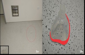
Post dural puncture headache was described as patient’s complaint of feeling bilateral throbbing headache experienced within 6-72 hours of administration of spinal anaesthesia, which improves on lying down. Criteria included history of spinal anaesthesia, duration, frontal or occipital pain, aggravated by standing or movement and relieved on lying down.

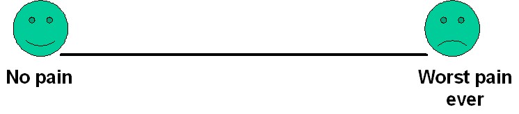
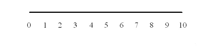
The Visual Analogue Scale used as tool to assess the severity of pain. The following diagram was printed on an A4 sheet ensuring that the lines are exactly 10 cm in length. The print out was then folded at the dotted line. Patient was asked to mark the line according to his/ her pain by moving from no pain to worst pain without showing the numbered side. After that the VAS score was measured by unfolding the numbered side and recording the corresponding score.

image 1


image 2

PDPH was measured in terms of analogue scale

Pain score: Mild 1-3, moderate 3-6, severe 6-9, unbearable 10

Statistical analysis: All the collected data was entered into SPSS version 16. Quantitative variables i.e. age and VAS score were presented by mean ±SD. Qualitative variable i.e. post dural puncture headache was presented as frequency and percentage. Chi-square test was applied for comparison of post dural puncture headache in both groups. A p value of ≤0.05 was taken as statistically significant.

RESULTS

In our study, total 120 female patients, who met the inclusion criteria without falling into any of the exclusion criteria, were included. They were randomly allocated to one of the two groups (Group A & Group B of 60 patients each) by draw (lottery) method. None of the patients were dropped out or lost from the study at any stage.

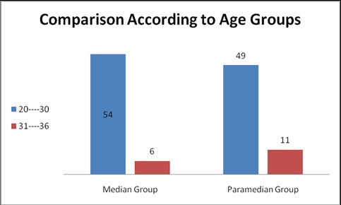
Patients in both groups were similar regarding age distribution. In Group A, (median approach), there were 54 patients (90%) in age group 20-30 years & 06 patients (10%) in age group 31-36 years. In Group B, (paramedian approach), there were 49 patients (81.7%) in age group 20-30 years & 11 patients (18.3%) in age group 31-36 years. Statistically there was insignificant difference between the age of the patients among the two study groups i.e. p-value=0.302 (Figure 1)

The mean age of the total patients were noted as 26.92 ± 4.02 years with minimum age of 20 and maximum age of 36 years. In Group A, mean age of patients were noted as 26.82 ± 4.52 years with 20 & 36 years minimum and maximum ages respectively. In Group B, the mean age of the patients were noted as 26.77 ± 4.13 years with 20 & 36 years minimum and maximum ages respectively.

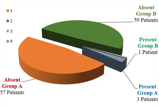
Out of total 120 patients, included in the study, 4 patients presented with PDPH. The overall frequency of PDPH in all patients under study was 3.33%. In Group A (Median group), 3 patients (out of 60) developed PDPH. Frequency of PDPH in Group A was 0.05 (5%). In this group 1st patient was of 20 years old, primipara,, developed PDPH within 24 hours that was of moderate degree, 2nd patient was 24 years old P2+0, developed PDPH of moderate degree after 24 hours and 3rd patient was 27 years old, P3+0 developed PDPH after 24 hours which was of mild degree.

In Group B (Paramedian group), only 1 patient (out of 60) developed PDPH. Frequency of PDPH in Group B was 0.0167 (1.67%). She was 25 years old, para 2+0, developed PDPH after 24 hours, which was moderate in degree.

In mild case, patient was treated by rest, plenty of fluids and simple analgesics containing caffeine. In moderate cases, patients were treated by bed rest, plenty of fluids, strong analgesics, and caffeine and muscle relaxants. Caffeine was given in the form of tablets, tea and coffee. In all patients, PDPH was relieved within 2-3 days without any complication.

PDPH was absent in 116 (96.67%) patients in which 57 (95%) patients belonged to Group A and 59 (98.33%) patients belonged to Group B. Statistically there was insignificant difference between the study groups i.e. p-value=0.30 (Table 1; Figure 2).The mean VAS of the total patients were noted as 2.54 ± 1.187 with minimum score of 1 and maximum score of 06 (Table 2).

Comparison According to Ages-figure-1

Figure 1: Comparison According to Ages (Group A Vs. Group B)

Table 1: Comparison of PDPH between study groups

PDPH Group Total
Group A Group B
Present 3(5%) 1(1.67%) 4(3.33%)
Absent 57(95%) 59(98.33%) 116(96.67%)
Total 60(100%) 60(100%) 120(100%)

Chi-square= 1.034; p -value= 0.30 (insignificant)

p -value= 0.24(insignificant)/Fischer exact test

Group A= Median Group; Group B=Paramedian Group

Figure 2: PDPH in Group A vs. Group B

PDPH in Group A vs. Group B-figure-2

Table 2: Descriptive statistics of VAS of the patients

VAS Total 120
Mean 2.54
SD 1.187
Minimum 01
Maximum 06

VAS = Visual analogue score

DISCUSSION

The results of our study showed that frequency of PDPH was less with the use of paramedian approach as compared to median approach, using 25 gauge pencil point needle in patients undergoing elective cesarean section under spinal anesthesia; but unfortunately the difference in the frequency of PDPH between the two groups was statistically insignificant. Group A patients had received spinal anesthesia with median approach while Group B patients received spinal anesthesia with paramedian approach. In Group A, 3 (out of 60) patients developed PDPH and the frequency of PDPH was 0.05 (5%). In Group B, 1 (out of 60) patients presented with PDPH and the frequency of PDPH was 0.0167 (1.67%). Though apparently there was reduction in the incidence of PDPH with the use of paramedian approach, the difference in the frequency of PDPH between the two groups was statistically insignificant i.e. p-value=0.30.

Some studies do favor our results whereas some do not.

Haider et al. on 50 patients undergoing different elective surgeries under spinal anesthesia found a statistically significant difference in the incidence of PDPH with median and paramedian approaches. They concluded that the paramedian approach using the Quincke level needle reduces the incidence of PDPH significantly.9

Mosaffa et al. concluded that there is no difference in PDPH incidence with median versus paramedian approaches, and therefore recommend the paramedian approach, especially for older patients with degenerative changes in the spine and intervertebral spaces, and those who cannot assume the proper position for the median approach; the easier positioning would result in less pain for the patient and a higher success rate for spinal anesthesia.11

Sadeghi et al. conducted a randomized double blind clinical trial of 125 patients scheduled for elective cesarean section who received spinal anesthesia with median or paramedian approach. Headache was evaluated for three days following surgery. The incidence of headache was 9.8% in paramedian group versus 9.4% in median group (p>0.05). The authors concluded that the use of paramedian approach in pregnant women who have difficulty in positioning is acceptable and without increasing risk of headache and hemodynamic changes.12

Janick et al. on 250 patients undergoing transurethral prostate surgery under spinal thesia reported a significantly higher rate of PDPH with the paramedian approach than with the median approach in relatively older patients, while no significant difference was observed in younger patients.13

Li JY et al. compared the technical difficulty and the incidence of post dural puncture headache (PDPH) between two approaches of spinal anesthesia i.e. median and paramedian approaches. Cesarean section was performed in 700 women under spinal thesia with either median or paramedian approach. It revealed that median approach was associated with a significantly greater success rate in the first attempt (231 of 350 patients) than paramedian approach. (205 of 350 patients) (p<0.05). The incidences of PDPH between median and paramedian approaches after single dural puncture is 4.33% (10 of 231 patients) and 0.97% (2 of 205 patients), respectively. They concluded that paramedian approach might significantly reduce the incidence of PDPH but it would need a more skillful hand to increase the successful rate.14

Muranaka et al.“ compared midline approach with paramedian approach for combined spinal-epidural thesia(CSEA) by needle through needle technique.70 patients undergoing elective gynecological surgery received CSEA with a 27 G Whitacre spinal needle, which protrudes 12 mm beyond the tip of the Tuohy needle. They concluded that the choice of midline or paramedian approach for CSEA did not affect the success rate of the subarachnoid puncture, but paramedian approach required longer protrusion length of the spinal needle than midline approach. To raise the success rate of subarachnoid puncture by paramedian approach, they recommended a long protruded spinal needle to raise the success rate by paramedian approach.15

Kumar and Mehta reported three cases in which patients with Ankylosing Spondylitis were successfully administered spinal thesia using a paramedian approach after failed attempts with a median approach.119 Nevertheless, Schelew et al., suggest that both; midline and paramedian methods may be attempted with success.16

Sivrikaya et al. study described that spinal anesthesia was successfully administered by the median approach in the lateral position on the first attempt, but preceded by two failed attempts by the median approach in the sitting position.17

Morgan et al. described that the median approach involves passage of needle through the supraspinal and interspinal ligaments and the ligamentum flavum, but the paramedian approach avoids the supra and interspinal ligaments and approaches the ligamentum flavum directly after passing through the para spinal muscles.18

Zhurda, Ahmed, & Ahsan-ul-Haq, in their studies concluded that paramedian approach would be an easier method of spinal anesthesia especially for older patients, who had sclerosed ligaments and degenerative changes in the spine and intervertebral spaces.19,20

Since female gender & pregnancy are already well known risk factors for PDPH, we specifically conducted our study only on obstetric (pregnant) female patients undergoing cesarean section under spinal anesthesia to exclude any confounding element between the two study groups.

In our study, we used 25-guage pencil-point (Whitacre) needles, not Quincke needles, because pencil-point needles are known to cause less PDPH than Quincke needles. Moreover, this is the type of needle are now routinely available for performing spinal anesthesia in our institute. Hence, our thetist have become used to handle these needles, without causing many complications. Another reason for conducting this study with 25-guage pencil-point needle is that only limited research was available on this topic particularly with 25 gauge pencil point needle. That is why, we used 25-guage pencil point needle to perform spinal anesthesia in all patients of both study groups.

The main limitation of our study is a smaller sample size due to which we couldn’t get statistically significant results. In fact, the results of our study have merely increased the need for conducting more studies with larger sample size to establish whether median or paramedian approach is better in reducing PDPH.

CONCLUSION

It is concluded through results of this study that paramedian approach is apparently better than median approach in terms of reducing frequency of PDPH in patients undergoing elective cesarean section under spinal anesthesia, though the results were clinically insignificant.

It is, therefore, recommended that more clinical trials with larger sample size are conducted so as to get statistically significant results to establish whether median or paramedian approach is better in reducing PDPH in patients undergoing cesarean section under spinal anesthesia.

Conflict of interest: Nil

REFERENCES

  1. Morgan Jr GE, Mikhail MS, Murray MJ. Clinical Anesthesiology. New York: McGraw-Hill Medical; 2013.
  2. Luck JF, Fettes PD, Wildsmith JA. Spinal anaesthesia for elective surgery: a comparison of hyperbaric solutions of racemic bupivacaine, levobupivacaine, and ropivacaine. Br J Anaesth 2008; 101:705-710. doi: 10.1093/bja/aen250. [PubMed] [Free full text]
  3. Brull R, MacFarlane AJR, Chan VWS. Spinal, epidural, and caudal anesthesia. In: Miller RD, ed. Miller’s Anesthesia. 8th ed. Philadelphia, (PA): Elsevier Saunders; 2015:chap 56.
  4. Kettner SC, Marhofer P Willschke H. Does regional anaesthesia really improve outcome? Br J Anaesth. 2011 Dec;107 Suppl 1:i90-5. doi: 10.1093/bja/aer340.doi: 10.1093/bja/aer340. [PubMed] [Free full text]
  1. Ali ZH. Effect of pre and post nursing intervention on the occurrence of tension headache among surgical patients undergoing spinal anesthesia. J Anesth Clin Res 2012;3:7-11.[Free full text]
  2. Hamzei A, Basiri-Moghadam M, Pasban-Noghabi S. Effect of dexamethasone on incidence of headache after spinal anesthesia in cesarean section. A single blind randomized controlled trial. Saudi Med J. 2012 Sep;33(9):948-53. [PubMed]
  3. Bezov D, Ashina S, Lipton R. Post-dural puncture headache: Part II–prevention, management, and prognosis. Headache. 2010 Oct;50(9):1482-98. doi: 10.1111/j.1526-4610.2010.01758.x. [PubMed]
  4. Ankorn C, Casey WF. Spinal Anaesthesia – a practical guide. Update in Anaesthesia. 2000; 12: 21-34 [Free full text]
  5. Haider S, Butt KJ, Aziz M, Qasim M. Post Dural Puncture Headache-A Comparison Of Midline And Paramedian Approaches. Biomedica. 2005;21:90–2.
  6. Ghaleb A. Postdural puncture headache. Anesthesiol Res Pract. 2010;2010. pii: 102967. doi: 10.1155/2010/102967. [PubMed] [Free full text]
  7. Mosaffa F, Karimi K, Madadi F, Hasan KS, Besheli LD, Eajazi A. post dural puncture headache: a comparison between median and paramedian approaches in orthopedic patients. Anesth Pain Med. 2011 Fall;1(2):66-9. doi: 10.5812/kowsar.22287523.2159.[PubMed] [Free full text]
  8. Sadeghi A, Razavi SS, Gachkar L, Ariana P, Ghahremani M. Comparison of the incidence of post spinal headache following median and para median approach in cesarean patients. J Iranian Soc. Anesthesiol Intens Care 2009;31:4-9.
  9. Janik R, Dick W. Post spinal headache. Its incidence following the median and paramedian techniques. Anesthetist 1992;41:137-41. [PubMed]
  10. Li JY, Tsai SC, Wang CH, Hui YL, Tan PC. Paramedian Approach Reduce the Incidence of post dural puncture headache. Chinese J. Pain 1995;5:71-76.
  11. Muranaka K, Mizutani H, Seo K, Yoshida M, Gohara T, Miyawaki H. A comparison between midline and para median approaches for combined spinal-epidural anaesthesia. Masui 2001;50:1085-8. [PubMed]
  12. Kumar CM, Mehta M. Ankylosing spondylitis: lateral approach to spinal anaesthesia for lower limb surgery. Can J. Anesth 1995;42:73-6. [PubMed]
  13. Sivrikaya GU, Hanci A, Dobrucali H, Yalcinkaya A. Cesarean section under spinal anaesthesia in a patient with ankylosing spondylitis. Middle East J Anesthesiol 2010;20:865-8. [PubMed]
  14. Morgan G, Mikhail M, Murray M, Larson Jr M. Anesthesia for orthopedic surgery. In: Morgan GE, Mikhail MS Clinical Anesthesiology 2nd ed. Stamford, Conn: Appleton & Lange 1996; p. 675-7.
  15. Zhurda T. Kërçi M, Bajraktari M, Muzha D, Kurti B. The advantages of para median approach for spinal anesthesia of elderly patients in urology surgery .Eur J Anaesthesiol 2010;47:130. [Free full text]
  16. Ahsan-ul-Haq M, Amin S, Javaid S. Paramedian technique of spinal anesthesia in elderly patients for hip fracture surgery. J Coll Physicians Surg Pak. 2005;15(3):160–1 [PubMed]

A prospective, cross-sectional survey of pre-operative fasting of pediatric surgical patients in a university hospital

$
0
0

Nouman I. Alvi, MRCA, FCARCSI

Assistant Professor, Department of Anesthesia & Pain Medicine, Aga Khan University Hospital, Karachi (Pakistan)

Correspondence: Dr Nouman I. Alvi, Department of Anesthesia & Pain Medicine, Aga Khan University Hospital, Karachi (Pakistan); E-mail: nouman.alvi@aku.edu

ABSTRACT

Background: Fasting of children before anesthesia is mandatory but blighted with logistical issues. There may be inadvertent prolonged fasting due to lack of individualized fasting plans for children coming for surgeries. We aimed to have a survey of state of preoperative fasting in our pediatric patients.

Methodology: A questionnaire based, prospective, cross-sectional survey was conducted in our Department for one month. A total of 102 children, up to age of 16 scheduled for pediatric surgery were included in the data collection. The questionnaires were to be filled either by anesthesia consultant or trainees. The information related to duration of fasting and any extra oral hydration, was attained from patients’ parents/guardians. The duration of fasting was then compared with the recommended one.

Results: In this study only 4% of children could be labelled as having the optimum fasting. Based on the current guidelines, in 96% of children, the guidelines were not followed.

Conclusion: Pediatric patients are being subjected to prolonged durations of fasting prior to anesthesia and surgery against the accepted norms and the guidelines.

Key words: Fasting, Duration; Hydration; Pediatric

Citation: Alvi NI. A prospective, cross-sectional survey of pre-operative fasting of pediatric surgical patients in a university hospital. Anaesth Pain & Intensive Care 2016;20(2):171-175

Received: 16 February 2016; Reviewed: 30 May 2016; Corrected: 31 May 2016; Accepted: 01 June 2016

INTRODUCTION

Fasting in children is a pre-requisite before anesthesia. To comply with optimum pre-anesthetic fasting in pediatric age group is a big challenge.1 Fasting duration is decided keeping the age and demands of children.1,2,3 However, it has been commonly observed that children are kept hungry, thirsty for periods which are often longer than the recommended ones.1,2,3

One has to weigh the risk of aspiration at the cost of dehydration, hypoglycemia, anxiety and crying due to excessive fasting durations.2 Unpredictable situations, changes in list order and queue jumping due to emergency cases further compound the dilemma. The available literature suggests that the utility of six hours fasting in pediatric population is not conclusively proven.3 There are studies which suggest that there is no increased risk of aspiration even if total fasting has been for only two or three hours.3,4

We follow international guidelines3, 23,25-28 which are taken as standard. Based upon previous departmental guidelines, booklets from other hospitals and anecdotal experience, our department has determined that the gold standard or standard practice of fasting was minimum six hours after solid foods, milk and greasy meals and a four hour fasting requirement for juices and syrupy fluids. The minimum fasting duration for clear liquids is two hours.

The objective of this survey was to measure and document the actual fasting times of children coming for anesthesia in our department and then compare the results with gold standards of this practice internationally.

METHODOLOGY

This prospective, cross-sectional survey was conducted on children enlisted for anesthesia for pediatric surgery at Aga Khan University Hospital Karachi (Pakistan). A questionnaire was designed with both English and University approved Urdu translation. A consent form in English and Urdu was used for patients and/or guardians. The study was formally approved by the Institutional Ethical Review Committee. Mode of sampling was consecutive non-purposive sampling method. The survey was scheduled to be conducted over a one month period. The sample size was calculated from the mean average, derived of anesthetic surgeries conducted over the past seven years in our hospital. Hospital network based software ‘SAHL’ was used to record the total number of patients during this period. The total elective cases since 2006 were found to be 7511 over 84 months; thus, giving a sample size of 90 as reflective of our typical monthly case load. Since the survey was planned for one month this number of cases was chosen. The inclusion criteria were as follows: neonates, infants and children up to an age of 16 years, scheduled for elective surgery. The patients outside this age group and coming for emergency surgery were excluded. The questionnaires were to be filled either by an anesthesia consultant or a trainee. The process entailed interviewing prospective patients/patient’s guardians in the reception area before the anesthesia. They were asked about their fasting duration; and any hydration through an extra oral route. The actual duration of fasting was compared against the recommended one and results were collated. Demographic data was obtained from the confidential folder. Results were collated using SPSS vs.19 (SPSS Inc. Chicago, IL).

RESULTS

A total of 102 responses were evaluated. The median age of the children was 24 (IQR= 28) months [Range: 2 to 120 months]. There were 90 patients of 102, who were eligible for solid intake. The fasting timing of these children was evaluated and the median solid fasting time was 12.0 (IQR =7) hours [range 6-48 hour]. Out of 90 cases, 4 (4.4%) children fasted solids up to 6 hrs, 34 (37.8%) fasted from 6 hrs to 10 hours and 52 (57.8%) had fasted more than 10 hrs (Table 1). Most of the children were fasted for more than 6 to 10 hrs, and more than 10 hrs in <3 yrs age groups.

Table 1: Fasting of Solids by children (with respect to age groups, n=90)

Hours since last solid Age Groups (Years) Total

n=90

≤ 1Years

n=20

>1 to 3 Years

n=44

>3Years

n=26

Up to 6h 0(0%) 0(0%) 4(15.4%) 4(4.4%)
>6 to 10h 8(40%) 16(36.4%) 10(38.5%) 34(37.8%)
>10hours 12(60%) 28(63.6%) 12(46.2%) 52(57.8%)

Total cases 102; 12 children had not weaned to solids so they were excluded.

The median fasting duration for juices/syrupy liquids was 8 hrs [range 4-24 hrs] (IQR =5). There were only 2 (2.9%) children who fasted sugary liquids up to 4 hrs, 12 (17.1%) children had fasted for 4 – 6 hrs and 56 (80%) fasted >6 hrs (Table 2).

Table 2: Fasting of sugary and syrupy liquids and semi solids by children (with respect to age groups, n=70)

Hours since last Age Groups (Years) Total

n=70

≤ 1Yrs

n=26

1 – 3 Yrs

n=30

>3 Yrs

n=14

Up to 4h 2(7.7%) 0(0%) 0(0%) 2(2.9%)
> 4 to 6h 8(30.8%) 4(13.3%) 0(0%) 12(17.1%)
> 6h 16(61.5%) 26(86.7%) 14(100%) 56(80%)

Total cases 102; 70 children were reportedly able to tolerate semi solids and 32 were not

Out of 102 responses, 98 children (96.1%) had taken clear liquids for a variable period of time. The median fasting time for clear liquids was 9 hrs (IQR=5) [range: 1 – 48 hrs]. Out of 98 children, 2 (2.9%) had fasted for clear liquids up to the recommended fasting time (2 hrs), while 14 (14.3%) had fasted for 2 – 6 hrs and 82 (83.7%) for >6 hrs.

Table 3: Fasting of clear liquids by the children with respect to age groups (n=98)

Hours since last clear liquids


Age Groups (Years) Total

n=98

≤ 1Years

n=28

1 to 3 Years

n=44

>3Years

n=26

Up to 2h 2(7.1%) 0(0%) 0(0%) 2(2.9%)
>2 to 6h 2(7.1%) 6(13.6%) 6(23.1%) 14(14.3%)
>6h 24(85.7%) 38(86.4%) 20(76.9%) 82(83.7%)

Total cases 102; 98 children were reportedly able to take liquid and 4 were not taken liquid

D

Four children had intravenous hydration. 2 of these were under 22 months and had no solids for the last 48 hrs, and two were < 8 months and had no liquids for 8 hrs.

In this survey only 4% of children had had the optimum fasting (p < 0.002) (Table 4).

Table 4: Optimum fasting. Data given as N (%).

Age Groups (Years) Optimum fasting P-value
Achieved Not achieved
≤ 1 0 30 (100) <0.002
>1 to 3 0 46 (100)
>3 4 (15.4) 22 (84.6)
Total 4 (3.9) 98 (96.1)

DISCUSSION

An overwhelming majority of our patients were fasting considerably longer than the recommended duration of fasting for anesthesia and surgery. The proportion of children who have inordinate or suboptimal fasting was very high i.e. 96% of the surveyed children. The only group of children which complied favorably with optimum guidelines was aged above 36 months.

More than half of the children had fasted for more than ten hours. While this was somewhat understandable (given the logistical issues) but not justifiable i.e. 83.7% of the children were still fasting for more than six hours for clear liquids. The recommended suggestion for that is two hours.

In Pakistan, perioperative hydration and fasting before anesthesia is sparsely probed topic.6,7. There is scarce literature on fasting in children undergoing surgery relevant specifically to Pakistan. One of the studies done at major teaching hospitals of Karachi displayed that Day case Surgery was the majority and preferred mode of surgery in pediatric age group. Nearly 8% of children undergoing such surgeries had issues with ‘feeding within the first 24 hours’ and nearly 14% children had vomiting (more than two episodes with 24 hours) post operatively.7 In light of these previous findings and our own survey; it can be extrapolated that children coming for anesthesia and surgeries are at very high risk for iatrogenic dehydration and associated complications e.g. renal failure, mental state changes, collapse, tachycardia, starvation etc.

In another study from Pakistan done in the early nineties, the mean fasting period in children was found to be 10.87 ± 2.68 hours.6. This study showed that the mean pre-anesthetic glucose levels were 4.8± 0.8 mmol/L. The glucose level though found to be within normal range but was only marginally normal and the most probable etiology would have been the prolonged fasting duration. Clearly the fasting durations (in that study) were much longer than the recommended periods. Our survey has found that the fasting durations are still very high, something which has unfortunately not changed in a long time.

As a result of this study we were able to have a snapshot of the actual prevalent status of pre-operative fasting in children. Interestingly the findings are very identical from published audits and audits10-13 from many other countries with similar socio economic conditions. The condition is not limited to developing countries; children have been found to be fasting for very long periods before surgeries, even in developed western countries.14 Several audits,10-13 which looked at actual fasting times in children’s hospitals, found that children had inadvertently fasted for unacceptably prolonged periods.

It is worth adding that this is an on-going problem in Anesthesia departments in many parts of the world14. There are multiple factors involved and some studies have suggested continuous programs as the tangible solution to this recurring problem. In this respect our findings are not vastly different from earlier studies in other countries with similar backgrounds or in developed countries.

At our institution we observe the old principle of 2:4:6, which inadvertently leads to prolonged fasting. Surveys and regional audits12-14 have shown that the tricky balance between recommended fasting durations (2:4:6) and risk of pulmonary aspirations has inadvertently made people err on the side of caution. This would explain why despite reasonable evidence, anesthesiologists still demand 2:4:6 rules for fasting in children. Although, the classical dogma of at least six hour full fasting is increasingly being challenged.1,3,7,15,16 This has been so because of observations made of residual gastric volume. Intake of clear fluids up to two hours prior to surgery does not negatively impact gastric residual volume of the gastric Ph.15,16 This findings underpins the new drive to stop the conventional six hour fasting.4

In our survey nearly 12% of the children were found to have on-going intravenous hydration. While IV hydration is better than nothing, but it carries the risk of: hyponatremia5, over hydration and electrolyte imbalances.8,9,10 Another area for future research which has been highlighted is, parents’ perceptions about fasting for their children. It has been observed in studies that parental educational was an important factor in prolonged fasting in children.17,18

Other benefits of saving children from inadvertent prolonged fasting was protection from hypotension at induction and administration of dextrose containing fluids.18 Children are also found to be less irritable after anesthesia if they had oral liquids up to two hours before induction.20,21

One of the potential limitations of this survey was selection of a period of one month for carrying out this survey. We chose this sample size because this was a number very close to an estimated average for our monthly pediatric cases for the past six years. Due to logistical constraints we could not collect data from patients coming to remote anesthesia locations in the hospital premises e.g. MRI and radiology suite.

Evidently the situation is less than ideal but we will endeavor to improve this with multidisciplinary cooperation. It is hoped that with better coordination and shared consensus among teams, things will change for good.

CONCLUSION

Pediatric patients are being subjected to inappropriately prolonged durations of fasting prior to anesthesia despite presence of departmental guidelines. Medical and nursing staff need to be continuously educated regarding adherence to an optimum period of fasting for the child’s age.

Acknowledgments: I would like to offer my sincerest gratitude to Mr. Amir Raza, our departmental statistician, without whose help, I would have been unable to present data in a proper format. His suggestions have been precious and his presence indispensable.

Conflict of interest: None

REFERENCES

  1. Hanna AH, Mason LJ. Challenges in paediatric ambulatory anesthesia. Curr Opin Anaesthesiol 2012; 25:315-20. doi: 10.1097/ACO.0b013e3283530de1. [PubMed]
  2. Veall GR, Floor K, Dorman T. Prolonged starvation in paediatric surgery. Anaesthesia. 1995; 50:458–60. [PubMed]
  3. Brady M, Kinn S, Ness V, O’Rourke K, Randhawa N, Stuart P. Preoperative fasting for preventing perioperative complications in children. Cochrane Database Syst Rev. 2009 Oct 7;(4):CD005285. doi: 10.1002/14651858 [PubMed]
  4. Cook-Sather SD, Harris KA, Chiavacci R, Gallagher PR, Schreiner MS. A liberalized fasting guideline for formula-fed infants does not increase average gastric fluid volume before elective surgery. Anesth Analg. 2003 Apr;96(4):965-9, table of contents. [PubMed]
  5. Reducing the risk of hyponatraemia when administering intravenous infusions to children. NPSA 2007; 22:1-10. [PubMed]
  6. Mandhan P, Shah A, Khan AW, Muniruddin, Hasan N. Outpatient pediatric surgery in a developing country. J Pak Med Assoc 2000; 50:220-4. [PubMed] [Free full text]
  7. Shah M, Zahoorullah, Haq TU, Akhtar T. The effect of pre-anaesthetic fasting on blood glucose level in children undergoing surgery. J Pak Med Assoc. 1990; 40:243-5. [PubMed]
  8. Kannan L, Lodha R, Vivekanandhan S, Bagga A, Kabra SK, Kabra M. Intravenous fluid regimen and hyponatraemia among children: a randomized controlled trial. Pediatr Nephrol. 2010; 25:2303–9. doi: 10.1007/s00467-010-1600-4. [PubMed]
  9. American Society of Anesthesiologists committee on standards and practice parameters. Practice guidelines for preoperative fasting and the use of pharmacologic agents to reduce the risk of pulmonary aspiration: Application to healthy patients undergoing elective procedures. An updated report. Anesthesiology. 2011;114:495–511. [PubMed]
  10. Saqr L, Chambers WA. Preventing excessive pre-operative fasting: national guideline or local protocol? . Anaesthesia, 61: 1–3. [PubMed]
  11. Arun BG, Korula G. Preoperative fasting in children: An audit and its implications in a tertiary care hospital. J Anaesthesiol Clin Pharmacol. 2013 Jan-Mar; 29(1): 88–91. doi: 10.4103/0970-9185.105810. [PubMed] [Free full text]
  12. Adudu OP, Egwakhide EOA, Adudu OG. Parents and patients’ compliance to revised preoperative fasting guidelines in Benin, Nigeria. Paediatr Anaesth. 2008 Oct;18(10):1013-4. doi: 10.1111/j.1460-9592.2008.02607.x.[PubMed]
  13. Pollach G, Kapenda R, Anusa B, Waluza E, Namboya F. Excessive fasting times: still an underaddressed challenge for African pediatrics and anesthesia? J Ped H Med Ther. April 2014;5:9-13. [Free full text]
  14. Emerson BM, Wrigley SR, Newton M. Pre-operative fasting for paediatric anaesthesia A survey of current practice. Anaesthesia. 1998 Apr;53(4):326-30. [PubMed] [Free full text]
  15. Splinter WM, Stewart JA, Muir JG. Large volumes of apple juice preoperatively do not affect gastric fluid pH and volume in children. Can J Anaesth 1990; 37:36–9. [PubMed]
  16. Splinter WM, Schaefer JD, Zunder IH. Clear fluids three hours before surgery does not affect the gastric fluid contents of children. Can J Anaesth 1990; 37:498–501. [PubMed]
  17. Bates J.Reducing fast times inpaediatricday surgery. Nurs Times. 1994 Nov 30-Dec 6;90(48):38-9. [PubMed]
  18. Klemetti S, Suominen T. Fasting in paediatric ambulatory surgery. Int J Nurs. Pract 2008; 14:47-56. doi: 10.1111/j.1440-172X.2007.00666.x. [PubMed]
  19. Murat I, Dubois MC. Perioperative fluid therapy in pediatrics. Paediatr Anaesth 2008; 18:363-70. doi: 10.1111/j.1460-9592.2008.02505.x [PubMed]
  20. Klemetti S, Kinnunen I, Suominen T, Antila H, Vahlberg T, Grenman R, et al. The effect of preoperative fasting on postoperative pain, nausea and vomiting in pediatric ambulatory tonsillectomy. Int J Pediatr Otorhinolaryngol 2009; 73:263-73. doi: 10.1016/j.ijporl.2008.10.014. [PubMed]
  21. Radke OC, Biedler A, Kolodzie K, Cakmakkaya OS, Silomon M, Apfel CC. The effect of postoperative fasting on vomiting in children and their assessment of pain. Paediatr Anaesth 2009; 19:494-9. doi: 10.1111/j.1460-9592.2009.02974.x. [PubMed]
  22. Engelhardt T, Wilson G, Horne L, Weiss M, Schmitz A. Are you hungry? Are you thirsty?–fasting times in elective outpatient pediatric patients. Paediatr Anaesth 2011; 21:964-8. doi: 10.1111/j.1460-9592.2011.03573.x [PubMed]
  23. Cook-Sather SD, Litman RS. Modern fasting guidelines in children. Best Pract Res Clin Anaesthesiol. 2006; 20:471-81. [PubMed]
  24. Phillips S, Daborn AK, Hatch DJ. Preoperative fasting for paediatric anaesthesia. Br J Anaesth1994; 73:529–36. [PubMed] [Free full text]
  25. Smith I, Kranke P, Murat I, Smith AF, O’Sullivan G, Søreide E, et al. Perioperative Fasting in Adults and Children: Guidelines from the European Society of Anaesthesiology. Eur J Anaesthesiol. 2011 Aug;28(8):556-69. doi: 10.1097/EJA.0b013e3283495ba1. [PubMed]
  26. American Academy of Pediatrics; American Academy of Pediatric Dentistry, Coté CJ, Wilson S; Work Group on Sedation. Guidelines for monitoring and management of pediatric patients during and after sedation for diagnostic and therapeutic procedures: an update. Pediatrics. 2006 Dec;118(6):2587-602. [PubMed] [Free full text]
  27. Pediatric Anesthesia NPO Guidelines [Internet]. Icahn School of Medicine at Mount Sinai. [Accessed June 1, 2016]. Available from:http://icahn.mssm.edu/departments-and-institutes/anesthesiology/divisions/pediatric-anesthesia/npo-guidelines
  28. Preparing for Surgery: Adolescents | Johns Hopkins Children’s Center [Internet]. [Accessed June 1, 2016]. Available from:http://www.hopkinsmedicine.org/johns-hopkins-childrens-center/patients-and-families/your-visit/abcs-surgery/adolescents.html

Comparison of i-gel™, LMA Proseal™ and LMA Classic™ in spontaneously breathing pediatric patients

$
0
0

Yogita Dwivedi, MD1, Amrita Gupta2, Uma Srivastava3, Keshav Dev Jagar4, Atiharsh Mohan2, Sandeep Mangla4
1Assistant Professor; 2Lecturer; 3Professor; 4Senior Resident
Sarojini Naidu Medical College, Mahatma Gandhi Road, Agra, Uttar Pradesh, (India)
Correspondence: Dr. Amrita Gupta, Lecturer, Sarojini Naidu Medical College, Mahatma Gandhi Road, Agra, Uttar Pradesh, (India); Mob: 09837077784; E-mail: amritagupta78@gmail.com

ABSTRACT
Introduction: LMA Proseal™ is considered the premier supraglottic airway device in children. I-gel circumvents the cuff related problems of second generation devices as its seal is made of thermoplastic elastomer. Its potential advantages include easy insertion, minimal tissue compression and good stability. We planned this study to assess the clinical performance of i-gel, LMA Proseal™ and LMA classic in children breathing spontaneously.

Methodology: 90 patients of ASA grade I and II, weighing between 10-25 kg, posted for elective surgery with a duration of less than 2 hrs, were randomly divided into three groups (30 each). Standard general anesthesia was administered to all children. Ease of insertion of the device and nasogastric tube, oropharyngeal seal pressure, hemodynamic parameters and intra- and postoperative complications were noted.

Results: The patients were comparable with respect to demographic data. Insertion was assessed as very easy in all three groups. Success rate of insertion in first attempt was >90% in each group. I-gel showed shortest mean time for insertion (16 ± 4 seconds). I-gel had highest seal pressure (25.2 ± 2.8). followed by LMA Proseal™ (22.6 ± 2.8) and Classic LMA (16.8 ± 2.6).

Conclusion: I-gel is comparable to LMA Proseal™ and Classic LMA in clinical performance. I-gel had highest oropharyngeal seal pressure and required least time for insertion. Therefore, it can be reliably used in pediatric anesthesia.

Key words: Supraglottic devices; Anesthesia, Pediatric; Ventilation; Airway; Airway management

Citation: Dwivedi Y, Gupta A, Srivastava U, Jagar KD, Mohan A, Mangla S. Comparison of i-gel™, LMA Proseal™ and LMA Classic™ in spontaneously breathing pediatric patients. Anaesth Pain & Intensive Care 2016;20(2):176-181

Received: 4 March 2016; Reviewed: 12, 30 March 2016; Corrected: 31 March, 6 June 2016; Accepted: 6 June 2016

INTRODUCTION

Introduction of new supraglottic devices (SGD’s) has changed the era of airway management in children due to ease of insertion, easy learning curve and ease to ventilate at peak airway pressure without gastric distention. Previous randomised studies1,2 have established the safety of Proseal LMA (PLMA) in children. It is now considered premier SGD in children and has set a bench mark in second generation devices. I-gel circumvents the cuff related problems of above devices as its anatomic seal is made of thermoplastic elastomer. The airway seal improves as it slowly adapts to the temperature of body. Its elliptical shape minimise axial rotation and improves stability.3,4,5 Despite its advantages there are a few studies comparing Classic LMA (CLMA) and PLMA with i-gel in children.
The aim of our study was to compare the clinical performance between CLMA, PLMA Proseal and i-gel in spontaneously ventilating children. Our primary objective was to compare oropharyngeal seal pressure and secondary objective was to compare insertion parameters, mean duration of insertion, gastric tube placement and haemodynamic parameters.

MATERIAL AND METHODS
After approval from the hospital proforma committee and obtaining written and informed consent from patients,90 patients with ASA grade 1 and 2 weighing between 10-25 kg were included in this randomized prospective study to compare i-gel, PLMA and CLMA in spontaneously breathing children undergoing elective surgery of less than 2 hours duration (colostomy closure, herniotomy, circumcision, cataracts, upper and lower limb surgeries etc.) performed under general anesthesia in our department during the time period of January 2013 to August 2014.

Patients with irritable upper respiratory tract, risk of aspiration, trismus, limited mouth opening, were excluded from the study. Ninety patients were divided randomly into one of the three groups of 30 each by concealed chits. (Groups C, P and I)

A standard anesthesia protocol was followed for every case. All the children were kept nil per orally before surgery. After shifting the patient to operation theatre, standard monitors pulse oximeter, non-invasive blood pressure, 5 lead Electrocardiogram, EtCO2 was applied and baseline parameters was recorded. Patients were premedicated with inj glycopyrrolate 0.005 mg/kg, fentanyl 1-2 µg/kg and inj ondansetron 0.08 mg/kg before induction.

Anesthesia was induced with propofol 3 mg/kg IV along with sevoflurane in oxygen. Once an adequate depth of anesthesia was achieved, judged by loss of verbal contact, jaw relaxation and absence of movement on jaw thrust, the SGD was inserted with the standard routine technique (introducer for PLMA, single finger technique for CLMA and i-gel). PLMA of size 1.5 and 2 were used for patients weighing 5-10 and 10-20 kg respectively. I-gel size 1.5, 2 or 2.5 was used for patients weighing 5-12, 10-25 and 25-35 kg respectively. The device was inserted in sniffing position or a combination of maneuvers such as chin lift, jaw thrust, head extension and neck flexion as required. All the insertions were done by anesthesiologist with minimum one year of experience and who had inserted each of the device more than 25 times earlier. Insertion of device was recorded as very easy: when assistant help was not required), easy: when jaw thrust was needed by assistant, and difficult: when jaw thrust and deep rotation or second attempt was used for proper device insertion.
Once inserted into the pharynx, the cuff was inflated with air until effective ventilation was established or the maximum recommended inflation volume reached (60 cmH2O). Fixation of the devices was done according to the manufacturer’s instructions. Effective ventilation was judged by observation of chest wall movement, auscultation of bilateral breath sound and a square wave capnography trace. Anesthesia was maintained with 1-2% sevoflurane and 70% nitrous oxide in oxygen. Insertion time was noted from the time of picking of device and time to achievement of adequate airway as judged by adequate chest expansion, auscultation of bilateral breath sound and a square wave capnography trace. Number of insertion attempts was noted. Three attempts were allowed before insertion was considered a failure. If insertion failed, alternative device was used.

Once insertion was successful, the intra-cuff pressure was set at 60 cmH2O (for CLMA and PLMA) using a digital manometer (Mallinckrodt Medical, Ireland). This pressure was maintained throughout the surgery by regular cuff pressure monitoring. The oropharyngeal leak pressure was determined by closing the expiratory valve of the breathing system at a fixed gas flow of 3 lit/min and noting the airway pressure at which equilibrium was reached (maximum allowed, 40 cm H2O). At this point gas leak was heard from mouth, epigastrium. Manometric test was considered the most reliable test.

Any episodes of desaturation, coughing, bronchospasm or aspiration / regurgitation/ vomiting were documented. At the end of the procedure, the SGD was inspected for any blood stains, tongue-lip-dental trauma. Postoperative sore throat was noted.

The following hemodynamic parameters were recorded in all patients; heart rate (HR), mean arterial pressure (MAP) in mmHg, pulse oxygen saturation (SpO2) and EtCO2. Insertion of a nasogastric tube Fr 8-10 was attempted through the gastric port of PLMA and i-gel, before the commencement of surgery. Correct gastric tube placement was assessed by suction of fluid or detection of injected air by epigastric stethoscope. Three attempts were made before gastric tube insertion was considered a failure.

Statistical analysis; Statistical analysis was done, using SPSS software. To calculate sample size oropharyngeal sealing pressure was considered the primary variable with Type one error .05 and power of 0.8 considering a projected difference of 30% between the three groups. ANOVA test was used for demographic data (age, weight), oropharyngeal seal pressure (OSP) and hemodynamic data analysis. The insertion characteristics and complications were analyzed using Chi square test. Fischers test was used to analyze insertion attempts of gastric tube.

RESULTS

There was no statistical difference in demographic data between groups (Table I). No failure with insertion and gastric tube placement in all the three groups.
Table I: Demographic data

Parameter Group C
N= 30 Group I
N=30 Group P
N=30 Group C vs Group I
(P value) Group I vs Group P
(P value) Group P vs Group C
(P value)
AGE (YRS) 3.51 ± 1.37 3.13 ± 1.51 3.3 ± 1.46 0.947 1.000 1.000
WEIGHT (KG) 16.17 ± 2.98 15.67 ± 3.01 15.40 ± 3.42 0.520 0.747 0.356
(M/F) (14/16) (15/15) (13/17) NS NS NS

Majority of insertion attempts were very easy (83.3, 93.3 and 83.3 in Groups C, I and P respectively). Two difficult insertions were encountered in Group P and none in Group C and Insertion of device was successful on first attempt in 96% of patients and was comparable to Group P (93%) and Group C (91%). Mean insertion time of Group C, I, and P was 22.1 ± 4.4, 16 ± 4.1 and 19.4 ± 6.3 respectively. Group I showed the shortest time for insertion. In the present study mean oropharyngeal seal pressure in Group I (25 ± 2.3) was significantly higher than Group P (22 ± 6 .3) and Group C (16.8 ± 2.6). There was neither desaturation nor significant changes in blood staining and postoperative nausea and vomiting was observed in two cases in Group C and n
one in Group I and P (Table 2).

Table 2: Comparative data between Classic LMA, i-gel, LMA Proseal

Parameter Group C
N= 30 Group I
N=30 Group P
N=30 Group C vs Group I
(P value) Group I vs Group P
(P value) Group P vs Group C
(P value)
Ease of intubation
(VE / E / D / F) 25 / 5 / 0 / 0 28 / 2 / 0 / 0 25 / 3 / 2 / 0 .577 .732 .343
Mean insertion time (SECS) 22.13 ± 4.4 16 ± 4.11 19.4 ± 6.3 .000 .034 .128
Oropharyngeal seal pressure
(cmH2O) 16.8 ± 2.6 25.2 ± 2.8 22.6 ± 2.8 .000 .002 .000
Number of attempts
1st / 2nd / 3rd / failure 27 / 3 / 0 / 0 29 / 1 / 0 / 0 28 / 2 / 0 / 0 NS NS NS
Gastric tube placement
1st / 2nd / 3rd / failure – 27 / 2 / 1 / 0 28 / 1 / 1 / 0 – NS –
Nausea and vomiting 2 0 0 NS NS NS
Laryngospasm/bronchospasm 0 0 0 NS NS NS
Hypoxia(Oxygen Desaturation) 0 0 0 NS NS NS
Tongue-lip-dental trauma 1 0 0 NS NS NS
Blood staining of device 1 0 0 NS NS NS
Regurgitation 0 0 0 NS NS NS
Sore throat & hoarseness in PACU 0 0 0 NS NS NS
Comparative mean heart rate changes in all the three groups at different time intervals are given in Figure 1.

Figure 1: Showing comparative mean heart rate changes during various stages

Showing comparative mean heart rate changes -fgiure-1

Comparative mean arterial blood pressures in all the three groups at different time intervals are given in Figure 2.

Figure 2: Showing comparative mean arterial pressure during various stages

Showing comparative mean arterial pressure-figure-2

DISCUSSION

Oropharyngeal seal pressure is used to monitor airway seal which was the primary variable in the study. Pro seal LMA is better suited for pediatric airway than adults6 and its OSP is higher than CLMA. The mean oropharyngeal seal pressure of i-gel was 25.2 ± 2.8 cmH2O for size 1.5, 2.0, 2.5 which was significantly higher than PLMA 22.6 ± 2.8 (size 1.5, 2, 2.5). These results indicate that i-gel provides better seal than same sizes PLMA and CLMA (16.8 ± 2.6). Goldman et al1 reported OSP of 23 cmH2O for PLMA size 1.5, 2.5. Beylacq9 conducted observational study in children and reported an OSP of 25 cmH2O for i-gel in children. Similar results were obtained by Goyal et al10 who concluded that OSP of size 2 i-gel was 26 ± 2.6 cmH2O which was statistically higher than size 2 PLMA (23 ± 1.2 ). Similar results were obtained by Tokgoz11 and Das et all 12.

In patients with high airway pressure i-gel may provide a wide safety range for positive-pressure ventilation. The cuff of the size 1.5, 2 and 2.5 PLMAs differ from the adult sizes as they lack a dorsal cuff which plays a minor role in the improved seal13.Perhaps, wider proximal end and larger distal cuff helps in improved seal.

Shimbori et al found no significant difference in children and found OSP of PLMA 18 cmH2O and CLMA 19 cmH2O, whereas Krippacheril reported OSP of 23.1 and 23.26 cmH2O respectively for these devices.

The majority of insertion attempts were very easy in all three devices. There were two difficult insertions with PLMA. The placement of PLMA has been found to be more difficult in adults than children. The larger bowl of PLMA is more difficult to insert and is likely to fold over. The large tongue, floppy epiglottis, anterior larynx and presence of tonsillar hypertrophy makes PLMA difficult to insert in pediatric patients.15 I-gel and CLMA are easier to insert as they have small bowl and cuff size. Our results are in concurrence with previous studies.16
First attempt insertion rates were greater than 90% in all three groups. No patients required third attempt nor was there a failure to insert the device in any group. Our results are similar with previous studies.9,17,18,19 In manikin study using eight types of SGD overall success rate for insertion of i-gel was >90%.20
In our present study, the mean times for insertion in groups C, I and P were 22.13 ± 4.439 seconds, 16.03 ± 4.115 and 19.43 ± 6.393 seconds respectively. I-gel showed lshortest mean time for insertion. Shorter insertion times influence the feasibility of SGD for routine use.21 This can be attributed to the fact that I-gel has a more robust and streamlined design than the PLMA and LMA Classic, making it easier to hold and insert. Also it does not have a cuff so the time taken to inflate the cuff is saved in this device. This advantage might not have a marked clinical impact on routine elective surgeries but it definitely gains importance in situations of resuscitation and in difficult airway situations where achieving an effective airway quickly is of paramount importance.

Both i-gel™ and PLMA are more reliable than CLMA in terms of aspiration risk because they allow gastric drainage. Previous studies indicated that nasogastric tubes (N/G) of size 8-10 fr could be easily passed through the i-gel channel and gastric contents could be aspirated via the N/G.17,22 In present study success rate of gastric tube placement in 1st attempt was 90% and 93.33% for i-gel and PLMA respectively and the gastric tube was inserted in 100% of cases in 2nd attempt. This helps in preventing and decreasing air leak and thus decreasing postoperative nausea and vomiting. The successful placement of the gastric tube also aids in correct positioning of the PLMA.

Regarding the hemodynamic stability and effect of each of the SGDs, no statistically significant difference was reported when comparing heart rate and mean arterial blood pressure intraoperatively.23,24 Since placement of the CLMA and PLMA involves the inflation of the cuff in the hypopharynx, these are expected to produce a similar response. I-gel does not have an inflatable cuff, it has a cuff made of thermoplastic elastomer it still showed a similar response like the other two devices. As the receptors adapt to constant pressure on the pharyngeal wall these changes were expected to be transient as showed in the present study.

One of the most important parameters to be compared between three SGDs was perioperative complications. It was estimated that difference between CLMA, i-gel and PLMA regarding perioperative complications was not statistically significant except nausea / vomiting, and blood staining of device. Incidence of postoperative nausea vomiting was significantly higher in CLMA due to high incidence of gastric insufflation. There was no incidence of sore throat in any group. This observation of our study is supported by the study of Wong et al25 where they stated that if the intracuff pressure remains less than 60 cmH2O there is minimal chance of sore throat. None of the children had laryngospasm/bronchospasm, hypoxia, tongue-lip-dental trauma and sore throat and hoarseness in post anesthesia care unit in the present study.

LIMITATIONS

Our study has few limitations that need discussion. We included children with normal airway. Therefore, the results of this study cannot be extrapolated to patients with difficult airway. Although group assignment was random but the person collecting the data was not blind to study groups. Therefore an observer’s bias can exist.

CONCLUSION
Based on these finding we conclude that i-gel is comparable to PLMA and CLMA in clinical performance. There was no difference regarding ease of insertion, number of attempts for successful placement and perioperative complications. I-gel has a higher oropharyngeal seal pressure than CLMA and PLMA and time taken for insertion was also shorter. It has an added advantage of gastric channel, which is found only in PLMA and LMA supreme.

Thus i-gel is equally safe, efficient and cost effective in children compared with other pediatric supraglottic airway devices and can be reliably used routinely by anesthesiologist in pediatric patients.

Conflict of interest: None declared by the authors

Author contribution: YD, AG: Help in conduction of study. US: Help in manuscript editing (guide). AM: Help in statistics. KDJ & SM: Conducted the study

REFERENCES

  1. Goldmann K, Jakob C. A randomized crossover comparison of the size 2 1/2 laryngeal mask airway ProSeal versus laryngeal mask airway-Classic in pediatric patients. Anesth Analg 2005;100:1605-1. [PubMed]
  2. Goldmann K, Roettger C, Wulf H. The size 1.5 Proseal laryngeal mask airway in infants: a randomised,crossover investigation with the classic laryngeal mask airway. Anesth Analg 2006;102:405-10. [PubMed]
  3. Ramesh S, Jayanthi R. Supraglottic airway devices in children. Ind J Anaesth 2011;55:476-82. doi: 10.4103/0019-5049.89874. [PubMed] [Free full text]
  4. Mitra S, Das B, Jamil SN. Comparison of size 2.5 i-gelTM with proseal LMA in anaesthetised, paralyzed children undergoing elective surgery. North Am J Med Sci. 2012;4:453-7. [PubMed] [Free full text]
  5. Sharma S, Scott S, Rogers R, Popat M. The I-gel airway for ventilation and rescue intubation. Anaesthesia 2007;62:419-20. [PubMed] [Free full text]
  6. Gabbot DA, Beringer R. The iGELsupraglotticairway:A potential role for resuscitation? Resuscitation 2007;73:161-2. [PubMed]
  7. Sinha A, Sharma B, Sood J. ProSeal as an alternative to endotracheal intubation in pediatric laparoscopy. PediatrAnesth. 2007;17:327–32 [PubMed]
  8. Karippacheril JG, Varghese E. Crossover comparison of airway sealing pressures of 1.5 and 2 size LMA-ProSeal™ and LMAClassic™ in children, measured with the manometric stability test. PediatrAnesth. 2011;21:668–72. doi: 10.1111/j.1460-9592.2011.03554.x [PubMed]
  9. Beylacq L, Bordes M, Semjen F, Cros AM. The i-gel, a single use supraglottic airway device with a non-inflatable cuff and an esophageal vent: An observational study in children. Acta Anaesthesiol Scand 2009;53:376-9. [PubMed] doi: 10.1111/j.1399-6576.2008.01869.x.
  10. Goyal R, Shukla RN, Kumar G. Comparison of size 2 i-gel supraglottic airway with LMA-ProSeal™ and LMA-Classic™ in spontaneously of Anesthesiologists (ASA) Annual Meeting; 17–21 October, 2009. p. A147. [PubMed]
  11. Tokgoz O, Tufek A, BeyazSG, Yuksel MU, Celik F, AycanI O, Guzel A. Comparison of the efficacies of I-gel™ and LMA-ProSeal™ for airway management in pediatric patients. Turk J Med Sci. 2013;43:208-213.
  12. Das B, Mitra B, Jamil SN, Varshney RK; Comparison of three supraglottic devices in anesthetised paralysed children undergoing elective surgery. Saudi J Anaesth. 2012:6;224-228 . [PubMed] [Free full text]
  13. Lopez-Gil M, Brimacombe J, Garcia G. A randomised non-crossover study comparing the ProSeal and Classic laryngeal mask airway in anaesthetised children. Br J Anaesth 2005;95:827-830. [PubMed] [Free full text]
  14. Saran S Mishra, S K bandhe, A S Vasudev, A Elakkumanan, LB Mishra. Comparison of I gel supraglottic airway and LMA proseal in paediatric patients under controlled ventilation. J Anaesthesiol Clin Pharmacol. 2014;30:195-8 [PubMed] [Free full text]
  15. ClinicalTrials.gov [Internet] Comparison of I-gel to the Laryngeal Mask Airway. [updated 2011 March 11; cited 2012 Jan 22]. Available from: www.clinicaltrials.gov/ct2/show/NCT00706823.
  16. Janakiraman C, Chethan DB, Wilkes AR, Stacey MR, Goodwin N. A randomised crossover trial comparing the i-gel supraglottic airway and classic laryngeal mask airway. Anaesthesia 2009;64:674-8. doi: 10.1111/j.1365-2044.2009.05898.x. [PubMed] [Free full text]
  17. Richez B, Saltel L, Banchereau F, Torrielli R, Cros AM. A new single use supraglottic airway device with a noninflatable cuff and an esophageal vent: An observational study of the i-gel. Anesth Analg. 2008;106:1137-9. doi: 10.1213/ane.0b013e318164f062. [PubMed]
  18. Bopp C, Carrenard G, Chauvin C, Schwaab C, Diemunsch P. American Society of Anesthesiologists (ASA) Annual Meeting. A147. New Orleans, USA: 2009. Oct 17-21, The I-gel in paediatric surgery: Initial series.
  19. Shimbori H, Ono K, Miwa T, Morimura N, Noguchi M, Hiroki K. Comparison of the LMA-ProSeal and LMA-Classic in children. Br J Anaesth 2004;93:528-31. [PubMed] [Free full text]
  20. Jackson KM, Cook TM. Evaluation of four airway training manikins as patient simulators for the insertion of eight types of supraglottic airway devices. Anaesthesia. 2007;62:388-93. [PubMed] [Free full text]
  21. Lee JR, Kim MS, Kim JT, Byon HJ, Park YH, Kim HS, et al. A randomised trial comparing the i‑gelTM with the LMA classicTM in children. Anaesthesia. 2012;67:606‑11. doi: 10.1111/j.1365-2044.2012.07072.x. [PubMed] [Free full text]
  22. Gibbison B, Cook TM, Seller C. Case series: Protection from aspiration and failure of protection from aspiration with the i-gel airway. Br J Anaesth 2008;100:415-7. doi: 10.1093/bja/aem396 [PubMed] [Free full text]
  23. Jindal P, Rizvi A, Sharma JP. Is I-gel a new revolution among supraglottic airway devices?–a comparative evaluation. Middle East J Anaesthesiol. 2009 Feb;20(1):53-8. [PubMed]
  24. Helmy AM, Atef HM, El-Taher EM, Henidak AM. Comparative study between I-gel, a new supraglottic airway device, and classical laryngeal mask airway in anesthetized spontaneously ventilated patients. Saudi J Anaesth 2010;4:131-6. doi: 10.4103/1658-354X.71250. [PubMed] [Free full text]
  25. Wong JG, Heaney M, Chambers NA, Erb TO, von Ungern-Sternberg BS. Impact of laryngeal mask airway cuff pressures on the incidence of sore throat in children. PediatrAnesth. 2009;19:464–9. doi: 10.1111/j.1460-9592.2009.02968.x [PubMed]

Effect of intravenous clonidine on hemodynamic changes in laparoscopic cholecystectomy: a randomized control study

$
0
0

Pravin Ubale, MD1, Indrani Hemantkumar, MD, DA, DNB2

Associate Professor; Professor

Dept. Of Anaesthesiology, Topiwala National Medical College & BYL Nair Charitable Hospital, Dr. A. L. Nair Road, Mumbai, Maharashtra 400008, (India)

Correspondence: Dr. Pravin Ubale, Anand Bhavan , B–16, 4th Floor, TNMC & BYL Nair Hospital, Mumbai Central, Mumbai, Maharashtra 400008, (India); Tel: 9322211472; E-mail : drpravinubale@gmail.com

Abstract

Background: Laparoscopic surgery is associated with significant hemodynamic and pathophysiological changes due to creation of pneumoperitoneum. Clonidine is known to inhibit catecholamine and vasopressin release during pneumoperitoneum. This randomized, double-blinded, controlled study was conducted to evaluate the effect of administration of intravenous clonidine for the control of hemodynamic responses during the laparoscopic surgery and also to evaluate requirement of propofol during laparoscopic surgery.

Methodology: 60 patients undergoing elective laparoscopic cholecystectomy were randomized into Group-C (clonidine group) and Group-S (saline group). In clonidine group patients received 3µg/kg of clonidine diluted in 10 ml saline over 10 minutes, while in saline group patients received 10 ml saline. Induction of anesthesia was same in both groups. Heart rate, systolic, diastolic blood pressure and mean arterial pressure were measured before premedication, before induction, after intubation, before CO2 insufflation, after insufflation and then subsequently at 15 min interval till desufflation and after extubation. Propofol requirement was calculated in both groups.

Statistical Analysis: Unpaired ‘T’ test was used to compare both groups. Decision of applying unpaired t-test was based on normality test (Shapiro-Wilk).

Results: Heart rate, systolic, diastolic and mean arterial blood pressures were significantly less in clonidine group as compared to control group. Intraoperatively there was significant heart rate variation in control group 82.93 ± 6.53/min to 96.13 ± 6.80/min than in clonidine group 86.30 ± 9.12/min to 73.13 ± 8.51/min (P < 0.001). Mean blood pressure varied from 94.51 ± 4.82 mmHg to 102.18 ± 5.56 mmHg in control group while in clonidine group it varied from 94.14 ± 7.82 mmHg to 72.62 ± 1.87 mmHg. (P < 0.001). Propofol requirement was significantly less in clonidine group.

Conclusion: Administration of clonidine attenuates hemodynamic response to pneumoperitoneum and reduces the requirement of propofol.

Key words: Clonidine; Propofol; Laparoscopic surgery; Pneumoperitoneum

Citation: Ubale P, Hemantkumar I. Effect of intravenous clonidine on hemodynamic changes in laparoscopic cholecystectomy: a randomized control study. Anaesth Pain & Intensive Care 2016;20(2):182-186

Received: 21 March 2016; Reviewed: 30 March, 24 April 2016; Corrected: 5 April, 4 June 2016; Accepted: 10 June 2016

INTRODUCTION

Laparoscopic cholecystectomy has become gold standard surgical option for cholelithiasis. It is a technique with cosmetic advantage, reduction of hospital stay, less postoperative pain and less morbidity; that make it the procedure of choice for gall stone disease. However, in terms of anesthetic care, an increased airway pressure, increased blood pressure values (systolic, diastolic and mean arterial pressures), decreased ventilatory capacities as well as associated hypercarbia have been the problems to be addressed. Pneumoperitoneum affects several homeostatic systems leading to alteration in acid base balance, cardiovascular and pulmonary physiology and stress response.1 The cardiovascular changes associated with pneumoperitoneum include an increase in mean arterial pressure, decrease in cardiac output and increase in systemic vascular resistance, which in turn compromise tissue perfusion.2 Clonidine, an imidazoline derivative is a selective alpha-2 adrenergic agonist and a potent antihypertensive drug which produces a fall in the heart rate, blood pressure, systemic vascular resistance (SVR) and cardiac output. Clonidine inhibits the release of catecholamines and vasopressin and thus modulates the hemodynamic changes induced by pneumoperitoneum in laparoscopic surgery.3,4 Intravenous clonidine has been used in the past as premedicant in neurosurgical patients, cataract surgeries and orthopedic procedures requiring application of tourniquet but very few studies are available which have used intravenous clonidine as premedicant for preventing adverse hemodynamic changes during laparoscopic cholecystectomy.5 Considering all these observations the present study is designed to evaluate the type and extent of hemodynamic changes, efficacy of clonidine in prevention of hemodynamic changes as well as to find out total propofol requirements during laparoscopic surgery.

METHODOLOGY

After approval from the institutional ethics committee and written informed consent from the patients this prospective, randomized double blind study was conducted from January 2013 to February 2014. Sixty patients, ages 18-65 years, ASA physical status I-II, BMI < 30 kg/m2, undergoing elective laparoscopic cholecystectomy under general anesthesia, were randomly divided into two groups, Group-C (clonidine group) and Group-S (saline group), using computer generated randomization list. Patients having gross renal, hepatic dysfunction, hypertension, patients on methyldopa, β receptors blockings drugs, benzodiazepines, MAO inhibitors and patients with anticipated difficult intubation were excluded from the study. Group allocation was done by an anesthesiologist who was not the part of study design. Drugs were administered by the anesthesiologist who was not the part of data collection and analysis. In clonidine group patients received 3 µg/kg of intravenous clonidine diluted to 10 ml normal saline over 10 minutes as premedication, while in saline group patient received 10 ml normal saline over 10 minutes. In case of hypotension mephentermine 6 mg and for bradycardia atropine 0.6 mg was to be used as a rescue treatment. In the operating room, intravenous cannula was secured and basic monitors were attached. Monitoring included heart rate (HR), systolic blood pressure (SBP), diastolic blood pressure (DBP), mean arterial pressure (MAP), pulse-oximetry (SpO2), and end tidal CO2 (EtCO2). Baseline (before premedication) values were noted. Patients were then premedicated with inj. glycopyrrolate 4 µg/kg, inj. ranitidine 1 mg/kg, inj. midazolam 0.03 mg/kg, and inj. fentanyl 2 µg/kg. Ringer lactate 10 ml/kg was infused during premedication. The baseline parameters before induction were then noted. General anesthesia was induced with thiopentone sodium 4-7 mg/kg. Orotracheal intubation was facilitated by 0.1 mg/kg vecuronium. Intubation was done with the appropriate sized endotracheal tube by trained anesthesiologists in all cases. Propofol infusion was started at 5 mg/kg/hr after inj. vecuronium and titrated to maintain mean arterial pressure between 60 to 80 mmHg. Time was noted when propofol infusion was started. Anesthesia was maintained with oxygen, nitrous oxide and propofol infusion. Neuromuscular relaxation was maintained by intermittent boluses of vecuronium 0.03 mg/kg according to TOF response. Pneumoperitoneum was created using CO2 by electronic insufflators. All patients were operated in 15 head-up tilt. Intraabdominal pressure was kept at 12 mmHg. Intermittent positive pressure ventilation (IPPV) was delivered with tidal volume and respiratory rate adjusted to maintain EtCO2 between 35 and 45 mmHg. Data recorded was heart rate, systolic blood pressure, diastolic blood pressure, mean arterial pressure, EtCO2, SpO2 after intubation, before insufflations, after insufflation and then every 15 min interval till desufflation and after extubation. Propofol infusion was stopped after desufflation and amount of propofol was noted. At the completion of surgery, residual neuromuscular block was antagonized with neostigmine and atropine, and each patient was extubated when he/she was fully wake, and transferred to recovery room. In the postanesthesia care unit (PACU) they were monitored for complications or adverse events like bradycardia and hypotension.

Statistical Analysis: After data collection, data entry was done in Microsoft Excel software. Data analysis was done with the help of PSPP software. Power of the study with the available mean and standard deviation for variables in our study was calculated and found to be more than 80%. Sample size of 30 patients in each group were enrolled in our study. Quantitative data was presented with the help of mean and standard deviation. Comparison among study groups was done with the help of unpaired T test. P value < 0.05 was taken as a level of significance.

RESULTS

Demographic data when analyzed did not show any significant difference in age, weight, among study groups and thus the two groups were comparable (P > 0.05).

Mean duration of pneumoperitoneum in clonidine group was 87.50 min vs. 83.33 min in control group, which was statistically not significant (P > 0.05). Mean duration of propofol use was 104.50 min in clonidine group vs, 99.67 in control group, which was statistically not significant (P > 0.05).

Comparison of HR showed significant difference at all intervals except at before premedication and before induction with a P value of 0.106 and 0.381 respectively. Mean HR varied from 86.30 ± 9.12 / min to 73.13 ± 8.51 / min in clonidine group while in control group it varied from 82.93 ± 6.53 / min to 96.13 ± 6.80 / min (P < 0.001). There was significant rise in HR after intubation and after insufflations and throughout the perioperative period. HR remained stable throughout the perioperative period in the clonidine group which was significant (P < 0.05). (Graph 1).

Graphical representation of heart rate-graph-1

Graph 1: Graphical representation of heart rate between the groups

Comparison of SBP showed significant differences at all intervals except before premedication (P = 0.236). Mean SBP varied from 122.70 ± 11.04 to 99.73 ± 4.16  mmHg in clonidine group while in control group it varied from 125.53 ± 6.80 to 136.67 ± 7.71 (P < 0.001). There was significant rise in SBP after intubation and after insufflations and remained on the higher side in control group. While in clonidine group it remained significantly stable after intubation and after insufflations and throughout the perioperative period (P < 0.05) (Graph 2).

Graphical representation of SBP between the groups-graph-2

Graph 2: Graphical representation of SBP between the groups

Comparison of DBP shows significant difference at all intervals except before premedication (P = 0.601). Mean DBP varied from 79.87 ± 7.23 to 59.07 ± 3.35 in clonidine group while in control group it varied from 79.00 ± 5.40 to 84.93 ± 5.75 (P < 0.001). There was significant rise in DBP

after intubation and after insufflations and remained on the higher side in control group. While in clonidine group diastolic blood pressure remains stable throughout the intraoperative and postoperative period (P < 0.05) (Graph 3).

Graphical representation of Diastolic-graph-3

Graph 3: Graphical representation of Diastolic BP between the groups

There was significant difference in MAP in the groups at all intervals except, before premedication (P = 0.828). MAP varied from 94.14 ± 7.82 to 72.62 ± 1.87 in clonidine group while in control group it varied from 94.51 ± 4.82 to 102.18 ± 5.56 (P < 0.001). MAP remains on the lower side in clonidine group after intubation and after insufflations and throughout the intraoperative and postoperative period in the clonidine group while in control group it remained on the higher side (P < 0.05) (Graph 4).

Graphical representation of Mean Arterial Pressure-graph-4

Graph 4: Graphical representation of Mean Arterial Pressure between the groups


Propofol requirement between two groups-graph-5

Graph 5: Propofol requirement between two groups

The SpO2 in both groups remained in the range of 98 to 100% and there was no statistical difference found in between the two groups (P > 0.05). Normocapnia was maintained throughout the surgery in both groups and there was no statistically difference in EtCO2 levels between the two groups (P > 0.05).

Total propofol requirement was significantly less in clonidine group than control group (4.57 vs, 6.71 mg/kg/hr) which was statistically significant (P < 0.05) (Figure 5).

DISCUSSION

Laparoscopic cholecystectomy has gained popularity in modern clinical practice as compared to open cholecystectomy. Operations that once required long hospitalization are now being performed on a short-stay basis. The advantages are; small incisions, reduced postoperative pain and discomfort, shorter hospital stay, early ambulation and early return to work. Anesthetic management of these patients is complicated by the major physiologic effects of pneumoperitoneum and patient positioning. The extent of cardiovascular changes associated with pneumoperitoneum includes an increase in arterial pressures, decrease in cardiac output, increase in SVR and PVR which in turn compromise tissue perfusion.

Reid and Brace first described the hemodynamic response to laryngoscopy and intubation due to intense sympathetic discharges caused by stimulation of larynx. Various pharmacological agents like adrenoreceptor blocker,6 betablockers,7 lidocaine,8 pregabalin,9 and magnesium sulphate10 have been used to attenuate hemodynamic response during pneumoperitoneum. Inj. propofol, an ultrashort acting agent, also obtunds the hemodynamic response associated with laparoscopy. However, the dose requirements may be high to achieve this goal. Clonidine, an imidazoline derivative is a selective alpha-2 adrenergic agonist. It produces a fall in the heart rate and blood pressure associated with decrease in SVR and cardiac output. Clonidine is known to have longer recovery profile as it has a half life of 9-12 hours, and is thus suitable for anesthesia for major surgical procedures and not recommended for ambulatory anesthesia. Recent α2-adrenoceptor agonists with short duration of action (dexmedetomidine and mivazerol) are adapted for the administration of anesthesia for short stay procedures and in patients at high risk for coronary artery disease during surgery. The α2-adrenoceptor agonists have an analgesic action at several sites of the peripheral and central nervous system. It also prolongs effect of epidural or intrathecal local anesthetics and opioids.11 Clonidine, an α2 agonist inhibits the release of catecholamine and vasopressin and thus modulates the hemodynamic changes induced by pneumoperitoneum during laparoscopy.5

The present study has evaluated the effect of intravenous clonidine during laparoscopic cholecystectomy surgery under general anesthesia. The hemodynamic pressor response was effectively attenuated and the requirement of propofol was significantly reduced by clonidine.

In our study, clonidine 3 µg/kg IV has attenuated the stress response by pneumoperitoneum. Our results show that the hemodynamic parameters remain stable throughout intraoperative period, also the requirement of propofol is reduced, providing stable hemodynamic and protection against stress response.

Clonidine has been used in various doses (from 2 to 8 µg/kg) to attenuate hemodynamic responses to pneumoperitoneum in laparoscopic cholecystectomy. Malek et al12 used 150 μg of clonidine as IV infusion and intramuscularly while Sung et al13 and Yu et al14 used 150 μg of oral clonidine as premedication for maintenance of hemodynamic stability during pneumoperitoneum. They found that clonidine provided reasonably well hemodynamic stability at these doses without adverse effects. Ray et al15 and Altan et al16 used IV 3 μg/kg clonidine 15 min prior to induction, followed by continuous infusion and found significant incidence of bradycardia and hypotension. We used 3 μg/kg clonidine in premedication infused over 10 min. No intraoperative infusion was given, hence incidence of hypotension and bradycardia were insignificant in our study.

LIMITATIONS

Our study has few limitations. In our study design, we did not include observation of postoperative shivering, nausea, vomiting and pain relief. Our aim of the study was to find out effect of clonidine on hemodynamic effect in laparoscopic surgery in the dose we used. We did not compared drugs from a similar group drugs with similar effect to that clonidine like beta blockers.

CONCLUSION

Clonidine 3 μg/kg intravenously in premedication is effective in preventing the hemodynamic stress response during laparoscopic cholecystectomy. It is also cost effective in terms of reduction in the requirement of propofol.

Acknowledgment: We express our sincere thanks to our Head of Department and hospital ethics committee members for making this study possible.

Conflict of Interest: None

Author contribution: PU: Conduction of the study; IHK: Manuscript editing

REFERENCES

  1. Hong JY, Chung KH, Lee YJ. The changes of ventilatory parameters in laparoscopic cholecystectomy. Yonsei medical journal 1999; 40(4):307-312. [PubMed] [Free full text]
  2. Cunningham AJ, Turner J, Rosenbum S. Transoesophageal echocardiographic assessment of haemodynamic function during laparoscopic cholecystectomy. Br JAnaesth 1993;70:621. [PubMed]
  3. Joris J, Chiche JD, Lamy M. Clonidine reduced haemodynamic changes induced by pneumoperitoneum during laparoscopic cholecystectomy. Br J Anaesth. 1995; 74 (suppl): A124.
  4. Joris JL, Chiche JD, Canivet JL, Jacquet NJ, Legros JJ, Lamy ML. Haemodynamic changes induced by laparoscopy and their endocrine correlates: Effects of clonidine. J Am Coll Cardiol. 1998;32:1389–96.[PubMed] [Free full text]
  5. Laisalmi M, Koivusalo AM, Valta P, Tikkanen I, Lindgren L. Clonidine provides opioid-sparing effect, stable hemodynamics, and renal integrity during laparoscopic cholecystectomy. Surg Endosc. 2001;15:1331–5. [PubMed]
  6. Taittonen M, Kirvelä O, Aantaa R, Kanto J. Cardiovascular and metabolic responses to clonidine and midazolam premedication. Eur J Anaesthesiol. 1997;14:190–6. [PubMed]
  7. Koivusalo A M, Scheinin M, Tikkanen I, Yli- Suomu T, Ristkari S, Laakso J, et al. Effects of esmolol on hemodynamic response to Co2 pneumoperitoneum for laparoscopic surgery. Acta Anaesthesiol Scand.1998; 42: 510-7. [PubMed]
  8. De Oliveira GS Jr, Fitzgerald P, Streicher LF, Marcus RJ, McCarthy RJ. Systemic Lidocaine to improve postoperative quality of recovery after ambulatory laparoscopic surgery. Anesth Analg 2012; 115: 262-7. doi: 10.1213/ANE.0b013e318257a380. [PubMed]
  9. Gupta K, Sharma D, Gupta PK. Oral premedication with pregabalin or clonidine for hemodynamic stability during laryngoscopy and laparoscopic cholecystectomy: A comparative evaluation. Saudi J. Anaesth 2011; 5: 179-84. [PubMed] [Free full text]
  10. Jee D, Lee D, Yun S, Lee C. Magnesium sulphate attenuates arterial pressure increase during laparoscopic cholecystectomy. Br J Anaesth. 2009 Oct; 103(4):484-9. doi: 10.1093/bja/aep196. [PubMed] [Free full text]
  11. Maze M, Tranquilli W. Alpha-2 adrenoceptor agonists: defining the role in clinical anesthesia. Anesthesiology 1991; 74(3): 581-605. [PubMed] [Free full text]
  12. Yu HP, Hseu SS, Yien HW, Teng YH, Chan KH. Oral clonidine premedication preserves heart rate variability for patients undergoing laparoscopic cholecystectomy. Acta Anaesthesiol Scand 2003; 47(2): 185-190. [PubMed] [Free full text]
  13. Sung CS, Lin SH, Chan KH, Chang WK, Chow LH, Lee TY. Effect of oral clonidine premedication on perioperative hemodynamic response and postoperative analgesic requirement for patients undergoing laparoscopic cholecystectomy. Acta Anaesthesiol Sin 2000; 38(1): 23-29. [PubMed]
  14. Mutzbauer TS, Obwegeser JA, Gratz KW. Clonidine in oral medicine, literature review and our experience. Schweiz Monatsschr Zahnmed 2005; 115(3): 214-218. [PubMed]
  15. Ray M, Bhattacharjee DP, Hajra B, Pal R, Chatterjee N. Effect of clonidine and magnesium sulphate on anaesthetic consumption, haemodynamics and postoperative recovery: A comparative study. Indian J Anaesth. 2010;54:137–41. [PubMed] [Free full text]
  16. Altan A, Turgut N, Yildiz F, Türkmen A, Ustün H. Effects of magnesium sulphate and clonidine on propofol consumption, haemodynamics and postoperative recovery. Br J Anaesth 2005; 94:438-41.  [PubMed] [Free full text]

Ultrasound guided brachial plexus block

$
0
0

Kewal Krishan Gupta, MD1, Joginder Pal Attri, MD2*, Amanjot Singh, MD1

1Assistant Professor, Department of Anesthesiology & ICU, Guru Gobind Singh (GGS) Medical College & Hospital, Faridkot, Punjab, (India)

2Associate Professor, Department of Anesthesiology & ICU, Government Medical College (GMC) Amritsar, Punjab, (India)

Correspondence: Dr. Kewal Krishan Gupta, House No. 204, Medical Campus,  Faridkot- 151203, Punjab, (India); Tel: +91-9988316306; E-mail: doc_krishan31@yahoo.co.in

ABSTRACT

Upper limb procedures are commonly carried out under brachial plexus block alone or in combination with general anesthesia. . The brachial plexus block can be performed by either of the techniques – blind; nerve stimulator (NS)-guided or ultrasound (US)-guided technique. But the introduction of ultrasound has revolutionized the puncture techniques dramatically since last decade. For successful and safe block, direct visualization for diffusion areas of drugs is recommended than targeting the nerves directly. The aim of this article is to review the different ultrasound-guided approaches used for brachial plexus block.

Key words: Brachial plexus block; Ultrasound; Nerve stimulator

Citation: Gupta KK, Attri JP, Singh A. Ultrasound guided brachial plexus block. Anaesth Pain & Intensive Care 2016;20(2):187-192

INTRODUCTION

In anesthesia practice, efforts to incorporate ultrasound are fundamentally rooted in the goals of improving patient safety and interventional anesthesia efficacy. Currently a focus for anesthesia education is the use of ultrasound techniques for vascular access and regional anesthesia, but the introduction of this technology presents novel challenges of acquiring new knowledge and skill sets to achieve these goals. The use of imaging modalities to speed performance and improve the success rate of regional anesthesia is currently under trial.1,2

Proper identification of the nerve bundle is very important to inject local anesthetic. Various methods like paresthesia technique, nerve stimulator and ultrasound are being used to identify the nerve bundle. The oldest one is the paresthesia technique with complications like inadequate block, failed block, in-advertent vascular puncture and temporary damage of the nerve. To locate the nerve bundle with nerve stimulator, the equipment necessary is compact and relatively inexpensive. In skilled hands, it has been observed that high success rates are achievable when performing peripheral blocks guided by surface anatomy and neurostimulation.4,5 The disadvantages of nerve stimulator include patient discomfort requiring sedation5, lack of evoked response even when the needle to nerve distance is minimized, and complications due to penetration of adjacent vascular or other structures by the stimulating needle.

ADVANTAGES OF USG

Due to number of advantages (Table 1), Ultrasound has largely superseded others techniques within last few years. There is now level 1b evidence available demonstrating that ultrasound-guidance improves both the quality and the speed of block onset.6

Table 1: Advantages of ultrasound

Direct real time image of neural structures

Direct visualization of surrounding structures (blood vessels, bones) facilitating nerve identification

Correct needle placement

Direct observation of local anesthetic spread

Less chances of intraneuronal / intravascular injection

Decreased complications (e.g. pleural puncture)

Rapid onset of block and use of less local anesthetic dosage

Avoidance of fracture-site pain with nerve stimulation

Compensation for anatomical variation with different approaches (not landmark dependent)

However, to execute these advantages into a safe and effective regional anesthesia, an anesthesiologist must have a thorough knowledge of relevant sonoanatomy and better understanding in basic principles of ultrasound with related applications. Along with this, regular practice with ultrasound is also essential.

In this article, we will mainly review the various ultrasound-guided approaches used for brachial plexus block along with their relevant sonoanatomy and problem. In addition, a brief analysis of the literature about brachial plexus block is also provided.

TECHNICAL ASPECTS OF ULTRASOUND

A detailed discussion regarding physics and technical intricacies of ultrasound imaging is beyond the scope of this article; however certain basic aspects that strongly influence image quality are worth highlighting. Higher frequency probes can produce stunningly detailed images of superficial structures, including the brachial plexus at the interscalene, supraclavicular, axillary, and mid-humeral levels.7 However, higher spatial resolution is achieved at the expense of lower penetration, making deeper structures such as the brachial plexus at the infraclavicular level more difficult to visualize with a high frequency than with a low- or mid-frequency probe.8, Needle visualization is only possible when ultrasound is reflected from the needle back to the probe. Needle size, as well as the angle of introduction, also play an important role in determining the amount of ultrasound reflected back to the probe, with larger needles and shallower angles of introduction allowing better visualization.9 When viewed with ultrasound, nerves can be hyper- or hypoechoic, depending on the level and angle at which they are viewed, the density of surrounding structures, and individual variations.

  1. Interscalene brachial plexus block

Surgeries involving shoulder, proximal humerus and lateral clavicle are reliably performed under interscalene brachial block anesthesia. At interscalene level, target nerve roots of brachial plexus are sandwiched between the anterior and the middle scalene muscles. To perform this block, patient is positioned supine with head slightly elevated and turned towards opposite side of the block. The operator sits on the side of operative limb with ultrasound machine on opposite side of the patient table to align eye, probe, needle and screen in one direction.

A High frequency linear probe (8-13 MHz) is used to scan the neck transversely between the level of cricoid cartilage and supraclavicular fossa. At the interscalene level, the brachial plexus roots often appear as hypoechoic nodules arranged like peas in a pod between the anterior and middle scalene muscles (Figure 1). The lack of a vascular landmark may make localization of the nerve roots or trunks more difficult, especially for the beginner with ultrasound. So it may be useful to visualize the brachial plexus at the supraclavicular level, and then follow the nerves back up into the interscalene groove (Traceback approach). The block needle is then carefully inserted either parallel (in-plane technique) or perpendicular (out-of-plane technique) to the probe. For single injection nerve block, in-plane approach is commonly recommended but for siting a catheter between the two muscles for postoperative infusion analgesia, an out-of-plane approach (in caudal direction) may be useful. Earlier, higher drug volume (20-40 ml) was used for nerve stimulator or paraesthesia guided block. However, the advent of ultrasound has decreased the minimum effective drug volume i.e. 10-15 ml. Visualization of the nerve roots bathed in local anesthetic in an interscalene groove distended by the injected solution reliably predicts success. The rare, but disastrous pulmonary and neurological complications associated with old technique of interscalene brachial plexus block have made ultrasonography an attractive guidance modality.10,11

Interscalene region sonoanatomy showing-figure-1


Figure 1- Interscalene region sonoanatomy showing brachial plexus roots (N) as hypoechoic nodules between two muscles.

SCM= Sternoclediomastoid muscle, ScA= Anterior scalene muscle, ScM= Medial scalene muscle, N= nerve root.

  1. Supraclavicular Brachial plexus block

Supraclavicular route of brachial plexus block is consider as ‘The Spinal of Arm’, in which local anesthetic agent is delivered at a point where the three trunks are compactly arranged and carry entire sensory, motor and sympathetic innervations of the upper extremity.12 This block is routinely performed to provide surgical anesthesia and analgesia for procedures from mid-arm proximally to the hand distally. Preparation of the patient is done in the same position as for the interscalene approach.

Here a linear ultrasound high-frequency probe is kept above the clavicle to scan the supraclavicular fossa in a coronal-oblique plane, to obtain the relevant short-axis view of the subclavian artery, first rib, pleura, and tightly packed nerve plexus, which are typically seen as a bunch of grapes lying cephalodorsally to the subclavian artery (Figure 2). While injecting the drug, ensure anterior displacement of nerve plexus and distribution of local anesthetics all around the nerves. .The multiple injection technique has been found comparable to the single-shot technique (at the junction between the artery and the first rib) which is easier to perform.13,14 However, many studies fail to demonstrate the reduction in minimum effective volume of local anesthetic to produce adequate blockade with US guided supraclavicular block. Almost all anesthesiologist are still using 30 mL of local anesthetic with a recent study reporting use of 32 ml as the minimum effective volume in 90% of patients.15 Although adjacent structures such as the lung and pleura are clearly visualized and, therefore, easy to avoid as long as the location of the needle tip is known. Subramanyam et al. found that the reluctance of the operators to place the needle tip close to the pleura for fear of pneumothorax have resulted in slower onset of ulnar nerve blockade and ulnar nerve sparing still remains an issue with this technique.16 Technical difficultly encountered in this approach is due to the presence of the supraclavicular depression, which has made manipulation of the probe and puncture needle more complex and demands solid experience with ultrasound guidance.

Supraclavicular region sonoanatomy-figure-2


Figure 2: Supraclavicular region sonoanatomy showing brachial plexus as bunch of grapes superolaterally to subclavian artery.

SA= Subclavian artery, FR= First rib.

  1. Infraclavicular plexus block

As it was one of the less favored approach for block because of the complexity of anatomical landmarks and high incidence of vascular puncture, these days introduction of ultrasound has increased its popularity. Surgery involving lower arm, forearm and hand can reliably performed under this block. This approach is also preferred for placing the catheter for continuous infusion analgesia. Anatomically, the brachial plexus gets divided into 3 cords at this level which are compactly organized medially, laterally and inferiorly to the axillary artery. The patient is positioned supine with arm adducted and the operator stands on the head side of patient. The probe is placed in infaclavicular region, medial to the coracoid process and parallel to the rostrocaudal axis. In view of the increased depth of brachial plexus at this level with respect to other techniques, low frequency probe (5 to 7.5 MHz) may be preferred. On ultrasound, the three hyperechoic round cords of the plexus is seen around the axillary artery with axillary vein on medial side (Figure 3). The block needle is inserted below the clavicle by in-plane technique and the local anesthetic is injected postero-lateral to the axillary artery (5-7 o’clock position) ensuring a good spread (U shape) around the artery for successful block. This hypothesis has been supported by multiple clinical trials.17,18 A fascial click felt just before reaching the posterior aspect of the artery confirms right position of needle tip. Most authors use 30 mL of local anesthetic for this block. Major limitations for US guided infraclavicular block include technical difficulty in visualizing the plexus with block needle at this depth with high frequency probes and chances of vascular puncture due to anatomical variations.

Infraclavicular region sonoanatomy showing-fgiure-3

Figure 3: Infraclavicular region sonoanatomy showing cords of brachial plexus as hyperechogenic structures around axillary artery.

PMM= Pectoralis major, PMiM= Pectoralis minor, AA= Axillary artery, AV= Axillary vein, MC= Medial cord, PC= Posterior cord, LC= Lateral cord.

  1. Axillary block

This approach is routinely indicated for operative procedures involving elbow, forearm, and hand. Anatomically, in this region axillary artery is surrounded by the median, radial and ulnar nerves and the musculocutaneous nerve is usually found between coracobrachialis and the biceps bachialis muscle (Figure 4). The patient lays supine with arm extended and abducted by 90 degree. The anesthesiologist sits above the patient’s arm and the high frequency probe is placed perpendicularly to the arm’s axis to obtain short axis view of neurovascular structure. Rapid and precise depositions of local anesthetic on either side of the artery (vascular landmark technique) or directly next to individually identified nerves have shown good results. Success rate with two injections technique (one around the musculocutaneous nerve and another posterior to the axillary artery) has been compared and found equivalent to four injections technique by recent studies.19,20 A study by Gonzalez et al. found that the minimum effective volume to block the musculocutaneous nerve is 5.5 ml with a further 23.5 ml for perivascular injection to block the remaining nerves in 90% of patients.21 Due to increased ability of the operator to identify the anatomical variation with ultrasound in this region, has resulted in better success rate with this block. Beginner with ultrasound should practice this approach, as the possibility of lesser serious complications compared with other proximal approaches and the need for the multiple needle manipulation to block the individual nerves provides valuable practical experience. Major pitfalls of this approach include sparing of radial nerve due to poor visibility and inadvertent i.v local anesthetic injection due to loss of visualization of needle tip. Therefore repeated aspiration and incremental injection should always be practiced.

Axillary region sonoanatomy showing-figure-4

Figure 4: Axillary region sonoanatomy showing different nerves of brachial plexus around axillary artery.

AA= Axillay artery, UN= Ulnar verve, RN= Radial nerve, MN= Median nerve, MC= Musculocutaneous nerve.

DISCUSSION

The choice of technique for brachial plexus block varies depending upon the site of surgery, experience of the anesthesiologist and the patient’s clinical status. One has to remember that nerves are not blocked by the needle but by the local anesthetic. The older techniques used for nerve blocks have consistently failed to meet this perfectly logical requirement. As US guidance provides real-time image of the block needle, the brachial plexus, and its anatomical relationship to the surrounding vital structures; it has not only increased the success rates, but also has reduced the complication rates. Most of the studies show use of US guidance for performing brachial plexus block, results in near 100% success with or without complications. Though still in its infancy, US guided regional anesthesia has already demonstrated advantages over neurostimulation, whether it be in terms of block execution times or block quality.22

Mithun Duncan et. al concluded high success rate in US and NS group guidance for performing supraclavicular brachial plexus blocks and a decreased incidence of complications that are associated with the blind technique. The US-guided technique also have shown an edge over the NS-guided technique.23 Use of neurostimulation in conjuction with ultrasound has been practiced by some anesthesiologist but there is little evidence that this approach has improved the block quality in patients. Different studies have evaluated the minimum local anesthetics doses for individual blocks and found that lesser drug volume is required with ultrasound guidance in comparison to other traditional methods, although this finding was inconsistent with infraclavicular and supraclavicular approaches. Ferraro demonstrated that with the use of ultrasound it is possible to perform the brachial plexus block achieved by axillary approach with a minimum effective volume of 0.5% bupivacaine with 1:200,000 epinephrine (1.56 mL) for each nerve in hand surgery.24

N.S. Sandhu et al. demonstrated that ultrasound guided perineural deposition of drug, has improved the success rate and decreased the complications of infraclavicular brachial plexus block.8 An another study on infraclavicular block had also reported improved success rate (almost 100%) with the use of ultrasound guidance .25

Despite these potential benefits, studies on the comparison of long-term complications such as nerve injury and local anesthetic toxicity with ultrasound guidance compared with other standard techniques are still sparse. Although a recent prospective analysis by Sites et al. of 12668 ultrasound-guided nerve blocks over an 8-yr period had reported only one case of inadvertent intravascular injection of local anesthetic complicating into seizures.26 According to another study, the learning curves for neurostimulation guided blocks are long and it estimated that >60 brachial plexus blocks need to be performed to achieve a loosely defined success rate of 87%. In contrast, US guidance allows relatively inexperienced practitioners to achieve high success rates after only a few blocks.27

The dissemination of ultrasound guidance technique is mainly limited by the cost and the availability of ultrasound device. But no doubt, it is a onetime capital expense, that will be used over a large number of patients, becoming cost effective, especially when the time saved for each procedure and patient safety are taken into account.28

CONCLUSION

The rapid learning, less complication rate, low level of patient discomfort and more success rates with US-guided blocks have increased its use among anesthesiologist, all over the world. The benefits of direct visualization of targeted nerve structure and the distribution of local anesthetic are significant. In addition, uses of lower volume of drugs with ultrasound have increased patient’s safety profile. Although the learning curve is short, proficiency with US guided block is not instantaneous. So we recommend, novice anesthesiologists wishing to use ultrasound must give themselves, their hands, and their eyes time to practice the technique.

Conflict of interest: None declared by the authors

Author contribution: All of the authors took part in preparation of the manuscript

REFERENCES

  1. Mukherji SK, Wagle A, Armao DM, Dogra S. Brachial plexus nerve block with CT guidance for regional pain management: initial results. Radiology. 2000;216:886-90. [PubMed] [Free full text]
  2. Nishiyama M, Naganuma K, Amaki Y. A new approach for brachial plexus block under fluoroscopic guidance. Anesth Analg 1999;88:91-7. [PubMed]
  3. La Grange P, Foster PA, Pretorius L. Application of the doppler ultrasound blood flow detector in supraclavicular brachial plexus block. Br J Anaesth 1978; 50:965-7. [PubMed] [Free full text]
  4. Carles M, Pulcini A, Macchi P, Duflos P, Raucoules AM, Grimaud D. An evaluation of the brachial plexus block at the humeral canal using a neurostimulator (1417 patients): the efficacy, safety, and predictive criteria of failure. Anesth Analg. 2001;92:194-8. [PubMed]
  5. Fanelli G, Casati A, Garancini P, Torri G. Nerve stimulator and multiple injection technique for upper and lower limb blockade: failure rate, patient acceptance, and neurologic complications. Study Group on Regional Anesthesia. Anesth Analg 1999;88:847-52. [PubMed]
  6. Liu SS, Ngeow J, John RS. Evidence basis for ultrasound-guided block characteristics: onset, quality, and duration. Reg Anesth Pain Med. 2010 Mar-Apr;35(2 Suppl):S26-35.doi: 10.1097/AAP.0b013e3181d266f0. [PubMed]
  7. Perlas A, Chan VW, Simons M. Brachial plexus examination and localization using ultrasound and electrical stimulation: a volunteer study. Anesthesiology. 2003;99:429-35. [PubMed] [Free full text]
  8. Sandhu NS, Capan LM. Ultrasound-guided infraclavicular brachial plexus block. Br J Anaesth. 2002;89(2):254–9. [PubMed] [Free full text]
  9. Schafhalter-Zoppoth I, McCulloch CE, Gray AT. Ultrasound visibility of needles used for regional nerve block: an in vitro study. Reg Anesth Pain Med 2004;29:480-8. [PubMed]
  10. Childs SG. Tension pneumothorax: a pulmonary complication secondary to regional anesthesia from brachial plexus interscalene nerve block. J Perianesth Nurs 2002;17:404-10. [PubMed] [Free full text]
  11. Benumof JL. Permanent loss of cervical spinal cord function associated with interscalene block performed under general anesthesia. Anesthesiology 2000;93:1541-4. [PubMed] [Free full text]
  12. Morgan, GE; Mikhail, MS; Murray, MJ (2006). “Peripheral nerve blocks”. In Morgan, GE; Mikhail, MS; Murray, MJ. Clinical Anesthesiology (4th ed.). New York: McGraw-Hill Medical. pp. 283–308.
  13. Tran DQ, Munoz L, Zaouter C, Russo G, Finlayson RJ. A prospective, randomized comparison between single- and double injection, ultrasound-guided supraclavicular brachial plexus block. Reg Anesth Pain Med 2009;34: 420-4. doi: 10.1097/AAP.0b013e3181ae733a. [PubMed]
  14. Roy M, Nadeau MJ, Cote D, Levesque S, Dion N, Nicole PC et al. Comparison of a single- or double-injection technique for ultrasound-guided supraclavicular block: a prospective, randomized, blinded controlled study. Reg Anesth Pain Med 2012; 37: 55-9. doi: 10.1097/AAP.0b013e3182367b97. [PubMed]
  15. Tran de QH, Dugani S, Correa J, Dyachenko A, Alsenosy N, Finlayson RJ. Minimum effective volume of lidocaine for ultrasound-guided supraclavicular block. Reg Anesth Pain Med 2011; 36: 466–9. doi: 10.1097/AAP.0b013e3182289f59. [PubMed]
  16. Subramanyam R, Vaishnav V, Chan VWS, Brown SD, Brull R. Lateral versus medial needle approach for ultrasound-guided supraclavicular block: a randomised controlled trial. Reg Anesth Pain Med 2011; 36: 387–92. doi: 10.1097/AAP.0b013e318217ab1f. [PubMed]
  17. Desgagnes MC, Levesque S, Dion N, Nadeau MJ, Cote D, Brassard D et al. A comparison of a single or triple injection technique for ultrasound-guided infraclavicular block: a prospective randomized controlled study. Anesth Analg 2009; 109: 668-72. [PubMed]
  18. Tran DQ, Bertini P, Zaouter C, Munoz L, Finlayson RJ. A prospective, randomized comparison between single- and double injection ultrasound-guided infraclavicular brachial plexus block. Reg Anesth Pain Med 2010; 35: 16-21. doi: 10.1097/AAP.0b013e3181c7717c.[PubMed]
  19. Imasogie N, Ganapathy S, Singh S, Armstrong K, Armstrong P. A prospective, randomized, double-blinded comparison of ultrasound- guided axillary brachial plexus blocks using 2 versus 4 injections. Anesth Analg. 2010; 110: 1222-6. [PubMed]
  20. Bernucci F, Gonzalez AP, Finlayson RJ, Tran DQ. A prospective, randomized comparison between perivascular and perineural ultrasound-guided axillary brachial plexus block. Reg Anesth Pain Med 2012; 37: 473-7. doi: 10.1213/ANE.0b013e3181cb6791 [PubMed]
  21. Gonzalez AP, Bernucci F, Pham K, Correa JA, Finlayson RJ, Trande QH. Minimum effective volume of lidocaine for double-injection ultrasound guided axillary block. Reg Anesth Pain Med 2013; 38: 16–20. doi: 10.1097/AAP.0b013e3182707176. [PubMed]
  22. Williams SR, Couinard P, Arcand G, Harris P, Ruel M, Boudreault D, et al. Ultrasound guidance speeds execution and improves the quality of supraclavicuar block Anesth Analg 2003;97:1518–23. [PubMed]
  23. Duncan M, Shetti AN, Tripathy DK, Roshansingh D, Krishnaveni N. A comparative study of nerve stimulator versus ultrasound-guided supraclavicular brachial plexus block. Anesth Essays Res 2013;7(3):359-64. doi: 10.4103/0259-1162.123235. [PubMed] [Free full text]
  24. Takeda A, Ferraro LHC, Rezende AH, Sadatsune EJ, Falcão LFR, Tardelli MA. Minimum effective concentration of bupivacaine for axillary brachial plexus block guided by ultrasound. Revista Brasileira de Anestesiologia. 2015;65(3):163-9. [Free full text ]
  25. Ootaki C, Hayashi H, Amano M. Ultrasound-guided infraclavicular brachial plexus block: anlternative technique to anatomical landmark-guided approaches. Reg Anesth Pain Med. 2000;25(6):600–4. [PubMed]
  26. Sites BD, Taenzer AH, Herrick MD, Gilloon C, Antonakakis J, Richins J, et al. Incidence of local anaesthetic systemic toxicity and postoperative neurologic symptoms associated with 12,668 ultrasound-guided nerve blocks: an analysis from a prospective clinical registry. Reg Anesth Pain Med. 2012; 37: 478–82. [PubMed] doi: 10.1097/AAP.0b013e31825cb3d6.
  27. Konrad C, Schupfer G, Wietlisbach M, Gerber H. Learning manual skills in anesthesiology: Is there a recommended number of cases for anesthetic procedures? Anesth Analg 1998;86:635-9. [PubMed]
  28. Mehta SS, Shah S. Comparative study of supraclavicular brachial plexus block by nerve stimulator vs ultrasound guided method. NHL Journal of Medical Sciences 2015;4:49-52. [Free full text]

An overview of treatment options for postoperative nausea and vomiting after laparoscopic surgical procedures

$
0
0

Hüseyin C. Turgut1, Mustafa Arslan2

1Anesthesiology & Reanimation Specialist, Department of Maxillofacial Surgery, Faculty of Dentistry, Gazi University, Ankara (Turkey)

2Department of Anesthesiology & Reanimation, Faculty of Medicine, Gazi University, Ankara (Turkey)

Correspondence: Mustafa Arslan, Department of Anesthesiology & Reanimation, Faculty of Medicine, Gazi University, Ankara 06510 (Turkey); Tel: 90 312 202 41 66; (GSM) 90 533 422 85 77; E-mail: mustarslan@gmail.com

SUMMARY

Background: Postoperative nausea and vomiting (PONV) is a well-known entity following surgical procedures and may result in serious complications include aspiration of gastric contents, prolonged recovery period, impaired surgical wound healing. Laparoscopic surgery alone is a known risk factor for PONV and different treatment options with various agents are preferred for PONV prophylaxis and treatment.

Aim: We aimed to review advantages and disadvantages of various drugs and combination regimens for prophylaxis and treatment of PONV after different types of laparoscopic procedures.

Methodology: We made a comprehensive PubMed search using search terms PONV, laparoscopic surgery, prophylaxis, treatment, drug, without considering publication time period.

Findings: Relatively traditional anti-emetics, including anticholinergics, antihistamines and phenothiazines, have more prominent side effect profiles. Using different receptor antagonists (serotonin 5-HT3, neurokinin, dopamine receptor antagonists) especially when combined with agents of same group or from various different groups, e.g. dexamethasone – a strong corticosteroid, naloxone – an opioid receptor antagonist, or propofol – an intravenous anesthetic and hypnotic, effective anti-emesis can be achieved.

Conclusion: Combinations of antiemetic agents of different groups is more effective in prevention of postoperative nausea and vomiting.

Key words: PONV; Laparoscopic Surgery; Serotonin; Neurokinin; Opioid, Receptor Antagonist; Corticosteroid; Propofol; Anesthesia, Inhalation

Citation: Turgut HC, Arslan M. An overview of treatment options for postoperative nausea and vomiting after laparoscopic surgical procedures. Anaesth Pain & Intensive Care 2016;20(2):193-200

Received: 13 December 2015; Reviewed: 5 January, 11 April 2016; Corrected: 18 April, 31 May 2016; Accepted: 5 June 2016

INTRODUCTION

Postoperative nausea and vomiting (PONV) is a common complication of surgery and anesthesia protocols and reported incidence of PONV ranges between 12 and 38%.1 However, in special patient populations, incidence may be as high as 70%.2 PONV results in important undesirable clinical conditions such as prolonged hospital stay with patient discomfort, increased intracranial pressure, provoked bleeding, dehydration, electrolyte imbalance, impaired wound healing and stretching surgical sutures as well as aspiration of gastric contents that may result in serious pulmonary complications.3 Early PONV is described as nausea and vomiting in first two hours at postoperative period while late PONV is nausea and vomiting in first 24 hours postoperatively.4 There are many different agents and protocols in literature regarding PONV prophylaxis and treatment. However, there is no consensus on any one or more treatment modalities.

RISK FACTORS FOR PONV

Several risk factors for PONV have been identified such as female gender, previous PONV and/or motion sickness history, non-smoking status, certain agents used in perioperative period (volatile anesthetics, nitrous oxide, opioids, ketamine, parasympathomimetic drugs (neostigmine >2.5 mg), longer operating time, intraabdominal surgeries including gynecologic and laparoscopic surgeries.5-7

PHYSIOLOGICAL MECHANISMS

Central or peripheral emetogenic signals are generted by various receptors which are primary targets of anti-emetic drugs.8-10 Chemoreceptor trigger zone (CTZ) is placed at area postrema under the 4th ventricle and important for identifying noxious chemicals like volatiles, opioids and other emetogenics in body fluids, e.g. blood and cerebrospinal fluid. Serotonin type-3 (5-HT3), histamine type-1 (H1), muscarinic cholinergic type-1 (M1), dopamine type-2 (D2), neurokinin type-1 (NK1), and opioid receptors are located in CTZ. Toxins or drugs cause strong impulses at CTZ and, as an afferent center, newly generated afferent impulses from CTZ arrive nucleus tractus solitaries (NTS) in the brainstem. NTS is a key center for PONV and receives vagal impulses generated in vestibular and gastrointestinal system.11 And finally activated central pattern generator for vomiting center in lateral reticular formation of medulla oblongata results in vomiting.

Serotonin is the key neurotransmitter in gastrointestinal system and binds visceral 5-HT3 receptors at gastrointestinal canal activates vagal impulses result in CTZ activation and nausea vomiting.11 These receptors are primary targets of 5-HT3 receptor blocking agents.

Physiological Changes during Laparoscopic Surgery

Laparoscopic surgery has gained popularity among several surgical approaches and has many advantages, including less postoperative pain and hospital stay with early mobilization. Minimal wound size results in early wound healing with lower complication rates.12 In order to have a sufficient surgical sight and manipulation pneumoperitoneum is essential in laparoscopic surgery. However, various systemic changes occur dependent to the type of gas used and level of the intraabdominal pressure. Cardiopulmonary effects, systemic carbon dioxide absorbtion and venous gas embolism are major problems in laparoscopic surgery.13

Physiological changes occur related to pneumoperitoneum and patient position. Carbon dioxide (CO2) is the most commonly used gas and several chemical effects of CO2 may emerge during laparoscopic surgery.

In cardiovascular system increased sympathetic discharge, hypercarbia and decreased venous return lead to tachycardia. Additionally, sympathetic stimulation emerges secondary to decreased venous return and peritoneum stretching. Hypercarbia and acidosis lead to cardiac rhythm disturbances, including premature ventricular contractions, ventricular tachycardia and fibrillations. Vagal stimulations may lead to bradyarrhythmias. This vagal stimulation due to pneumoperitoneum, in addition to risk factors related to surgery itself, may cause PONV.

Physiological changes in respiratory system are primarily related with increased intraabdominal pressure. Elevated diaphragm and collapsed lung bases result in decreased functional residual capacity (FRC), ventilation perfusion mismatch and intrapulmonary shunting. Clinical outcomes of these physiological changes are hypoxemia and increased alveolar arterial oxygen gradient.

TREATMENT CHOICES FOR PONV

Various drug regimens are effective modalities in PONV. Its important to keep in mind that more than one receptors, serotonin (5-HT3), dopamine (D2), Mu (M1), histamine (H1) and NK1 (neurokinin), play integral role in PONV so that an agent blocks a special type of receptor may be inadequate for PONV prevention and treatment.14-16

Anticholinergics and Antihistamines

Anticholinergic agents (most commonly used agent is scopolamine) block muscarinic receptors and inhibit cholinergic impulses from the vestibular nuclei to the vomiting center 17. Anticholinergic agents have unfavorable adverse event profile that limits their use. Anti-cholinergic side effects include dry mouth and drowsiness, disorientation, memory disturbances, dizziness and hallucinations.18

Gan et al19 compared transdermal scopolamine and ondansetron combination with ondansetron alone in outpatient laparoscopic surgery or breast augmentation and showed less PONV incidence with decreased side effects in combination therapy.

Histamine-1 (H1) receptors induce nausea and vomiting via NTS. Antihistamines, such as cyclizine, dimenhydrinate, diphenhydramine, hydroxyzine, meclizine and promethazine, inhibit acetylcholine at vestibular apparatus and H1 receptors in NTS. Similar to anticholinergics, antihistamines have common side effects including dry mouth, constipation, and less commonly mental changes such as confusion; also secondary to muscarinic inhibition blurred vision and urinary retention.18

In a combination therapy investigation metoclopramide plus dimenhydrinate regimen resulted in effective PONV protection in laparoscopic gynecologic surgery although the findings of this study are questionable because of non-randomized and uncontrolled study design.20

Phenothiazines

Chemoreceptor trigger zone (CTZ) is rich in D2 receptors and receptor antagonists include the phenothiazines (e.g. chlorpromazine, fluphenazine), benzamides (e.g. domperidone, metoclopramide) and butyrophenones (e.g. droperidol, haloperidol) inhibit D2 receptors in CTZ.18 Serious and common adverse effect profiles of phenothiazines caused limited usage area for these drugs; while benzamides (especially metoclopramide) have extensive usage. Most common side effects of benzamides are sedation, restlessness, diarrhea, CNS depression and agitation. On the other hand several uncommon but serious side effects of these drugs, e.g. hypotension, supraventricular tachycardia, extrapyramidal side effects and neuroleptic malignant syndrome, have been reported.

Metoclopramide is the most commonly used agent in this group and various studies reported different results when compared with different agents in protection PONV during perioperative period of laparoscopic surgeries. In a study, similar protection rates for PONV with metoclopramide and ondansetron have been reported in laparoscopic cholecystectomy (LC).21 In contrast, Naguib et al22 showed adequate protection with ondansetron while no protection with metoclopramide at postoperative period of LC. In another study similar PONV protection rates were reported with metoclopramide 20 mg versus ondansetron 8 mg administered just before end of LC.23 Ko-Iam et al24 showed better protection rates with metoclopramide 5 mg plus dexamethasone 4 mg than metoclopramide 10 mg alone administered at 30 minutes before anesthesia induction. In accordance with previous study Nesek et al25 reported significantly effective protection profile with metoclopramide 10 mg + dexamethasone 8 mg than metoclopramide 10 mg alone.

Serotonin receptor antagonists (ondansetron, granisetron, tropisetron, dolasetron, and ramosetron)

Serotonin receptors are found in CTZ and/or vomiting center, thus serotonin plays important role in PONV.26 The most involved and effective receptor type is the 5-HT3 subtype that agents that work via receptor antagonism have significant effectiveness both for PONV protection and treatment.26 Usefulness of these drugs is particularly important in PONV due to their action profile, because the effectiveness is significant during early phase of PONV. In this group of anti-emetics ondansetron, granisetron, dolasetron, ramosetron and tropisetron are available. Particularly granisetron and dolasetron are highly specific for 5-HT3 receptor. All the agents in this group except granisetron are metabolized by the cytochrome P-450 (CYP) enzyme 2D6. And so patients with more than three CYP2D6 gene and/or ultra-metabolizer genotypes are resistant to ondansetron prophylaxis for PONV.27,28 Side effects of drugs in this group include headache, somnolence, ataxia, asymptomatic QTc interval prolongation, constipation, diarrhea, muscle pain and dizziness.26

Various studies investigating 5-HT3 RAs concluded different results in terms of PONV prophylaxis and treatment. In a study comparing various 5-HT3 receptor antagonists and metoclopramide in LC showed better PONV protection with ondansetron 4 mg than metoclopramide 10 mg while similar effectiveness levels with tropisetron 5 mg, granisetron 3 mg when compared with metoclopramide 10 mg.22 Similarly Farhat et al29 showed better PONV prophylaxis with ondansetron 4 mg than metoclopramide 10 mg administered at anesthesia induction in LC. In contrast two different placebo controlled studies showed equal efficacy with different doses of ondansetron and metoclopramide during LC.21,23 Another study showed better PONV prophylaxis with ondansetron 8 mg plus dexamethasone 8 mg than ondansetron alone in LC.30 In a prospective randomized and double blinded study three different 5-HT3 RAs (ondansetron 4 mg, ramosetron 0.3 mg and palonosetron 75 µg) were compared and better protection was found with palonosetron 75 µg.31 Bhattacharjee et al32 reported higher effectiveness levels with palonosetron 75 µg than granisetron 2.5 gr when administered before anesthesia induction in LC. Another study showed continuous infusion of ramosetron at postoperative period of laparoscopic gynecologic surgery was superior than single doses of either palanosetron or ramosetron.33 Ryu et al34 investigated different administration protocols of ramosetron and concluded that combination of oral and intravenous (0.1 mg and 0.3 mg respectively) administration provided better PONV protection. Also there are various studies reporting different results with 5-HT3 RA and corticosteroid regimens in PONV protection after different laparoscopic protocols.35-37.

Opioid Receptor Antagonists

Opioids have important modulatory effects on gastrointestinal system, including both inhibitory and excitatory effects. Opioids are not neurotransmitters in gastrointestinal system but there are at least 3 different opioid receptors — μ, δ and κ.38 Morphine and other exogenous opioid receptor agonists primarily effects intestinal motility via cholinergic transmission while decrease gastrointestinal motility and gastric emptying via Mu receptors.39

Reduced nausea and vomiting with lowered additional anti-emetic usage has been shown with low-dose naloxone (0.25 μg/kg/h) compared with placebo in adult patients,40 and significantly decreased opioid-related adverse effects such as nausea and vomiting in children and adolescents.41 In a study conducted in adult patients undergoing laparoscopic gynecological surgery two different combination regimens – naloxone+droperidol and naloxone+dexamethasone – were found significantly more effective than naloxone alone.42

Corticosteroids

There are increasing number of studies investigating the role of corticosteroids on PONV. Although the exact mechanism is unknown, anti-inflammatory or membrane-stabilising effects, both peripherally and/or centrally, are thought to be possible pathways on PONV protection.43 In summary decreasing available neurotransmitter levels44 and reducing the release of prostaglandin E,45 both well-known effects of corticosteroids, are possible steps induced by corticosteroids at cellular level. Increased gastric acid secretion, gastrointestinal distress, psychiatric disturbances, hyperglycemia with increased insulin resistance, immunosuppression, flushing and osteoporosis are common side effects of corticosteroids.

Bisgaard et al46 showed effective protection of PONV in LC with preoperatively administered dexamethasone 8 mg plus 4 mg ondansetron combination compared with placebo. Similarly in another study 8 mg dexamethasone added to 4 mg ondansetron provided more effective PONV protection compared with 4 mg ondansetron alone,30 In a study comparing palonosetron 0.075 mg + 8 mg dexamethasone with palonosetron alone in LC, combination therapy was found significantly more effective than palonosetron alone.35 However, another study couldn’t find any difference between 0.075 mg palonosetron+8 mg dexamethasone and palonosetron alone in LC.36

Amer et al47 compared metoclopramide 10 mg with metoclopramide 5 mg + dexamethasone 4 mg (administered 30 minutes before anesthesia induction) and concluded that combination therapy resulted significant PONV protection after LC. Similarly dexamethasone 8 mg added metoclopramide 10 mg was found more effective than metoclopramide alone for PONV after LC 25. Another study compared dexamethasone 8 mg with metoclopramide 10 mg alone has shown better PONV prophylaxis with dexamethasone after LC 48. In addition to listed treatment options above, various agents were investigated in different studies. Daabiss et al 49 showed that dexamethasone 5 mg plus ephedrine 0.5 mg/kg IM given ten minutes before the end of the LC was superior than control (saline) and dexamethasone 5 mg alone in protection PONV. Another randomized and placebo controlled study showed that methylprednisolone (125 mg iv) and methylprednisolone + etoricoxib (125 mg iv +120 mg orally) combination significantly reduced the incidence and severity of PONV 50.

Neurokinin (NK) Receptor Antagonists

The peptides belong to tachykinin family are widely distributed in different locations in the body and are excitatory neurotransmitters that have important roles within intercellular signaling pathways. Substance P is a well-known member of this family and has important role in afferent pathways of emesis.10 Enterochromaffin cells in gastrointestinal system and sensory neurons are thought to be the sources of substance P.10 Tachykinin peptides exert their activity via G-protein-coupled receptor subtypes found in the peripheral or central nervous tissue — NK1, NK2 and NK3. The NK1 receptors are distributed in the area postrema so that suspected roles of NK1 receptors in PONV (especially secondary to surgical trauma) are being investigated. In addition to NK1, NK2 receptors are located in gastrointestinal system and have important roles in visceral sensitivity, inflammation, regulation of motor functions and secretions. However, the exact mechanism of NK receptor antagonists in protection of PONV has not been identified.10 One of the potential advantage that the NK1 receptor antagonists have -compared with 5-HT3 receptor antagonists- is the protection of both acute and delayed emesis.10 Several studies conducted in laparoscopic gynecological surgery showed significant PONV protection with orally administered aprepitant (a novel NK1 receptor antagonist) compared with 5-HT3 RAs or controls.51-53

Propofol

Propofol is primarily an anesthetic agent with strong narcotic and hypnotic properties; however, clinical usage area is gradually increasing that includes antiemesis. Although the mechanisms of antiemetic properties has not been completely understood, a serotonin antagonistic effect and/or a blocking effect of glutamate and aspartate (excitatory amino acids in central nervous system) secretion are potential anti-emetic effects of propofol.54, 55

There are various studies comparing anti-emetic properties of propofol in combination with different agents or alone in laparoscopic surgery. Kim et al54 showed better PONV protection with low dose propofol infusion (0.5 and 1 mg/kg) 15 minutes before the anesthesia cessation compared with placebo in laparoscopic assisted vaginal hysterectomy. In contrast Scuderi et al55 showed equal PONV control with 0.1 mg/kg bolus administration followed by 0.1 mg/kg/hr propofol infusion compared with placebo in laparoscopic gynecological surgery. In another study conducted in laparoscopic prostatectomy, lower PONV incidence with profopol compared with desflurane was reported.56 Song et al57 investigated anti-emetic effects of propofol in two different inhalation anesthesia protocols and showed better PONV control with propofol (0.5 mg/kg) administered at the end of the LC in sevoflurane + N2O group than that in desflurane + N2O group. In another study sub-hypnotic dose of propofol (1 mg/kg/hr during operation) and dexamethasone (8 mg before anesthesia induction) were found equally effective compared to control (10% intralipid) in LC.58 Arslan et al59 compared sub-hypnotic dose (0.5 mg/kg) of propofol bolus combined with dexamethasone 8 mg versus propofol plus metoclopramide (0.2 mg/kg) at the end of the LC. They found better PONV protection with propofol + dexamethasone rather than propofol + metoclopramide administration. Also protective and anti-oxidant role of propofol has been shown against hypoperfusion–reperfusion phenomenon occurs in laparoscopic surgery.60

DIFFERENT ANESTHESIA PROTOCOLS & PONV

TIVA vs. Inhalation Anesthetics

Total intravenous anesthesia (TIVA) is a relatively new protocol for anesthesic management of patients. TIVA is generally accepted as a well-tolerated technique with rapid and early recovery with minimal residual anesthesia effects. Beyond its advantages listed above, low incidence of PONV, as compared to inhalational agents, have been reported in numerous studies. Propofol with remifentanil is the most common technique however various combinations of other drugs (dexmedetomidine, ketamine, midazolam) may be preferred. In a study conducted in laparoscopic gynecologic surgery, propofol + remifentanil combination was compared with sevoflurane + N2O + palonosetron 75 µg. Despite anti-emetic prophylaxis with palonosetron in second study group the authors reported similar PONV incidence between groups.56 Similar results were achieved when ondansetron was used for PONV protection.61 Another study comparing TIVA (propofol) vs. sevoflurane anesthesia indicated lower PONV incidence at postoperative first hour in TIVA group after laparoscopic gynecologic surgery.62 Akkurt et al63 showed better PONV protection with TIVA (propofol + alfentanyl (2-2.5 mg/kg and 20 μg/kg respectively) than inhalation anesthesia with desflurane + alfentanyl (4-6% and 20 μg/kg respectively).

CONCLUSION AND RECOMMENDATIONS

In this review we focused on PONV prophylaxis and treatment choices during laparoscopic surgical procedures. Based on different results regarding effectiveness of various anti-emetic agents presented in large number of different studies cited in the article, our investigation suggests that;

  1. Antiemetic prophylaxis after laparoscopic surgery is ineffective when a single antiemetic drug is used.
  2. Better antiemetic prophylaxis is achieved with combination regimens because different drugs act on different types of receptors and multi-receptor antagonism results in decreased PONV incidence and more effective treatment.
  3. There is insufficient evidence to recommend the most superior single antiemetic drug or a combination regimen for prophylaxis and treatment of postoperative laparoscopic surgery.
  4. A favorable side-effect profile of the selected agent and additional risks related to laparoscopic procedure should be kept in mind when selecting an agent for PONV prophylaxis.

REFERENCES

  1. Gan TJ. Mechanisms underlying postoperative nausea and vomiting and neurotransmitter receptor antagonist-based pharmacotherapy. CNS Drugs. 2007;21(10):813-33. [PubMed]
  2. Apfel CC, Laara E, Koivuranta M, Greim CA, Roewer N. A simplified risk score for predicting postoperative nausea and vomiting: conclusions from cross-validations between two centers. Anesthesiology. 1999 Sep;91(3):693-700. [PubMed] [Free full text]
  3. Macario A, Weinger M, Carney S, Kim A. Which clinical anesthesia outcomes are important to avoid? The perspective of patients. Anesth Analg. 1999 Sep;89(3):652-8. [PubMed]
  4. Apfel CC, Roewer N, Korttila K. How to study postoperative nausea and vomiting. Acta Anaesthesiol Scand. 2002 Sep;46(8):921-8. [PubMed]
  5. Habib AS, Gan TJ. Pharmacotherapy of postoperative nausea and vomiting. Expert Opin Pharmacother. 2003 Apr;4(4):457-73. [PubMed]
  6. Habib AS, Gan TJ. Evidence-based management of postoperative nausea and vomiting: a review. Can J Anaesth. 2004 Apr;51(4):326-41. [PubMed]
  7. Gan TJ, Meyer T, Apfel CC, Chung F, Davis P J, Eubanks S, et al. Consensus guidelines for managing postoperative nausea and vomiting. Anesth Analg. 2003 Jul;97(1):62-71 [PubMed]
  8. Hornby PJ. Central neurocircuitry associated with emesis. Am J Med. 2001 Dec 3,111 Suppl 8:106-112. [PubMed]
  9. Fortney JT, Gan TJ, Graczyk S, Wetchler B, Melson T, Khalil S, et al. A comparison of the efficacy, safety, and patient satisfaction of ondansetron versus droperidol as antiemetics for elective outpatient surgical procedures. S3A-409 and S3A-410 Study Groups. Anesth Analg. 1998 Apr;86(4):731-8. [PubMed]
  10. Saito R, Takano Y, Kamiya HO. Roles of substance P and NK(1) receptor in the brainstem in the development of emesis. J Pharmacol Sci. 2003 Feb;91(2):87-94. [PubMed] [Free full text]
  11. Andrews PL, Naylor RJ, Joss RA. Neuropharmacology of emesis and its relevance to anti-emetic therapy. Consensus and controversies. Support Care Cancer. 1998 May;6(3):197-203. [PubMed]
  12. O’Malley C, Cunningham AJ. Physiologic changes during laparoscopy. Anesthesiol Clin North America. 2001 Mar;19(1):1-19. [PubMed]
  13. Volz J, Koster S, Weiss M, Schmidt R, Urbaschek R, Melchert F, et al. Pathophysiologic features of a pneumoperitoneum at laparoscopy: a swine model. Am J Obstet Gynecol,. 1996 Jan;174(1 Pt 1):132-40. [PubMed]
  14. Apfel CC, Korttila K, Abdalla M, Kerger H, Turan A, Vedder I, et al. A factorial trial of six interventions for the prevention of postoperative nausea and vomiting. N Engl J Med. 2004 Jun 10;350(24):2441-51. [PubMed] [Free full text]
  15. Tramer MR, Reynolds DJ, Moore RA, McQuay HJ. Efficacy, dose-response, and safety of ondansetron in prevention of postoperative nausea and vomiting: a quantitative systematic review of randomized placebo-controlled trials. Anesthesiology. 1997 Dec;87(6):1277-89. [PubMed] [Free full text]
  16. Henzi I, Walder B, Tramer MR. Metoclopramide in the prevention of postoperative nausea and vomiting: a quantitative systematic review of randomized, placebo-controlled studies. Br J Anaesth. 1999 Nov;83(5):761-71. [PubMed] [Free full text]
  17. Kranke P, Morin AM, Roewer N, Wulf H, Eberhart LH. The efficacy and safety of transdermal scopolamine for the prevention of postoperative nausea and vomiting: a quantitative systematic review. Anesth Analg. 2002 Jul;5(1):133-43. [PubMed]
  18. Kranke P, Morin AM, Roewer N, Eberhart LH. Dimenhydrinate for prophylaxis of postoperative nausea and vomiting: a meta-analysis of randomized controlled trials. Acta Anaesthesiol Scand. 2002 Mar;46(3):238-44. [PubMed]
  19. Gan TJ, Sinha AC, Kovac AL, Jones RK, Cohen SA, Battikha JP, et al. A randomized, double-blind, multicenter trial comparing transdermal scopolamine plus ondansetron to ondansetron alone for the prevention of postoperative nausea and vomiting in the outpatient setting. Anesth Analg. 2009 May;108(5):1498-504 doi: 10.1213/ane.0b013e31819e431f.. [PubMed]
  20. Fatima N, Khan AR, Nasir KK. Role of metochlopramide and dimenhydrinate in prevention of postoperative nausea and vomiting in laparoscopic gynaecological surgery. JPMI 2008;22:136-9.
  21. Wilson EB, Bass CS, Abrameit W, Roberson R, Smith RW. Metoclopramide versus ondansetron in prophylaxis of nausea and vomiting for laparoscopic cholecystectomy. Am J Surg. 2001 Feb;181(2):138-41. [PubMed]
  22. Naguib M, el Bakry AK, Khoshim MH, Channa AB, el Gammal M, el Gammal K, et al. Prophylactic antiemetic therapy with ondansetron, tropisetron, granisetron and metoclopramide in patients undergoing laparoscopic cholecystectomy: a randomized, double-blind comparison with placebo. Can J Anaesth. 1996 Mar(3);43:226-31. [PubMed]
  23. Quaynor H, Raeder JC. Incidence and severity of postoperative nausea and vomiting are similar after metoclopramide 20 mg and ondansetron 8 mg given by the end of laparoscopic cholecystectomies. Acta Anaesthesiol Scand. 2002 Jan;46(1):109-13. [PubMed]
  24. Ko-Iam W, Sandhu T, Paiboonworachat S, Pongchairerks P, Junrungsee S, Chotirosniramit A, et al. Metoclopramide, versus its combination with dexamethasone in the prevention of postoperative nausea and vomiting after laparoscopic cholecystectomy: a double-blind randomized controlled trial. J Med Assoc Thai, 2015 Mar;98(3):265-72. [PubMed]
  25. Nesek-Adam V, Grizelj-Stojcic E, Rasic Z, Cala Z, Mrsic V, Smiljanic A. Comparison of dexamethasone, metoclopramide, and their combination in the prevention of postoperative nausea and vomiting after laparoscopic cholecystectomy. Surg Endosc. 2007 Apr;21(4):607-12. [PubMed]
  26. Lee W.S, Lee KB, Lim S, Chang YG. Comparison of palonosetron, granisetron, and ramosetron for the prevention of postoperative nausea and vomiting after laparoscopic gynecologic surgery: a prospective randomized trial. BMC Anesthesiol. 2015 Sep;15(3):121. doi: 10.1186/s12871-015-0102-0 [PubMed] [Free full text]
  27. Janicki PK. Cytochrome P450 2D6 metabolism and 5-hydroxytryptamine type 3 receptor antagonists for postoperative nausea and vomiting. Med Sci Monit, 2005 Oct;11(10):322-8. [PubMed]
  28. Candiotti KA, Birnbach DJ, Lubarsky DA, Nhuch F, Kamat A, Koch WH, et al. The impact of pharmacogenomics on postoperative nausea and vomiting: do CYP2D6 allele copy number and polymorphisms affect the success or failure of ondansetron prophylaxis? Anesthesiology. 2005 Mar;10(3):543-9. [PubMed] [Free full text]
  29. Farhat K, Pasha AK, Kazi WA. Comparison of Ondansetron and Metoclopramide for PONV Prophylaxis in Laparoscopic Cholecystectomy. . J Anesth Clinic Res, 2013;4:1-4. [Free full text]
  30. Elhakim M, Nafie M, Mahmoud K, Atef A. Dexamethasone 8 mg in combination with ondansetron 4 mg appears to be the optimal dose for the prevention of nausea and vomiting after laparoscopic cholecystectomy. Can J Anaesth. 2002 Nov;49(9):922-6. [PubMed]
  31. Kim SH, Hong JY, Kim WO, Kil HK, Karm MH, Hwang JH. Palonosetron has superior prophylactic antiemetic efficacy compared with ondansetron or ramosetron in high-risk patients undergoing laparoscopic surgery: a prospective, randomized, double-blinded study. Korean J Anesthesiol. 2013 Jun;64(6):517-23. [PubMed] [Free full text]
  32. Bhattacharjee DP, Dawn S, Nayak S, Roy PR, Acharya A, Dey R. A comparative study between palonosetron and granisetron to prevent postoperative nausea and vomiting after laparoscopic cholecystectomy. J Anaesthesiol Clin Pharmacol. 2010 Oct;26(4):480-3. [PubMed] [Free full text]
  33. Kim SH, Oh CS, Lee SJ. Efficacy of palonosetron and ramosetron on postoperative nausea and vomiting related to intravenous patient-controlled analgesia with opioids after gynecological laparoscopic surgery (double-blinded prospective randomized controlled trial). J Anesth. 2015 Aug;29(4):585-92. doi: 10.1007/s00540-015-1981-4 [PubMed]
  34. Ryu JH, Jeon YT, Hwang JW, Oh AY, Moon JY, Ro YJ, et al. Intravenous, oral, and the combination of intravenous and oral ramosetron for the prevention of nausea and vomiting after laparoscopic cholecystectomy: a randomized, double-blind, controlled trial. Clin Ther. 2011 Sep;33(9):1162-72. [PubMed]
  35. Bala I, Bharti N, Murugesan S, Gupta R. Comparison of palonosetron with palonosetron-dexamethasone combination for prevention of postoperative nausea and vomiting in patients undergoing laparoscopic cholecystectomy. Minerva Anestesiol. 2014 Jul;8(7):79-84. [PubMed]
  36. Blitz JD, Haile M, Kline R, Franco L, Didehvar S, Pachter HL, et al. A randomized double blind study to evaluate efficacy of palonosetron with dexamethasone versus palonosetron alone for prevention of postoperative and postdischarge nausea and vomiting in subjects undergoing laparoscopic surgeries with high emetogenic risk. Am J Ther. 2012 Sep;19(5):324-9. [PubMed] doi: 10.1097/MJT.0b013e318209dff1
  37. Hessami MA, Yari M. Granisetron versus dexamethasone in prophylaxis of nausea and vomiting after laparoscopic cholecystectomy. Anesth Pain Med. 2012 Fall;2(2):81-4. [PubMed] [Free full text] doi: 10.5812/aapm.6945
  38. Kojima Y, Takahashi T, Fujina M, Owyang C. Inhibition of cholinergic transmission by opiates in ileal myenteric plexus is mediated by kappa receptor. Involvement of regulatory inhibitory G protein and calcium N-channels. J Pharmacol Exp Ther, 1994 Feb;268(2):965-70. [PubMed]
  39. Tsuchida D, Fukuda H, Koda K, Miyazaki M, Pappas TN, Takahashi T. Central effect of mu-opioid agonists on antral motility in conscious rats. Brain Res. 2004 Oct 22;1024(1-2):244-50. [PubMed]
  40. Gan TJ, Ginsberg B, Glass PS, Fortney J, Jhaveri R, Perno R. Opioid-sparing effects of a low-dose infusion of naloxone in patient-administered morphine sulfate. Anesthesiology. 1997 Nov;87(5):1075-81. [PubMed] [Free full text]
  41. Maxwell LG, Kaufmann SC, Bitzer S, Jackson EV Jr. McGready J, Kost-Byerly S, et al. The effects of a small-dose naloxone infusion on opioid-induced side effects and analgesia in children and adolescents treated with intravenous patient-controlled analgesia: a double-blind, prospective, randomized, controlled study. Anesth Analg, 2005 Apr;100(4):953-8. [PubMed]
  42. Kasagi Y, Hayashida M, Sugasawa Y, Kikuchi I, Yamaguchi K, Okutani R, et al. Antiemetic effect of naloxone in combination with dexamethasone and droperidol in patients undergoing laparoscopic gynecological surgery. 2013 Dec;27(6):879-84. doi: 10.1007/s00540-013-1630-8. [PubMed]
  43. Wang JJ, Ho ST, Uen YH, Lin MT, Chen KT, Huang JC, et al. Small-dose dexamethasone reduces nausea and vomiting after laparoscopic cholecystectomy: a comparison of tropisetron with saline. Anesth Analg. 2002 Jul;95(1):229-32. [PubMed]
  44. Golden GA, Mason PE, Rubin RT, Mason RP. Biophysical membrane interactions of steroid hormones: a potential complementary mechanism of steroid action. Clin Neuropharmacol. 1998 May-Jun;21(3):181-9. [PubMed]
  45. Floman Y, Zor U. Mechanism of steroid action in inflammation: inhibition of prostaglandin synthesis and release. Prostaglandins. 1976 Sep;12(3):403-13. [PubMed]
  46. Bisgaard T, Klarskov B, Kehlet H, Rosenberg J. Preoperative dexamethasone improves surgical outcome after laparoscopic cholecystectomy: a randomized double-blind placebo-controlled trial. Ann Surg. 2003 Nov;238(5):651-60. [PubMed] [Free full text]
  47. Amer M, Uddin S, Rasheed F. Comparison of Use of Metoclopramide Alone and in Combination with Dexamethasone for Prevention of Post Operative Nausea and Vomiting in Laparoscopic Cholecystectomy. Epub: pjmhsonline.com, 2012. [Free full text]
  48. Asadollah S, Vahdat M, Yazdkhasti P, Nikravan N. The influence of dexamethasone on postoperative nausea and vomiting in patients undergoing gynecologic laparoscopic surgeries: a randomised, controlled, double blind trial. J Turk Soc Obstet Gynecol. 2014;11:219-23. [Free full text]
  49. Daabiss M. Ephedrine-Dexamethasone Combination Reduces Postoperative Nausea and Vomiting in Patients Undergoing Laparoscopic Cholecystectomy . Int J Anesth. 2007;18:1-6. [Free full text]
  50. Gautam S, Agarwal A, Das PK, Agarwal A, Kumar S, Khuba S. Evaluation of the Efficacy of Methylprednisolone, Etoricoxib and a Combination of the Two Substances to Attenuate Postoperative Pain and PONV in Patients Undergoing Laparoscopic Cholecystectomy: A Prospective, Randomized, Placebo-controlled Trial. Korean J Pain. 2014 Jul;27(3):278-84. doi: 10.3344/kjp.2014.27.3.278 [PubMed] [Free full text]
  51. Moon HY, Baek CW, Choi GJ, Shin HY, Kang H, Jung YH, et al. Palonosetron and aprepitant for the prevention of postoperative nausea and vomiting in patients indicated for laparoscopic gynaecologic surgery: a double-blind randomised trial. BMC Anesthesiol. 2014 Aug 10;14:68. doi: 10.1186/1471-2253-14-68 [PubMed] [Free full text]
  52. Jung WS, Kim YB, Park HY, Choi WJ, Yang HS. Oral administration of aprepitant to prevent postoperative nausea in highly susceptible patients after gynecological laparoscopy. J Anesth. 2013 Jun;27(3):396-401. doi: 10.1007/s00540-012-1529-9. [PubMed]
  53. Kakuta N, Tsutsumi YM, Horikawa YT, Kawano H, Kinoshita M, Tanaka K, et al. Neurokinin-1 receptor antagonism, aprepitant, effectively diminishes post-operative nausea and vomiting while increasing analgesic tolerance in laparoscopic gynecological procedures. J Med Invest. 2011 Aug;58(3-4):246-51. [PubMed] [Free full text]
  54. Kim EG, Park HJ, Kang H, Choi J, Lee H J. Antiemetic effect of propofol administered at the end of surgery in laparoscopic assisted vaginal hysterectomy. Korean J Anesthesiol. 2014 Mar;66(3):210-5. doi: 10.4097/kjae.2014.66.3.210 [PubMed] [Free full text]
  55. Scuderi PE, D’Angelo R, Harris L, Mims GR, Weeks DB, James RL. Small-dose propofol by continuous infusion does not prevent postoperative vomiting in females undergoing outpatient laparoscopy. Anesth Analg, 1997;84:71-5. [Free full text]
  56. Yoo YC, Bai SJ, Lee KY, Shin S, Choi EK, Lee JW. Total intravenous anesthesia with propofol reduces postoperative nausea and vomiting in patients undergoing robot-assisted laparoscopic radical prostatectomy: a prospective randomized trial. Yonsei Med J. 2012;53:1197-202. doi: 10.3349/ymj.2012.53.6.1197.[PubMed] [Free full text]
  57. Song D, Whitten CW, White PF, Yu SY, Zarate E. Antiemetic activity of propofol after sevoflurane and desflurane anesthesia for outpatient laparoscopic cholecystectomy. Anesthesiology. 1998 Oct;89(4):838-43. [PubMed] [Free full text]
  58. Celik M, Dostbil A, Aksoy M, Ince I, Ahiskalioglu A, Comez M et al. Is infusion of subhypnotic propofol as effective as dexamethasone in prevention of postoperative nausea and vomiting related to laparoscopic cholecystectomy? A randomized controlled trial. Biomed Res Int, 2015; 2015:349806 doi: 10.1155/2015/349806 [PubMed] [Free full text]
  59. Arslan M, Cicek R, Kalender HU, Yilmaz H. Preventing postoperative nausea and vomiting after laparoscopic cholecystectomy: a prospective, randomized, double-blind study. Curr Ther Res Clin Exp. 2011 Feb;72(1):1-12. [PubMed] [Free full text]
  60. Yagmurdur H, Cakan T, Bayrak A, Arslan M, Baltaci B, Inan N, et al. The effects of etomidate, thiopental, and propofol in induction on hypoperfusion-reperfusion phenomenon during laparoscopic cholecystectomy. Acta Anaesthesiol Scand, 2004 Jul;48(6):772-7. [PubMed]
  61. Purhonen S, Koski EM, Niskanen M, Hynynen M. Efficacy and costs of 3 anesthetic regimens in the prevention of postoperative nausea and vomiting. J Clin Anesth, 2006 Feb;18(1):41-5. [PubMed]
  62. Shinn HK, Lee MH, Moon SY, Hwang SI, Lee CS, Lim HK, et al. Post-operative nausea and vomiting after gynecologic laparoscopic surgery: comparison between propofol and sevoflurane. Korean J Anesthesiol. 2011 Jan;60(1):36-40. doi: 10.4097/kjae.2011.60.1.36 [PubMed] [Free full text]
  63. Akkurt BC, Temiz M, Inanoglu K, Aslan A, Turhanoglu S, Asfuroglu Z, et al. Comparison of recovery characteristics, postoperative nausea and vomiting, and gastrointestinal motility with total intravenous anesthesia with propofol versus inhalation anesthesia with desflurane for laparoscopic cholecystectomy: A randomized controlled study. Curr Ther Res Clin Exp. 2009 Apr;70(2):94-103. doi: 10.1016/j.curtheres.2009.04.002. [PubMed] [Free full text]

A comparative study of intraperitoneal ropivacaine and bupivacaine for postoperative analgesia in laparoscopic cholecystectomy: a randomized controlled trial

$
0
0

Rajesh Kumar Meena1*, Kavita Meena2, Sandeep Loha1, Shashi Prakash3

1Assistant professor; 2Senior resident; 3Associate professor

Department of Anaesthesiology, Institute of Medical Sciences, Banaras Hindu University (BHU), Varanasi, Uttar Pradesh 221005, (India)

Correspondence: Dr Rajesh Kumar Meena, Department of Anaesthesiology, Institute of Medical Sciences, Banaras Hindu University, Varanasi, Uttar Pradesh 221005, (India): E-mail: drrajaiims86@gmail.com: Phone: 0 9455231072

ABSTRACT

Introduction: Laparoscopic cholecystectomy is now the gold standard for treatment of symptomatic gallstones. After this surgery patients suffer visceral and shoulder pain secondary to peritoneal insufflation. Use of intraperitoneal and port site instillation of local anesthetics has been used to reduce postoperative pain and decreases the need for intravenous opioids. Studies regarding comparison of intraperitoneal use of ropivacaine and bupivacaine to reduce postoperative pain are few. This study compared the efficacy of ropivacaine and bupivacaine in reducing postoperative pain after laparoscopic cholecystectomy.

Methodology: After ethical committee’s clearance and informed consent 100 patients with symptomatic cholelithiasis, aged 20-70 years, of either gender, ASA status I to III and within ± 20% of ideal body weight, scheduled for laparoscopic cholecystectomy were included. . Patients were randomized into two groups with 50 patients in each group.

Group-B: Patients received 0.5% bupivacaine in a dose of 2 mg/kg diluted in normal saline to make a solution of 50 ml.

Group-R: Patients received 0.75% ropivacaine in a dose of 2 mg/kg diluted in normal saline to make a solution of 50 ml.

Drug was instilled intra-peritoneal through in situ placed infra-umbilical trocar before extubation. NIBP, HR, SpO2, VAS, verbal rating scale (VRS) and rescue analgesia were recorded immediately postoperatively and then regularly every hour for the next 12 hours.

Results: HR, SBP and DBP were comparatively lower in Group-R than in Group-B. The VAS score was significantly lower in Group-R from postoperative 5th hr to 12th hr. Rescue analgesia was given when VAS was > 40. VRS score was significantly lower in Group-R from postoperative 7th hr, showing longer duration of analgesia in this group. The rescue analgesia requirement was also less in Group-R.

Conclusion: We conclude that the instillation of bupivacaine and ropivacaine intraperitonelly is an effective method of postoperative pain relief in laparoscopic cholecystectomy. It provides good analgesia in immediate postoperative period with ropivacaine providing longer duration of analgesia.

Key words: Laparoscopic cholecystectomy; Intraperitoneal; Ropivacaine; Bupivacaine

Citation: Meena RK, Meena K, Loha S, Prakash S. A comparative study of intraperitoneal ropivacaine and bupivacaine for postoperative analgesia in laparoscopic cholecystectomy: a randomized controlled trial. Anaesth Pain & Intensive Care 2016;20(3):295-30

Received: 10 April 2016; Reviewed: 6 May 2016; Corrected: 23 My 2016; Accepted: 16 June 2016

INTRODUCTION

Laparoscopic cholecystectomy (LC) is now the gold standard treatment for symptomatic gallstones and is the commonest operation performed laparoscopically world-wide. The indications for its use in the treatment of gallstone are the same as open operation although the cholecystectomy rate has increased, since the introduction of laparoscopic technique.1

Although pain following LC is less intense than open surgery it can occur due to stretching of parietal peritoneum from insufflations of gas intraperitoneally, release of inflammatory mediators and irritation produced by blood. This can delay the patient’s autonomy; lengthen the hospital stay, and increase morbidity and costs. Multi modal analgesic techniques are therefore necessary to provide effective postoperative analgesia.2

Administration of intraperitoneal local anesthetic (LA), either during or after surgery, is used by many surgeons as a method of reducing postoperative pain. This technique was first evaluated in patients undergoing gynecological laparoscopic surgery by Narchi et al.3 Its application in LC was initially examined in a randomized trial in 1993 by Chundrigar et al.4 Since then, several trials evaluating the efficacy of intraperitoneal LA in LC have been published worldwide.5

The LA has been administered in different doses and at different sites with varying success.6 intraperitoneal administration of local anesthetic has not only proven to be effective in the relief of postoperative pain, but also reduces nausea and vomiting .7

Intraperitoneal use of LA decreases incidence of postoperative pain and the need for intravenous opioids. There have been encouraging results in recent studies using bupivacaine with NSAIDS and opioids.8

The objective of our study was to compare the efficacy of intraperitoneal bupivacaine and ropivacaine for postoperative pain relief and to observe for side effects.

METHODOLOGY

This randomized, blinded study included 100 patients with uncomplicated, symptomatic cholelithiasis admitted to general surgery department of IMS, BHU. Informed consent was obtained. All the investigated patients were managed by experienced surgeons. The study was approved by the institutional ethics committee of the institute. Patients were randomly divided into two groups. Inclusion criteria were age between 20-70 years, either gender, ASA physical status I to III, scheduled for LC. Patients with following underlying co-morbidities were excluded; coagulopathy, infection at local site, congestive heart failure, uncontrolled diabetes mellitus, respiratory distress, systemic infection, allergy to drugs used, emergency operation, history of malignancy, regular use of NSAIDS or any other analgesic, history of alcohol or drug abuse, confirmed local anesthetic toxicity, chronic pain syndrome, neurological disease and treatment with steroids prior to surgery.

At the time of pre-anesthetic check-up patient’s age, gender, height, weight, and relevant history were recorded. Patients were examined for airway assessment, blood pressure (systolic, diastolic and mean), heart rate, and other relevant systems. Patients were also instructed on the use of VSA.

Investigations included hemoglobin, urea, creatinine, total leucocytes count, fasting blood sugar, ECG, and chest x-ray. In the ope­rating room, baseline heart rate, non-invasive arterial blood pressure, pulse oximetry and respiratory rate were recorded. 18G peripheral venous cannula was inserted on the dorsal side of the patient’s left hand, and 5 ml/kg Ringer’s lactate was preloaded. Patients were randomized into one of the two groups using a computer generated table of random numbers. Drug solution was prepared by a doctor who was not directly participating in the study. Drug was filled in pre-coded 50 ml syringe. Blinded solution was prepared in perioperative period procedure. Blinding was continued in postoperative period. Dose was chosen on bases of previous studies.

Group-B: Patients received 0.5% bupivacaine in a dose of 2 mg/kg diluted in normal saline to make a solution of 50 ml.

Group-R: Patients received 0.75% ropivacaine in a dose of 2 mg/kg diluted in normal saline to make a solution of 50 ml.13

All patients received ondansetron (0.1 mg/kg) intravenously half an hour prior to induction of anesthesia and fentanyl (2 µg/kg) intravenously just before induction. Surgery was carried out under general anesthesia with propofol (1-2.5 mg/kg) and vecuronium (0.12 mg/kg) to facilitate tracheal intubation. Anesthesia was maintained on 60% N2O in oxygen with 0.5 to 1% isoflurane. Adequate muscle relaxation was achieved with intermittent doses of vecuronium bromide (0.01 mg/kg). Ventilation (tidal volume 6-8 ml/kg) was adjusted to maintain end tidal carbon dioxide between 35 and 40 mmHg. Patients were placed in 15-20° reverse Trendelenburg position during surgery. During laparoscopy, intra-abdominal pressure was maintained at 12 mmHg. All surgeries were performed by the same experienced surgeon. The CO2 was carefully evacuated at the end of surgery by manual compression of the abdomen with open trocars. The drug was instilled intra-peritoneally through the infra-umbilical incision before removal of trocar at end of the surgery, by an experienced surgeon. Trendelenburg position was used to facilitate dispersion of drug solution in sub hepatic region. Patients were shifted to recovery room only after complete recovery from anesthesia. All patients were monitored for next 12 hours in post anesthesia care unit.

Non-invasive blood pressure, heart rate and peripheral oxygen saturation were recorded immediate postoperatively and then regularly every hour till next 12 hours. The following verbal rating pain scale was used

Verbal Rating Pain Scale (VRS)

Score 0:           no pain and patient sedated

Score 1:           patient awake and no pain on coughing

Score 2:           pain on coughing but not on deep breathing

Score 3:           pain on deep breathing but not at rest

Score 4:           slight pain at rest

Score 5:           severe pain at rest.

The degree of postoperative pain was assessed using both visual analogue scale (VAS) and VRS on arrival in the recovery room, immediately after surgery and thereafter one hourly till 12 hours postoperatively. Patients having VAS > 40 mm after surgery were administered a bolus of diclofenac aqueous (75 mg) IV as rescue analgesia. Ondansetron (0.1 mg/kg IV) was administered on complaint of nausea. Time to first analgesic requirement, total analgesic consumption in the first 12 hours postoperatively and occurrence of adverse events were also recorded. Patients were regularly asked about pruritus and shoulder pain, and blood pressure was monitored for episodes of hypotension (MAP < 60 mmHg), heart rate (HR) was monitored for episodes of bradycardia (HR < 60). Total duration of surgery was recorded in all the cases. All perioperative complications like biliary spillage, hemorrhage, intra-operative bradycardia, hypotension and hypertension were recorded.

Statistical analysis was done using SPSS for Windows version 16.0 software. For non-continuous data Chi-square test was used. The mean and standard deviation of the parameters studied during observation period were calculated for two treatment groups and compared using Student’s t-test. The critical value of ‘p’ indicating the probability of significant difference was taken as < 0.05. 

 RESULTS

Table 1 shows that mean age, height, weight and duration of surgery in the two groups which was comparable

Table 1: Demographic distribution

Variables Group-B

(Mean ± SD)

Group-R

(Mean ± SD)

p-value
Age( yr) 41.58 ± 14.574 43.64 ± 13.815 0.470
Height( cm) 162.76 ± 9.428 164.36 ± 8.647 0.379
Weight( kg) 65.24 ± 11.698 67.28 ± 10.581 0.363
Duration of surgery( min) 33.74 ± 10.766 30.30 ± 6.011 0.051
 Sex (M/F) 16/34 21/39 0.300

Table 2 shows the comparison of mean heart rate in two groups at different intervals which showed that they were statistically significant (p < 0.05) from post-operative 1st hr to 9th hr. Afterwards, they were comparable and statistically non-significant. Heart rate was comparatively lower in Group-R than in Group-B in postoperative period.

Table 2: Comparison of heart rate in two groups (per min)

Variables Group-B

(Mean ± SD)

Group-R

(Mean ± SD)

t-value p-value
HR-baseline 81.58 ± 7.659 83.64 ± 9.501 -1.194 0.236
HR-Immediate postop period 85.92 ± 7.174 81.64 ± 14.470 1.874 0.064
HR-1 80.94 ± 7.797 73.76 ± 13.602 3.238 0.002
HR-2 84 ± 9.640 76.30 ± 14.305 3.189 0.002
HR-3 88.34 ± 12.047 75.04 ± 15.712 4.750 <0.001
HR-4 79.02 ± 6.906 72.74 ± 13.585 2.914 0.004
HR-5 79.36 ± 6.404 74.50 ± 12.500 2.447 0.016
HR-6 79.70 ± 7.560 73.00 ± 11.350 3.474 0.001
HR-7 81.06 ± 7.327 74.32 ± 11.133 3.576 0.001
HR-8 81.14 ± 7.467 75.32 ± 9.584 3.387 0.001
HR-9 81.20 ± 8.010 77.02 ± 11.188 2.148 0.034
HR-10 79.30 ± 5.219 76.38 ± 10.721 1.732 0.086
HR-11 79.88 ± 5.731 77.26 ± 10.762 1.519 0.132
HR-12 79.80 ± 6.752 78.82 ± 8.324 0.647 0.519

Table 3 shows the comparison of mean systolic blood pressure in two groups at different intervals which showed that they were comparable and statistically non-significant (p < 0.05) except in the immediate post-operative period. Systolic blood pressure was comparatively lower in Group-R than in Group-B in postoperative period.

Table 3: Systolic blood pressure distribution (mmHg)

Variables Group-B

(Mean ± SD)

Group-R

(Mean ± SD)

t-value p-value
SBP-baseline 123.00 ± 8.778 120.64 ± 14.787 0.970 0.334
SBP-Immediate postoperative period 132.00 ± 8.330 125.08 ± 12.873 3.191 0.002
SBP-1 126.10 ± 9.679 124.28 ± 12.749 0.804 0.423
SBP-2 121.36 ± 8.223 120.36 ± 12.753 466 0.642
SBP-3 121.80 ± 9.100 118.46 ± 10.979 1.656 0.101
SBP-4 120.24 ± 9.011 117.78 ± 0.332 1.269 0.208
SBP-5 120.32 ± 8.163 117.54 ± 11.022 1.433 0.155
SBP-6 119.74 ± 6.223 118.18 ± 9.983 0.938 0.351
SBP-7 120.34 ± 7.345 119.54 ± 7.702 0.532 0.596
SBP-8 120.06 ± 7.924 118.50 ± 9.511 0.891 0.375
SBP-9 121.50 ± 11.603 119.20 ± 9.315 1.093 0.277
SBP-10 117.88 ± 11.349 119.64 ± 10.129 -.818 0.415
SBP-11 119.82 ± 9.220 118.58 ± 9.498 0.662 0.509
SBP-12 117.88 ± 9.410 121.00 ± 10.844 -1.537 0.128

Table 4 shows the comparison of mean diastolic blood pressure in two groups at different intervals which showed that they were comparable and statistically non-significant (p < 0.05). Diastolic blood pressure was comparatively lower in Group-R than in Group-B in postoperative period

Table 4: Diastolic blood pressure (mmHg) distribution

Variables Group-B

(Mean ± SD)

Group-R

(Mean ± SD)

t-value p-value
DBP-baseline 79.62 ± 5.739 78.84 ± 5.508 0.693 0.490
DBP-Immediate postoperative period 85.42 ± 5.507 83.76 ± 7.397 1.273 0.206
DBP-1 80.04 ± 5.653 78.66 ± 10.481 0.819 0.415
DBP-2 81.28 ± 7.778 79.96 ± 10.292 0.724 0.471
DBP-3 82.42 ± 7.877 78.70 ± 10.066 2.058 0.042
DBP-4 78.04 ± 6.509 78.26 ± 8.238 -0.148 0.883
DBP-5 77.26 ± 7.376 77.52 ± 8.190 -0.167 0.868
DBP-6 77.10 ± 5.068 77.54 ± 6.575 -0.375 0.709
DBP-7 76.04 ± 6.803 78.02 ± 6.832 -1.452 0.150
DBP-8 77.04 ± 6.518 76.60 ± 8.236 0.296 0.768
DBP-9 77.00 ± 8.303 76.74 ± 7.491 0.164 0.870
DBP-10 76.80 ± 6.007 76.96 ± 7.295 -0.120 0.905
DBP-11 76.72 ± 5.782 76.16 ± 7.875 0.405 0.686
DBP-12 76.30 ± 7.080 76.16 ± 7.427 0.096 0.923

Table 5 shows that there was significant difference between the VAS score from 5th postoperative hr to 12th hr except in the 6th hr. This statistical difference was due to lower VAS score in Group-R.

Table 5: VAS distribution in two groups

Variables Group-B

(Mean ± SD)

Group-R

 (Mean ± SD)

t-value p-value
VAS-Immediate postoperative period 22.20 ± 5.067 23.20 ± 8.676 -.704 0.483
VAS-1 29.20 ± 4.445 27.40 ± 5.997 1.705 0.091
VAS-2 30.96 ± 8.002 28.80 ± 8.722 1.290 0.200
VAS-3 28.60 ± 9.260 26.60 ± 7.174 1.207 0.230
VAS-4 28.60 ± 4.953 27.40 ± 4.870 1.222 0.225
VAS-5 31.00 ± 4.629 27.80 ± 4.647 3.450 0.001
VAS-6 29.40 ± 4.243 27.80 ± 5.067 1.712 0.090
VAS-7 30.00 ± 2.020 27.40 ± 4.870 3.487 0.001
VAS-8 30.40 ± 3.476 26.80 ± 4.712 4.347 <0.001
VAS-9 30.80 ± 3.959 25.40 ± 5.789 5.445 <0.001
VAS-10 28.20 ± 3.881 23.40 ± 5.573 4.998 <0.001
VAS-11 23.60 ± 4.849 20.60 ± 4.243 3.293 0.001
VAS-12 21.40 ± 4.522 16.20 ± 4.903 5.513 <0.001

Table 6 shows that there was significant difference between these two groups in VRS score in immediate post-operative period, 1st hr, 3rd hr and then from 7th hr to 12th hr. This difference is due to the lower VRS score in Group-R.

Table 6: Verbal rating scale distribution

VRS time Group-B

(Mean ± SD)

Group-R

(Mean ± SD)

t-value p-value
VRS-Immediate postoperative period 1.92 ± .340 1.62 ± .567 3.205 0.002
VRS-1 2.04 ± .283 1.74 ± .565 3.359 0.001
VRS-2 2.08 ± .665 1.84 ± .817 1.611 0.110
VRS-3 1.88 ± .799 1.52 ± .646 2.477 0.015
VRS-4 1.82 ± .438 1.68 ± .471 1.540 0.127
VRS-5 2.06 ± .424 1.96 ± .283 1.387 0.169
VRS-6 1.94 ± .424 1.80 ± .495 1.519 0.132
VRS-7 2.02 ± .247 1.78 ± .465 3.226 0.002
VRS-8 1.90 ± .416 1.64 ± .485 2.876 0.005
VRS-9 2.06 ± .373 1.56 ± .501 5.657 <0.001
VRS-10 1.58 ± .499 1.32 ± .471 2.680 0.009
VRS-11 1.26 ± .443 1.00 ± .000 4.149 <0.001
VRS-12 1.10 ± .303 1.00 ± .000 2.333 0.022

The number of patients requiring rescue analgesia was comparable in both groups and was non-significant. There was a statistical difference between the groups at the 9th hour. Table 7)

Table 7: Number of patients requiring rescue analgesics

Postoperative

time interval

Group-B (n=50) Group-R (n=50) c2 p-value
No. % No. %
Immediate period 1 2 0 0 1.010 0.315
1st hour 5 10 3 6 0.543 0.461
2nd  hour 17 34 16 32 0.045 0.832
3rd  hour 22 44 13 26 3.560 0.059
4th  hour 3 6 2 4 0.211 0.646
5th  hour 4 8 1 2 1.895 0.169
6th  hour 1 2 2 4 0.344 0.558
7th  hour 1 2 1 2 0.000 1.000
8th  hour 1 2 0 0 1.010 0.315
9th  hour 7 14 0 0 7.527 0.006
Total doses of rescue analgesia required 60 38

The time required for rescue analgesia was less in bupivacaine group than with ropivacaine, which means Group-R has a longer action for relief of pain. Also the total analgesia required is with ropivacaine less but was statistically insignificant Table 8)

Table 8: Time to 1st analgesic requirement

Variables Group-B

(Mean ± SD)

Group-R

(Mean ± SD)

t-value p-value
Time to 1st Analgesic Requirement 117.55 ± 46.856 131.03 ± 33.795 -1.429 0.157
Total Analgesia Consumption (mg) 97.34 ± 46.693 83.82 ± 24.528 1.540 0.128


DISCUSSION

In comparisons to open cholecystectomy, LC is associated with less intense pain.9,10,11

In the present study, heart rates were lower in Group-R than in Group-B and that too for a longer time probably due to more dense and prolonged analgesia. Incidence of bradycardia was significantly higher with ropivacaine compared to bupivacaine, which was statistically significant. Gupta et al did same study with fentanyl and bupivacaine but the incidence of bradycardia was not increased.8The reason for this difference in incidence between the two studies could not be ascertain

Blood pressures (systolic, diastolic, and mean) were comparable and statistically insignificant in both the study groups, the reason being the rescue analgesia given on demand whenever VAS scores reached 40. Studies done by Gupta et al, Tae Han Kim et al, Goldstein et al also revealed the same findings, moreover none of the agents used intraperitoneally were described as causing rise in blood pressure.8,13,14

Our study (Table 5) showed that the analgesic effect was more pronounced with ropivacaine in the 7th hr. The difference in VAS score increased from 7th hr similarly, VRS scores in Group-B and in Group-R were significantly reduced in the immediate postoperative period and at first hr respectively. At 3rd hour VRS scores showed significantly less pain in patients receiving ropivacaine. VRS scores at the end of 7th hour showed a significant difference with ropivacaine (2.02 ± 0.247 in Group-B and 1.78 ± 0.465 in Group-R).Therefore, VAS and VRS were more in Group-B than in Group-R at all-time intervals.

Refaie et al12 and Scheinin et al15 also concluded that intensity of pain is reduced with bupivacaine compared to normal saline. Pain scores were 1.7 ± 0.2, 1.2 ± 0.1 and 0.9 ± 0.2 with bupivacaine at one, two and three hrs respectively vs. 1.9 ± 0.2, 3.2 ± 0.2 and 1.3 ± 0.3 in group with saline.12

Kim TH et al also concluded that intraperitoneal instillation of ropivacaine at the beginning of LC combined with normal saline infusion is an effective method for reducing pain after LC.13 Newcomb et al conducted a study to compare the efficacy of local anesthetic infiltration with or without preoperative non-steroidal anti-inflammatory drugs.16 They concluded that the use of preoperative rofecoxib, 0.5% bupivacaine infiltration, or both for postoperative analgesia did not decrease post-operative pain or decrease length of stay after LC compared with placebo. However, in our study intraperitoneal instillation of both bupivacaine and ropivacaine reduced the pain.

In 2007, Kucuk et al determined the effect of local anesthetic instillation and compared bupivacaine and ropivacaine in patients undergoing LC. The study showed that intraperitoneal instillation of 100 mg bupivacaine, 100 mg ropivacaine, or 150 mg ropivacaine at the end of a LC significantly reduced the morphine consumption during the first 24 h. For preventing postoperative pain. 150mg ropivacaine proved to be significantly more effective than either 100 mg bupivacaine or 100 mg ropivacaine.2 Ropivacaine proved more useful than bupivacaine in reducing the intensity of pain up to 12 hrs.

The number of patient requiring analgesia was not significantly different between the two groups up to 8th hr, which implies the pain relief was comparable between the two groups. In the 9th hr there was a significant difference between the two groups and from 10th hr onwards no patient required analgesia in either group. The number of patients receiving bupivacaine required more frequent dosing of analgesics and up to later periods of monitoring in postoperative hours, whereas requirement of second dose of analgesia got decreased and interval between two doses got increased considerably in patient receiving ropivacaine. A study done by Ashraf et al showed that the total analgesia requirement for patients with bupivacaine was lesser than with patients given normal saline,17 whereas Kang H et al compared ropivacaine with normal saline and showed better analgesia with ropivacaine.13

Time to first analgesic requirement was shorter with bupivacaine. The total analgesic dose consumption was also higher in this group. The differences in time to first analgesic requirement and total analgesic consumption were statistically insignificant (p < 0.05). This implies that the analgesia provided by ropivacaine is of longer duration and denser than bupivacaine. Total dose of analgesic consumption was higher in our study groups as compared to Gupta et al; this was probably due to tramadol given in premedication and longer duration of surgery in their study. Multiple doses of fentanyl and denser analgesia and sedation could have further lead to subsequent lesser dosing. In 2007, a similar study was conducted by Kucuk et al which showed that the intraperitoneal instillation of 100 mg bupivacaine, 100 mg ropivacaine, or 150 mg ropivacaine at the end of a LC significantly reduced the morphine consumption during the first 24 hrs.2 The instillation of ropivacaine 150 mg was more effective than bupivacaine 100 mg or ropivacaine 100 mg. Trikoupi et al also recorded the time of the first analgesic demand; the total amount of morphine received through PCA in the first 24 hours, and revealed similar results to us.18

Goldstein et al recorded that morphine consumption at wake-up and over the first 24 hr was significantly lower with bupivacaine and ropivacaine when compared with normal saline.14A study done by Rafaei et al revealed that the number of patients who needed postoperative analgesia in with bupivacaine was significantly lower than control.12 The morphine sparing effect of ropivacaine was significantly greater than that of bupivacaine. Park et al used fentanyl as rescue analgesia and concluded that fentanyl dose consumption was less in ropivacaine than normal saline.20 Sarvestani et al conducted a study using hydrocortisone which resulted in decreased pain and analgesic requirement.19

Complications                 

Ten percent of patients in the bupivacaine group had intra-operative complications. Incidence of bradycardia was more in Group-R (18%) than in Group-B (2%), and difference was statistically significant (p = 0.008).

Incidence of hypotension was more in patients receiving ropivacaine (6%) than bupivacaine (0%) but the results were not statistically significant (p = 0.079).

Incidence of emesis was equal in both the groups.

Incidence of pruritus was more with ropivacaine (12%) than with bupivacaine (4%), but difference was statistically insignificant (p = 0.140). Pruritus was self-limited.

The incidence of shoulder pain was less in our study perhaps because postoperative follow up was of shorter duration.

Limitations of the study

Patients were followed for 12 hour postoperatively which might have led us to overestimate rescue analgesic dose, as after 12 hours intensity of pain is decreased and less number of analgesic doses are required. Duration of analgesia provided could have been ascertained more precisely if study would have been longer.

We compared 2 µg/kg of bupivacaine and 2 µg/kg of ropivacaine. Cardiotoxicity and central nervous side effects of ropivacaine are less compared to bupivacaine in same plasma concentration.5,21,22 but, absorption after intraperitoneal instillation may be rapid, leading to plasma concentrations above the central nervous system toxicity threshold. We did not measure the plasma concentration of either drug. During general anesthesia, signs of neurological toxicity are masked, which calls for caution in dosing.

CONCLUSION

The results of our study show that intraperitoneal instillation of local anesthetic solution in laparoscopic cholecystectomy provides effective postoperative analgesia, and analgesia provided by ropivacaine is of longer duration as compared to bupivacaine.

Conflict of interest: None declared by the authors

Authors’ contribution: RKM – concept of study and manuscript editing

KM – conduction of study and data collection

SL – statistical analysis and literature search

SP – manuscript editing

REFERENCES

  1. Cuschieri A. Laparoscopic cholecystectomy. J R Coll Surg Edinb. 1999 Jun;44(3):187-92. [PubMed]
  2. Kucuk C, Kadiogullari N, Canoler O, Savli S. A placebo-controlled comparison of bupivacaine and ropivacaine instillation for preventing postoperative pain after laparoscopic cholecystectomy. Surg Today. 2007;37(5):396-400. [PubMed]
  3. Narchi P, Benhamou D, Fernandez H. Intraperitoneal local anaesthetic for shoulder pain after day-case laparoscopy. Lancet. 1991 Dec 21-28;338(8782-8783):1569–70. [PubMed]
  4. Chundrigar T, Hedges AR, Morris R, Stamatakis JD. Intraperitoneal bupivacaine for effective pain relief after laparoscopic cholecystectomy. Ann R Coll Surg Engl. 1993 Nov;75(6):437–9. [PubMed] [Free full text]
  5. Boddy AP, Mehta S, Rhodes M. The effect of intraperitoneal local anesthesia in laparoscopic cholecystectomy: a systematic review and meta-analysis. Anesth Analg. 2006 Sep;103(3):682-8. [PubMed]
  6. Gupta A, Thorn SE, Axelsson K, Larsson LG, Agren G, Holmström B, et al. Postoperative pain relief using intermittent injections of 0.5% ropivacaine through a catheter after laparoscopic cholecystectomy. Anesth Analg. 2002 Aug;95(2):450-6. [PubMed]
  7. Trikoupi A, Papavramidis T, Kyurdzhieva E, Kesisoglou I, Vasilakos D. Intraperitoneal administration of ropivacaine during laparoscopic cholecystectomy: 14AP12-5. Eur J Anaesth. 2010;27 :(47) :222. [Free full text]
  8. Gupta R, Bogra J, Kothari N, Kohli M. Postoperative analgesia with intraperitoneal fentanyl and bupivacaine: A randomized control trial. Can J Med. 2010;1(Suppl 1):1–9.
  9. Bisgaard T, Klarskov B, Kristiansen VB, Callesen T,Schulze S, Kehlet H, et al. Multi‐regional local anaesthetic infiltration during laparoscopic cholecystectomy in patients receiving prophylactic multi‐modal analgesia: a randomized, double‐blinded, placebo‐controlled study. Anesth Analg. 1999 Oct;89(4):1017–24. [PubMed]
  10. Joris J, Thiry E, Paris P, Weerts J, Lamy M. Pain after laparoscopic cholecystectomy: characteristics and effect of intraperitoneal bupivacaine. Anaesth Analg. 1995 Aug;81(2):379-84. [PubMed]
  11. Hendolin HI, Pääkkönen ME, Alhava EM, Tarvainen R, Kemppinen T, Lahtinen P. Laparoscopic or open cholecystectomy:a prospective randomised trial to compare postoperative pain, pulmonary function, and stress response. Eur J Surg. 2000 May;166(5):394-9. DOI: 10.1080/110241500750008961. [PubMed]
  12. Rafaie AMN, Khatab MM. Reduction of early postoperative pain after diagnostic laparoscopy with local bupivacaine: a randomized placebo controlled study. J Middle East Fertility Society. 2005;10(3):244-49.
  13. Kim TH, Kang H, Park JS, Chang IT, Park SG. Intraperitoneal Ropivacaine Instillation for Postoperative Pain Relief after Laparoscopic Cholecystectomy. J Korean Surg Soc. 2010;79:130-136. [Free full text]
  14. Goldstein A, Grimault P, Henique A, Keller M, Fortin A, Darai E. Preventing postoperative pain by local anesthetic instillation after laparoscopic gynecologic surgery: a placebo-controlled comparison of bupivacaine and ropivacaine. Anesth Analg. 2000 Aug;91(2):403-7. [PubMed]
  15. Scheinin B, Kellokumpu I, Lindgren L, Haglund C, Rosenberg PH. Effect of intraperitoneal bupivacaine on pain after laparoscopic cholecystectomy. Acta Anaesthesiol Stand. 1995 Feb;39(2):195-8. [PubMed]
  16. Newcomb W, Lincourt A, Hope W, Schmelzer T, Sing R, Kercher K, et al. Prospective, double-blinded, randomized, placebo-controlled comparison of local anesthetic and nonsteroidal anti-inflammatory drugs for postoperative pain management after laparoscopic surgery. Am Surg. 2007 Jun;73(6):618-24; discussion 624-5. [PubMed]
  17. Readman E, Maher PJ, Ugoni AM, Gordon S. Intraperitoneal ropivacaine and a gas drain: Effects on postoperative pain in laparoscopic surgery. J Am Assoc Gynecol Laparosc. 2004;11:486–91. [PubMed]
  18. Louizos AA, Hadzilia SJ, Leandros E, Kouroukli IK, Georgiou LG, Bramis JP. Postoperative pain relief after laparoscopic cholecystectomy: A placebo-controlled double-blind randomized trial of preincisional infiltration and intraperitoneal instillation of levobupivacaine 0.25% Surg Endosc. 2005;19:1503–6. [PubMed]
  19. Sarvestani AS, Amini S, Kalhor M, Roshanravan R, Mohammadi M, Lebaschi AH. Intraperitoneal hydrocortisone for pain relief after                 laparoscopic cholecystectomy. Saudi J Anaesth. 2013 Jan;7(1):14-7. doi: 10.4103/1658-354X…[Pubmed] [Free full text]
  20. Park YH, Kang H, Woo YC, Park SG, Baek CW, Jung YH, et al. The effect of intraperitoneal ropivacaine on pain after laparoscopic colectomy: a prospective randomized controlled trial. J Surg Res. 2011 Nov;171(1):94-100. doi10.1016/j.jss.2010.03.024. [PubMed]
  21. Barczynski M, Konturek A, Herman RM. Superiority of preemptive analgesia with intraperitoneal instillation of bupivacaine before rather than after the creation of pneumoperitoneum for laparoscopic cholecystectomy: a randomized, double-blind, placebo controlled study. Surg Endosc. 2006 Jul;20(7):1088-93. [PubMed]
  22. Labaille T, Mazoit JX, Paqueron X, Franco D, Benhamou D. The clinical efficacy and pharmacokinetics of intraperitoneal ropivacaine for laparoscopic cholecystectomy. Anesth Analg. 2002 Jan;94(1):100- [PubMed]
  23. Knudsen K, Beckman Suurkula M, Blomberg S, Sjovall J, Edvardsson N. Central nervous and cardiovascular effects of i.v. infusions of ropivacaine, bupivacaine and placebo in volunteers. Br J Anaesth. 1997 May;78(5):507- [PubMed] [Free full text]

OBITUARY – Professor A. P. Singhal

$
0
0


11


With great sorrow and grief, it is announced that Professor Dr. A. P. Singhal, former National President of Indian Society of Anaesthesiologists passed into eternal peace on 27th September 2016. He was not only a famous anesthesiologist but also a great teacher, scholar, academician and researcher, who lifted the subject of anesthesiology in India to new horizons. He joined the graduation in 1947 and obtained his Master of Surgery (MS) degree from famous G. R. Medical College, Gwalior, MP, (India). He earned his Diploma in Anesthesiology from Bombay and joined the Civil Medical Services in Madhya Pradesh (MP) state situated in central India. Following a Fellowship from McGill University, Montreal (Canada), he started neuro-anesthesia services at Gwalior which was the first in Central India. He put all his efforts to start postgraduation in medical colleges of Gwalior, and served as Head of Anesthesiology, Dean and Superintendent in Medical College Gwalior before retirement in 1990. He had more than 30 research publications in various national and international journals of repute on clinical, experimental and biochemical subjects in anesthesiology.

Under his expert guidance more than 140 students obtained their postgraduate degrees and are working on higher posts in India as well as abroad.

He was elected as President; Indian Society of Anaesthesiologists in 1980. Later on In 1999 ISA honored him by starting ‘Professor A. P. Singhal Lifetime Achievement Award’, which is awarded every year in ISA national conference.

Professor Singhal served the community as president of Rotary India and many other trusts and societies.

He is survived by his wife Mrs. Raj Singhal and two sons.

We prey to almighty God to give eternal peace to the departed soul and courage to his family to bear this great loss, Amin!

Prof. Dilip Kothari

2-A, J. A. Hospital Campus.

Lashkar, Gwalior – 474009 (India)

The 16th World Congress of Anaesthesiologists Conference 2016, Hong Kong – Reflection

$
0
0

Amreen Majeed Awan

Shaukat  Khanum Memorial Cancer Hospital & Research Centre, Lahore

I and  two other colleagues of mine  attended the  World Congress of Anaesthesiologists Conference as both paper presenters and  as post  graduate trainees of Shaukat Khanum  memorial Cancer  Hospital & Research  Centre, Lahore.  The  16th  World  Congress of Anaesthesiologists (WCA  2016)    continued  its   successful    international path  by providing a scientific  and  networking platform in the  exciting  city of Hong  Kong from  28 August  to  2

September, 2016.

WCA 2016  is the  premium event  that  ensures all areas of anaesthesiology and  its sub-specialities are presented to  a  truly  global  audience, with  over  9000  delegates attending the  congress. The  programme showcased the latest research and findings in anaesthesia, pain medicine and intensive care as well as delivered the benchmark for best practice.

The  7  day  conference  began   with   a  warm   welcome from  the  President of  World  Federation of  Society  of Anaesthesiologists (WFSA), Dr David Wilkinson,  whom  I met  twice before  in the  11th  South  Asian Association  for Regional Cooperation – Association of Anaesthesiologists (SAARC-AA), Nepal and 13th International Conference on Anaesthesia, Pain & Intensive Care (Apicon),  Pakistan.

The conference continued with an overwhelming opening ceremony with an astonishing new idea of playing music in  hospitals to  provide a relaxing  environment for  the patients and  their   attendants, after  which  they  called upon a group of musicians to play live for the audience. The recommendation (Grade A) states that: Use of music in  the  preprocedural period may reduce psychological anxiety and reduce the volume of sedative drugs required to manage anxiety  (Grade A means  Strong  support that merits  application). Even though their  music  was violin instrumental played  by a famous  band,  we  still missed our desi cliché music.

After the opening ceremony, the food served was amazing looking  which  apparently could  be eaten  only by locals. Not  that  I mean  it was  bad,  it was  just  not  meant for us.  Unluckily,  this  omen followed  us till the  last day of conference. Nothing  more  to say on that part.

Following  day  was  a big  day  for  us.  All decked up  to present our  papers. They  had  a total  of 11  theatres to present a whole  bunch of posters/e-posters and  papers. Each theatre was designated to a separate entity, mine was Airway and Respiration. I had the pleasure to be a part of a group with consultants from all over the world  including Australia, Egypt, India, Spain etc.  The discussions lead to

a few healthy  arguments and difference in opinions were gracefully  accepted among  each  other. Quite  a learning experience it was.

The same day we met Prof. Dr. Miller, author of the book Miller’s Anesthesia  who  was giving off signed  copies  of his latest edition. We requested to take pictures with him after which in a small talk he asked where  we were from. Upon  saying  Pakistan,  he  looked at  us  with  a surprise and asked “How did you get here? Which flight flies from Pakistan?” And we gave a detailed answer  to  his query. We also invited  him  to Pakistan  for our  conference and he gladly wrote  his phone number and email address on one  of the flexicare cards we had.

Third   and   the   fourth  days   were   dedicated  to   the promotions and  new  technologies in  Anaesthesia including various  companies flaunting and  advertising their  products and  latest  innovations. In the  afternoons and  evenings we  attended  workshops with  hands on experience  in  regional  and   peripheral  nerve   blocks, ACLS, thoracic anesthesia, apps  and  other information devices  for anesthesia, transcatheter aortic  valve replacement, cannot intubate cannot oxygenate emergency in  children, difficult  intravenous access  in pediatrics – interosseous approach, ultrasound tips and tricks, fibreoptic bronchoscopy, etc.

Fifth  and   the   sixth   days  we  attended  two   hours  of conference  in  the   mornings  and   left  for  sight-seeing and  other recreational activities  which  included visiting Peak  Victoria,  the  highest  peak  point with  an  amazing view of the  entire city. Then  we headed to  Disneyland Resort  where  everything was magical  (for  the  kids)  and huge   scary  rides   (for  us).   Our   next   destination  was Ocean  Park,  a bigger  and  better version  of Sozo  water world  in Lahore,  with  amazing  water  rides  and  thrilling experience, especially  with the roller-coaster.

Overall,  with  very expensive taxi fares  and  memorizing bus  and  subway  routes, holding the  map  every time  in our hands, was a new and extraordinary experience.

Attending   a  conference  is  a  professionally rewarding experience. In  addition to  socializing   with  colleagues from  other institutions and  a trip  to  a possibly  exotic locale,   the   main   reasons  to  attend  a  conference  are to hear  presentations and to converse with other researchers. By far, WCA is one  of the  best  conferences a medical  practitioner must  attend. Looking  forward  to attending the 17th WCA 2020, Prague.

Ultrasonography: Friendly tool for weaning from ventilatory support

$
0
0


Abdullah Arshad1, Inam Ul Haq2, Waqas Ahmad Kazi2

From the Dept of

1144 Medical Battalion, c/o Pak Army Code-45.

2Pakistan International Hospital, DHA – 2, Main G.T. Road, Islamabad (Pakistan).

Correspondence: Dr Abdullah Arshad, M.B.B.S., Graded Anesthetist, 144 Medical Battalion, c/o Pak Army Code-45;

E-mail: paediatric.anaesthesiologist@gmail.com

Key words: Diaphragm; Diaphragmatic dysfunction; Respiratory measurement; Respiratory muscles; Mechanical ventilation; Weaning failure; Ventilator weaning; Ultrasonography

Citation: Arshad A, Haq IU, Kazi WA. Ultrasonography: Friendly tool for weaning from ventilatory

support. Anaesth Pain & Intensive Care 2016;20(3):366-367

Failure to wean off from artificial ventilatory support is associated with an increased patient morbidity and mortality, with a big psychological impact on the relatives. It is defined as the incapacity to maintain or generate spontaneous breathing for at least two days, without any form of ventilatory support following the removal of an airway device such as an endotracheal tube.1 Ventilator-induced diaphragmatic dysfunction, co-existing thoracic and abdominal pathology, muscular dystrophy or respiratory muscle myopathy in the setting of nutritional deprivation or secondary to prolonged use of steroids and neuromuscular blockers, or co-existing cardiac disease are the most common inciting factors.2-4 Spontaneous breathing trials to wean off from mechanical ventilation may fail in approximately one fifth of the admitted patients. A valuable use of ultrasonography in the critical care setting is the assessment of the mechanical working of the diaphragm in such patients. The diaphragm plays a major role in enabling the individual patients to resume breathing spontaneously.

Ultrasonographically, the diaphragm is imaged as a three layered part containing two parallel echoic lines separated by a hypoechoic structure between them. Some authors have reported five instead of three diaphragmatic layers – two outer bright parallel layers of the parietal pleura and peritoneum with an irregular bright layer due to connective tissue and vessels within the echo poor diaphragm muscle layer.5

The diaphragm normally moves caudad and cephalad on inspiration and expiration respectively. When paralyzed, it remains either static or may exhibit paradoxical movement. In the clinical setting of weakness, the diaphragm moves in the correct direction of movement but with limited excursion. The latter can be evaluated with motion mode ultrasonography. Hemi-diaphragms can be ultrasonographically visualized through the liver or spleen windows5. Air containing pulmonary spaces can be an unfortunate hinderance.6 Prolonged diaphragmatic dysfunction is associated with exhaustion of respiratory muscle as a result of a loss of physiological equilibrium between respiratory demand and supply8. Reversibility and impact of diaphragmatic dysfunction are unknown9. A large number of diagnostic tools have been inculcated in the medical practice over time to study diaphragm dysfunction. These include procedures like phrenic nerve conduction study, fluoroscopy, and trans-diaphragmatic pressure measurement. These have some limitations, e.g. exposure to ionizing radiation, high costs, low availability, invasiveness, and the need for patient transportation and technically skilled operators.

Ultrasonography is a ubiquitous, cost-effective and portable technology. Motion mode ultrasonography has shown promising results in screening out patients at high probability of difficulty weaning.4 It allows electronic capture of structures lying cephalad and caudad with respect to the diaphragm. Assessment of diaphragmatic excursion by calculating hepatic or splenic downward displacements during spontaneous breathing trials, diaphragm motion and diaphragm thickening fraction by ultrasound have emerged as novel techniques.

Nevertheless, it is beset with some potential limitations viz-a-viz operator dependency and an absolute paucity of quantifiable reference indices for diaphragm parameters in patients with diaphragmatic dysfunction attributable to pulmonary or neuromuscular disease. Nonvisualization of a hemi-diaphragm has been reported as a procedure failure with frequencies ranging between 28–63%. Albeit, proper positioning utilizing a subcostal approach have marvelously reduced these to as low as 0.71%.4

Diaphragmatic excursion has a questionable role as regards to the functional evaluation of its contractile limits during assisted mechanical ventilation. It is greatest in the supine than in the sitting or standing positions4. More recently in spontaneous breathing trials, employment of motion mode ultrasonograms has revealed weaning failure owing to decreased diaphragm excursion in equal proportion to the rapid shallow breathing index cases.5 The cut off of diaphragm excursion for predicting weaning failure is 1.4 cm and 1.2 cm for the right and left hemidiaphragms respectively, and less excursion (<1 cm) is consistent with a greater chance of weaning failure6. Two potential impediments to successful visualization include the right lung’s downward excursion and the miniature window offered by the spleen.4

Ultrasonographic assessment of diaphragm thickening necessitates proper mandatory breath holding phases so as to acquire perfect views of the different layers of the diaphragm throughout the respiratory cycle. This task requires perfect mastery of skills which is almost impossible in ICU patients.6 Larger prospective multicenter studies at national level, may encompass parameters such as the age, weight and sex of the study subjects as well as to study the timings at which these noninvasive ultrasonograms should be undertaken post admission.

Conflict of Interest: There is no conflict of interest with any financial organization regarding the material discussed in the manuscript.

REFERENCES

  1. Ferrari G, De Filippi G, Elia F, Panero F, Volpicelli G, Aprà F. Diaphragm ultrasound as a new index of discontinuation from mechanical ventilation. Crit Ultrasound J. 2014;6(1):8. doi: 10.1186/2036-7902-6-8.
  2. Khanna AK. Weaning from prolonged mechanical ventilation: The complete picture. Indian J Anaesth. 2012;56(1):102– 3. doi: 10.4103/0019-5049.93566.
  3. Ahmadpour-Kacho M, Zahedpasha Y, Hadipoor A, Akbarian-Rad Z. Early surgical intervention for diaphragmatic paralysis in a neonate; report of a case and literature review. Iran J Pediatr. 2011;21(1):116-20.
  4. Boussuges A, Gole Y, Blanc P. Diaphragmatic Motion Studied By M-Mode Ultrasonography: Methods, Reproducibility, And Normal Values. Chest. 2009;135(2):391-400.
  5. Sarwal A, Walker FO, Cartwright MS. Neuromuscular Ultrasound for Evaluation of the Diaphragm. Muscle Nerve. 2013;47(3):319–29.
  6. Zanforlin A, Bezzi M, Carlucci A, Di Marco F. Clinical applications of diaphragm ultrasound: moving forward. Minerva Med. 2014 Nov 14.

Spotlight CT dedicated Cardiac Scanner

$
0
0

Arineta , an Isreali firm has designed a system specifically for cardiac imaging by exclusive focusing on heart and and thus offering high resolution, artifact free imaging at a lower cost.  

image-1

image-2       

It utilizes twin beam technology to analyse the images and accurately producing 3D images of heart.

Source: Arineta, Israel

Site: www.arineta.com

www.globes.co.il

MRI compatible subcutaneous implantable Defibrillator

Boston Scientific have introduced MRI compatible subcutaneous implantable defibrillator (S- ICD) That will allow the patients to undergo MRI in required indications as well as protect them from cardiac arrest during MRI. The system includes SMART pass technology to improve the accuracy of shock delivery and detect atrial fibrillation and report to cardiologist .

radius-wireless-patient-monitor-3

Source: Boston Scientific corporation, US

Site: www.bostonscientific.com

Radius Wireless patient monitor

Masimo has announced a wireless patient monitor device that can be worn on upper arm with an oximeter attached to finger and sensor to the neck allowing the patient to ambulate while being monitored continuosly. The device keeps track of ten parameters including total haemoglobin, SpO2  and pulse rate. It raises alarms at the nurse station in case parameters go beyond the set  limits.

33iamge-4

Source: Masimo, Irvine, CA

Site: www.masimo.com

Sequential Contraction Compression device

Flow Aid Medical technology has launched Sequential Contraction Compression Device that uses four electrodes stuck to the skin over the calf on each leg to generate muscle contractions to improve venous return from lower limbs. With compressions, it generates pressure waves within the veins and sequential repetitive nature of the electric current keeps continuous blood flow through the veins.

flow-image-5

Source: Flow Aid Medical Technology, New York

Site: flowaid.com

Minimally Invasive Peripheral Nerve Stimulator for Chronic and Acute Pain

SPR therapeutics has introduced Peripheral Nerve Stimulation (PNS) System, a minimally invasive and completely removable peripheral nerve stimulator indicated for chronic and acute pain. The device connects to a coiled electrode lead that is implanted percutaneously and is held in place by a patch for up to thirty days. The neurostimulator is about the size of a bandage, and it sends electrical signals toward a target nerve as distant as three centimeters away from the lead tip.

image-6

Source: SPR Therapeutics, Cleveland, Ohio.

Site: www.sprtherapeutics.com 

CricSpike to Speed Up Cricothyrotomies

image-7

CricSpike has been developed by Johns Hopkins University, a device for performing rapid cricothyrotomies for establishment of an airway, following traumatic battlefield injuries. It speeds up the process and leaves less room for error with the help of a delivery tip that penetrates the windpipe while preventing entry into the esophagus. The device comes with a large, easy to grab handle that is used to position the tip into the incision. It then breaks off to reveal the port through which an endotracheal tube is inserted.

Source: John Hopkin University,

Site: hub.jhu.edu

Compact Ultrasound

image-8

Healcerion, has developed an ultrasound system no bigger than the transducer itself. The SONON 300C uses a paired tablet or smartphone of your choice as the display, wirelessly transmitting the data to the smart device from where images can be analyzed and shared with others. It weighs only 13 ounces (360 grams), has a rechargeable lithium-ion battery, and has WiFi connectivity as well and sports 3G/LTE cellular capability. It utilizes convex array transducer.

Source: Healcerion

http://www.healcerion.com/


Smart Urimeter that measures variety of Parameters

The Accuryn catheter utilizes the urine drainage catheter in ICU patients to extract real time data on patient urine output, intra-abdominal pressure, and temperature. Filtering the intra-abdominal pressure data can provide the patient’s heart rate, respiratory rate, and relative stroke volume. The system features automatic clearing of the drain line to prevent backups and the associated catheter-borne urinary tract infections, as well as reduce the workload on nurses.The device also connects to in-hospital electronic medical records systems to store data on every patient.

Source: Potrero Medical, San Fransisco

Site: http://potreromed.com/accuryn-system/

sales@potreromed.com

Philips MR400 Patient Monitor for Use Near MRI Machines

Philips has introduced patient monitoring system for patients undergoing MRI. It works much like the monitors found in ICUs, but avoids image degradation and other interference arising from the large nearby magnet. The monitor tracks ECG, heart rate, blood oxygen saturation, CO2 levels, blood pressure, and temperature on a standard interface essentially the same as on patient monitors. It has integerated alarms that can be set for the various parameters to help detect apnea, improper heart beats, and desaturation. It can be used in both adult and pediatric patients while on or off of anesthesia, including those going through cardiac procedures and folks that are under critical care.

Source: www.philips.com/monitoring.


Too much transparency is too bad!!

$
0
0

Akshaya N. Shetti

Associate Professor, Department of Anesthesiology & Critical Care, RMC, PIMS, Loni (Bk), Maharashtra-413736 (India); Phone: 7507807673; E-mail: aksnsdr@gmail.com

Shetti AN. Too much transparency is too bad!!

Sir,

One of the key components in anesthesia delivery system is face mask. The anatomical face mask is also called as face piece and is designed to fit the contour of patient face and deliver the anesthetic gases. Wide ranges of masks are available in the market. Different varieties of masks have been evolved after understanding the difficulties of typical anatomical face mask.[1] Truly speaking these masks are typical anatomical masks with some modification to overcome the problem. [2] Transparent mask is one of the innovative ideas which are developed with the main intention of identification of patient condition viz, bleeding from the mouth, vomitus or oral secretions and cyanosis. The transparent mask though it has many advantages it is not free from disadvantages. Iatrogenic injury to patient due to use of damaged masks and or infections are common. Hence maximum care should be taken to prevent these. [2] In this report we share our experience.

A transparent silicon mask was fallen on the ground accidentally while shifting the patient from operation table to transportation trolley. Surprisingly it was not noticed by any one, as the attending anesthesiologist and attendants were busy in shifting the patient. While doing the anesthesia machine check for next case we could notice the face mask was missing. After 15minutes of search and close look in operation theatre we could identify the mask as it was fallen on the ground. The silicon transparent mask and the floor color were almost similar (Figure 1) and hence it was very difficult to identify with distance. The face mask consists of three parts namely, body, mount and the edge. We recommend the masks with either colored rim or colored mount (Figure 2 & 3) so that mask can be easily identified.

References

  1. Hinkle AJ. Scented masks in pediatric anesthesia.1987; 66(1):104-5. [PubMed] [Free full text]
  2. Jain A, Makkar JK, Batra YK. Easy way of improving seal with Rendell-Baker-Soucek mask: Old equipment revisited.Saudi Journal of Anaesthesia. 2011;5(2):237-238.
  3. Boyce JM, Pittet D. Healthcare Infection Control Practices Advisory Committee; HICPAC/SHEA/APIC/IDSA Hand Hygiene Task Force. Guideline for Hand Hygiene in Health-Care Settings. Recommendations of the Healthcare Infection Control Practices Advisory Committee and the HICPAC/SHEA/APIC/IDSA Hand Hygiene Task Force. Society for Healthcare Epidemiology of America/Association for Professionals in Infection Control/Infectious Diseases Society of America. MMWR Recomm Rep 2002;51:1-45. 

 difficulty-in-identifying-the

Figure 1: Difficulty in identifying the transparent mask on the floor.

   mask-for-easy-identification-2 colored-rim-or-the-ring-1

Figures 2a & 2b: Colored rim or the ring of the mask for easy identification

Congenital heart defects

$
0
0

Pooja Katyayan1, Gaurav Jain2

1Assistant Professor, Department of Anesthesiology, UP Rural Institute of Medical Sciences, Saifai, UP

2Assistant Professor, Department of Anesthesiology, Institute of Medical Sciences, Banaras Hindu University, Varanasi, Uttar Pradesh (India)

Section Editor: Prof Pranav Bansal, Course Director, (CS-II), Dept. of Clinical Skills, Medical University of the Americas, Potworks Estate, St. Kitts & Nevis, (West Indies); E-mail: pranavbansal1@gmail.com

Case scenario: A full term born, 6 month old child presented in outpatient department with chief complaints of respiratory distress, poor weight gain and recurrent chest infections since birth. On general examination, PR was 152/min, BP-80/48 mm Hg. Cardiac examination revealed an ejection systolic murmur at left lower sternal border, S2 split with accentuation of pulmonary component and apical diastolic rumble. ECG showed features of left atrial enlargement and and left ventricular hypertrophy. Chest x-ray showed cardiomegaly and increased pulmonary vascular marking.  Echocardiography revealed 12 mm ventricular septal defect with significant left to right shunt, aortic cusp prolapse and mild aortic insufficiency.

References:

  1. Hudson JK, Deshpande JK. Septal and Endocardial Cushion Defects. In: Lake CL, Booker PD. Pediatric Cardiac Anesthesia. 4th Philadelphia: William & Wilkins; 2005:330-344.
  2. Webb GD, Smallhorn JF, Therrien J, Redington AN. Congenital Heart Diseases. In: Braunwald E, Bonow RO, Mann DL, Zipes DP, Libby P. Braunwald’s Heart Disease: A Textbook of Cardiovascular Medicine. 9th Philadelphia: Elsevier Saunders; 2012:1421-1465.
  3. Lell WA, Pearse FB. Tetralogy of Fallot. In: Lake CL, Booker PD. Pediatric Cardiac Anesthesia. 4th Philadelphia: William & Wilkins; 2005:345-356.
  4. Greelay WJ, Berkowitz DH, Nathan AT. Anesthesia for Pediatric Cardiac Surgery. In: Miller RD, Eriksson LI, Fleisher LA, Weiner-Kronish JP, Young WL. Miller’s Anesthesia. 7th Philadelphia: Churchill Livingstone Elsevier; 2009:2599-2652.

Prepare today for tomorrow

$
0
0

Shahid A.Sami1, Fazal Hameed Khan2

1Senior Lecturer & Consultant Cardiothoracic Surgeon, Section of Cardiothoracic Surgery, Department of Surgery, Aga Khan University, Karachi (Pakistan)

2Professor Anesthesia & Interim Chair, Department of Emergency Medicine, Aga Khan University, Karachi (Pakistan)

Correspondence: Dr Shahid A. Sami, Senior Lecturer & Consultant Cardiothoracic Surgeon, Section of Cardiothoracic Surgery, Department of Surgery, Aga Khan University, Karachi (Pakistan); Phone: +92 34864708; E-mail: shahid.sami@aku.edu

ABSTRACT

There has been a rapid advancement in cardiac surgery and anesthesia recently .The burden of cardiothoracic disease in south East Asia especially in Pakistan is on the rise. To meet the challenges of future, the present day cardiologist, cardiothoracic surgeons and cardiothoracic anesthesiologists should be equipped with knowledge and expertise to train work force for future capable to treat the burden of cardiothoracic diseases. The future of cardiothoracic surgery and anesthesia is dependent on the advancements made in recent years in this specialty. It is time that the cardiothoracic anesthesiologists of the country embrace technologies such as transesophageal echocardiography and extracorporeal membrane oxygenation so that they can help in providing safe anesthesia and post-operative care for complex cardiothoracic operations.

Key words: Cardiothoracic Anesthesia; Challenges; Pakistan

Citation: Sami SA, Khan FM. Prepare today for tomorrow. Anaesth Pain & Intensive Care. 2016;20 Suppl 1:S1-S2

The history of cardiac surgery is not old. Rapid advancement in anesthesia, perfusion technology and surgical techniques saw exponential growth of cardiac surgery. An organ once thought to be untouchable is repaired, replaced or reperfused with reproducible, predictable and acceptable results against deadly diseases like coronary artery lesions and congenital heart diseases. However, established therapies are challenged by catheter based strategies for the treatment of congenital heart defects, valve replacement and coronary lesions. Cardiac surgeons, who are somewhat bewildered by catheter based therapies, have also started venturing in minimally invasive procedures.

In Pakistan, there is a large burden of cardiothoracic disease. The western countries have declining incidence of coronary artery disease but in South East Asia the incidence is not only increasing but affecting younger age population. The treatment of thoracic trauma, tuberculosis and many other thoracic diseases requires a number of hospitals with cardiothoracic surgery facilities, but unfortunately only few hospitals in larger cities of the country are equipped to deal with such cases. Lack of intensive care units and intensivists also bestows additional responsibilities on the shoulders of anesthesiologists.

The projected population of Pakistan will be 227 million by 2025, with 63% individuals below 30 years of age. Moreover, the health sector in Pakistan is currently facing multiple problems i.e. lack of resources, healthcare providers brain drain and a low health related expenditure (Pakistan is spending US$ 29.7 per person per anum, while Malaysia and Turkey are spending US$ 346 and US$ 696 respectively).1  To meet the challenges of future, the present day cardiologist, cardiothoracic surgeons and cardiothoracic anesthesiologist should be equipped with knowledge and expertise to train work force for future capable to treat such a large population. Anesthesiology is the backbone of cardiothoracic services. The establishment of Pakistan Association of Cardio-thoracic Anesthesiologists (PACTA) a few years back, was the first positive step made in the right direction by cognoscenti. The organized training program in cardiothoracic anesthesiology leading to fellowship by the efforts of PACTA and College of Physician & Surgeons of Pakistan has been fruitful and producing high caliber physicians in the sub-specialty of cardiothoracic anesthesiology.

The new cardiac therapies are bringing cardiologists, cardiac surgeons and cardiac anesthetists together in the form of ‘Heart Teams’.2 To work as a team member, leader or co-leader demands acquaintance with team dynamics. In the United Kingdom, the Royal College of Anesthetists offers a course in teamwork. The need of the time is to start leadership courses in Pakistan as well, to help nurture cardiothoracic anesthetists in line with modern standards. The Heart Team should strive to offer patient-centered care, maintain check and balance on quality of care and determine the best treatment for the patient. The team should continuously upgrade their knowledge and be current with new guidelines and developments. The famous ‘Bristol inquiry report’ identified that poor organization, failure of communication, lack of leadership and a ‘club like’ culture resulted in poor outcomes and increased mortality.

Cardiac surgical procedures, particularly coronary artery bypass grafting (CABG), are the most quantitatively studied therapies in the history of medicine. Each and every factor which may influence the outcome of surgery have been taken into account to analyze outcomes. Recently, the impact of anesthesiologists on CABG surgery outcomes was of interest on both sides of the Atlantic. A study in USA concluded that the rate of death or major complications among patients undergoing CABG varies markedly among anesthesiologists.3 The study from UK looked at the contribution of the anesthetist to risk‐adjusted mortality after cardiac surgery and found the anesthetist role as a negligible factor.4 Further studies and maintenance of a cardiac anesthesia database or its incorporation in existing cardiac surgery databases may be helpful in reaching to a final verdict.

In the prevailing environment of meager resources the cardiothoracic anesthesiologist’s awareness to health economics is important. Cost saving may be achieved by fast tracking, a better control of infection, streamlining clinical pathways and pre-operative recognition and elimination of factors which may delay patient discharge from intensive care unit and from hospital.

The landscape of cardiothoracic anesthesiology is changing. The physicians practicing this subspecialty are expected to go beyond the boundaries of a conventional anesthesiologist and to become proficient in research, academics, database and its use, continuous quality improvement, heath economics and development of new technologies. The future of cardiothoracic surgery and anesthesia is dependent on these advancements. It is time that all cardiothoracic anesthesiologists of the country embrace technologies such as transesophageal echocardiography (TEE) and extracorporeal membrane oxygenation (ECMO) and acquire the required level of proficiency, so that they can provide safe anesthesia and postoperative care for complex cardiothoracic operations, e.g. heart transplant etc.

Conflict of interest: None declared by the authors.

Authors’ contribution: SAS – Research, manuscript editing; FHK – Manuscript editing

REFERENCES

  1. Pakistan 2025 one nation-one vision. Available on http://pc.gov.pk/vision2025/Pakistan%20Vision-2025.pdf (Accessed on 19 September 2016).
  1. Holmes DR, Rich JB, Zoghbi WA, Mack MJ. The heart team of cardiovascular care. J Am Coll Cardiol. 2013; 61(9):903-7. [Free full text] doi:10.1016/j.jacc.2012.08.1034
  1. Glance LG, Kellermann AL, Hannan EL, Fleisher LA, Eaton MP, Dutton RP, et al. The impact of anesthesiologists on coronary artery bypass graft surgery outcomes. Anesth Analg. 2015;120(3):526-33. [Fre full text]
  1. Papachristofi O, Sharples LD, Mackay JH, Nashef SAM, Fletcher SN, Klein AA. The contribution of the anaesthetist to risk-adjusted mortality after cardiac surgery. Anaesthesia. 2016;71(2):138-46. [Free full text] doi: 10.1111/anae.13291/full

Establishing a congenital cardiac anesthesia service: challenges in a developing country

$
0
0

Fauzia Minai1

1Assistant Professor, Department of Anesthesiology, Aga Khan University, Karachi

(Pakistan)

Correspondence: Dr. Fauzia Minai, Assistant Professor, Department of Anesthesiology, Aga Khan University, Stadium Road, Box 3500, Karachi 74800, (Pakistan); Phone (office); +92 21 348 64715; Mobile: +92 300 2016910; E-mail: fauzia.minai@aku.edu

 ABSTRACT

Congenital heart disease is the commonest congenital birth defect seen in low and middle income countries and definitive care requires highly sophisticated equipment, drugs, and above all a specially trained professional teams. Financially viable and sustainable congenital heart programs are a big challenge in these countries although examples of creative solutions do exist.

Major challenges in establishing services are training, team building and staff retention. There is a lack of recognized fellowship programs as well as centers for training. Investment in a structured program is a cost effective solution for capacity building. One solution is to locate congenital cardiac service in a few strategic centers, with facilities of transport and accommodation which can then serve as recognized training centers. And which may cater to a number of peripheral referring centers.

Cost containment strategies such as clinical protocols and checklists, economical alternatives for expensive drugs, minimization of blood and blood product transfusion, prevention of infection, efficient turnover time, and fast track extubation to reduce ICU and hospital length of stay are important for cost effective care.

Key words:  Congenital cardiac anesthesia; Education;
Education, Professional; Training programs; Financial Management, Hospital; Financial Management; Program Sustainability

Citation: Minai F. Establishing a congenital cardiac anesthesia service: challenges in a developing country. Anaesth Pain & Intensive Care 2016;20 Suppl 1:S3-S5

Received: 16 My 2016; Reviewed: 20 June 2016; Accepted: 10 July 2016

Congenital heart disease is the most common congenital birth defect and the burden of disease is significant at a global scale. In a review of 35 publications, Bernier et al reported an incidence of 1.2 to 17 per 1,000 births, which is approximately one million patients annually.1 See comment in PubMed Commons belowA large part of this patient population remains underserved in low and middle income countries. The main reason being inability of the large majority of the population to afford healthcare in the private sector; public sector hospitals being underfunded for a material and manpower intensive infrastructure necessary for care of this unique subset of patients.2,3

‘Congenital Cardiac Anesthesia Service’  is necessarily a collaborative and integrated part of a multidisciplinary service structure in physical proximity, consisting of cardiology, cardiac surgery, operating rooms, cardiac intensive care unit, catheterization laboratory, magnetic resonance imaging suites and perioperative echocardiography services. Additional essential support services required are easily accessible pathological laboratory, radiology, blood bank, and ECMO service. Anesthesia service needs to be supported by adequate staffing, space, monitoring and equipment for appropriate cost effective post procedure care, which might range from same day discharge to highly sophisticated intensive care of variable duration.

Technological advancement in anesthesia care has progressed from administration of cyclopropane through a face mask held by a nurse for simple ligation of an uncomplicated patent ductus arteriosus in 1938. Present day perioperative care is highly sophisticated and caters for complex, staged intracardiac repairs on cardiopulmonary bypass, procedures requiring deep hypothermic circulatory arrest as well as heart transplants, in a diverse age group ranging from preterm babies to surviving adults. Versatile anesthesia techniques have to be tailored for these high risk patients for diagnostic imaging and interventional procedures in the cardiac catheterization laboratory and MRI suite.

Age appropriate anesthesia equipment, drugs and essential monitors in operating room (OR) and outside OR for congenital cardiac anesthesia are a costly affair. Creative solutions for long term financial support for the congenital heart program are the highest priority in developing countries because of limited government support. Healthcare in India, Guatemala, Nicaragua, China, Vietnam and Brazil are some of the examples of innovative financial models to supplement government resources.3,4 These models have to be improvised in the social, cultural and political context of each country. Some examples are funds from endowments by local and overseas philanthropists, CSR sector of industry, social security schemes, innovative health insurance schemes, donations from affording patients, payments scaled to the ability to pay from less affording patients, NGOs, and in our own context annual zakat funds. An effective marketing strategy is the key to sustain the flow of these resources.

In addition to advanced training in cognitive and psychomotor skills, developing shared mental models and strong nontechnical skills for effective communication and teamwork is pivotal to deliver comprehensive anesthesia care for patients with congenital heart disease.5,6

It follows that team consistency is also an important requirement for this subspecialty. One of the major challenges in low income countries, including Pakistan, is shortage of highly trained subspecialists; staff retention is a problem for multiple reasons, primarily related to financial and social security, and availability of more thriving areas of anesthesia practice.4 This is a factor disrupting team building and needs to be addressed actively.

Professional growth incentives for retaining adequately trained anesthesiology teams and attracting high caliber trainees are important to provide an adequately staffed service. It is important to create awareness through seminars and dedicated sessions in anesthesia conferences, as the need for a dedicated subspecialty training in congenital cardiac anesthesia is as yet under recognized all over the world, especially in developing countries.

There are currently no formal training guidelines or certification processes. Unstructured training by the apprenticeship model occurs with exposure to congenital cardiac patients during training for either adult cardiothoracic anesthesia or pediatric anesthesia.

The Congenital Cardiac Anesthesia Society set up a task force in 2008 to address the need for dedicated anesthesia training similar to training in other subspecialties of Congenital Heart Disease. The recommendations of this task force were published in 2010.7 This training guideline recommends basic training in anesthesia supplemented by a one or two years fellowship in pediatric anesthesia or adult cardiac anesthesia, followed by an elective time of one year in a recognized high volume congenital cardiac center with a structured program of didactic teaching and rotations in all departments.

Training abroad is expensive for candidates of developing countries. Investment in local structured and standardized training programs can ensure availability of high quality anesthesiology teams to meet the demands of a growing service.8

One of the skills receiving emphasis in the anesthesiologists’ armamentarium is perioperative echocardiography because of their continued availability and presence in OR.7 It removes dependence on cardiologists and sonographers to assess adequacy of repair and improves OR time utilization. Availability of equipment and training opportunities need to be addressed locally to save cost and build professional expertise.

To maximize resource allocation, manpower, training programs, continuing professional development and staff retention the professional societies of leading countries recommend care of congenital heart disease patients to be concentrated in few strategically placed centers catering to a number of referring centers with facilities of transport and accommodation.8,9 There is increasing recognition of volume outcome relationship in congenital cardiac surgery and minimum numbers of procedures per center to minimize mortality and morbidity.6 Such centers can be accredited by professional societies for recognized training in anesthesia.

Cost containment strategies are important for efficient utilization of limited manpower and consumables. Implementation of clinical protocols and checklists, economical alternatives for expensive drugs, minimization of blood and product transfusion, prevention of infection, efficient turnover time, and fast track extubation to reduce ICU and hospital length of stay are essential quality assurance tools for cost effective care.3-5 These need to be addressed by professional societies at the national level.

To summarize, the challenges in establishing congenital cardiac anesthesia services in developing countries are primarily related to service infrastructure, long term sustainable financial support for maintenance of a standardized integrated service, focus on cost containment strategies, availability of adequately trained professionals, structured training programs, and development of regional high volume centers with a referral network, team building and staff retention.

Conflict of interest: None declared by the author.

Author’s contribution: The author contributed in the literature search, data analysis and manuscript preparation, and accepts full responsibility for the material presented.

REFERENCES

  1. Bernier PL, Stefanescu A, Samoukovic G, Tchervenkov CI. The challenge of congenital heart disease worldwide: epidemiologic and demographic facts. Semin Thorac Cardiovasc Surg Pediatr Card Surg Annu. 2010;13(1):26-34.[Pubmed]
  1. Nair SG. Pediatric cardiac program in India: changing perspectives. Ann Card Anaesth. 2011;14(2):79-81. [Pubmed]
  1. Nguyen N, Pezzella AT. Pediatric cardiac surgery in low-and middle-income countries or emerging economies a continuing challenge. World J Pediatr Congenit Heart Surg. 2015;6(2):274-83. [Pubmed]  doi: 10.1177/2150135115574312.
  1. Larrazabal LA, Jenkins KJ, Gauvreau K, Vida VL, Benavidez OJ, Gaitán GA, et al. Improvement in congenital heart surgery in a developing country: the Guatemalan experience. Circulation. 2007;116(17):1882-7. [Pubmed] [Free Full Text]
  1. Wahr JA, Prager RL, Abernathy JH, 3rd, Martinez EA, Salas E, Seifert PC, et al. Patient safety in the cardiac operating room: human factors and teamwork: a scientific statement from the American Heart Association. Circulation. 2013;128(10):1139-69.

[PubMed] [Free full text] doi: 10.1161/CIR.0b013e3182a38efa

  1. Merry AF, Weller J, Mitchell SJ. Teamwork, communication, formula-one racing and the outcomes of cardiac surgery. The journal of extra-corporeal technology. 2014;46(1):7-14

[Pubmed] [Free Full Text]

  1. DiNardo JA, Andropoulos DB, Baum VC. A proposal for training in pediatric cardiac anesthesia. Anesth & Analg. 2010;110(4):1121-5. [Pubmed] doi: 10.1213/ane.0b013e3181acc87f
  1. Fenton KN, Castillo SH, Claro CD, Novick WM. Teamwork and program organization in developing countries. World J Pediatr Congenit Heart Surg. 2011;2(2):219-24.

[Pubmed]  doi: 10.1177/2150135110395334.

  1. Yacoub MH. Establishing pediatric cardiovascular services in the developing world: a wake-Up call. Circulation. 2007;116(17):1876-8. [Pubmed] [Free Full Text]

Perioperative coagulation monitoring: a new dimension

$
0
0

Mohammad Hamid

Associate Professor, Department of Cardiac Anesthesia,

Aga Khan University, Stadium Road, Karachi 74800, (Pakistan)

Correspondence: Dr Mohammad Hamid, Associate Professor, Department of Cardiac Anesthesia, PWII Floor, Aga Khan University, Stadium Road, Box 3500, Karachi 74800, (Pakistan); Phone: +92 300 2412205; E-mail: mohammad.hamid@aku.edu

ABSTRACT

Perioperative coagulation monitoring is essential to identify surgical patients who are likely to bleed and to guide hemostatic therapy accordingly. In addition, surgery induces hypercoagulable state and its monitoring may play a role in reducing the incidence of thrombotic or thrombo- embolic events. There is a wide spread use of antiplatelet drugs by cardiologists and the monitoring and management of platelet dysfunction also becomes a vital task for anesthesiologist. Routine preoperative coagulation investigations are static tests which looked at the various parts of coagulation cascade is isolation. These tests have several limitations including reporting delays and inability to detect platelet dysfunction. Point of care testing (POC), with the use of viscoelastic testing has emerged as an alternative option for patient management during surgery. This editorial highlights the new concepts in coagulation and monitoring strategies.

Key words: Coagulation; Point-of-Care; Platelets; Monitoring; Perioperative

Citation: Hamid M. Perioperative coagulation monitoring: a new dimension. Anaesth Pain & Intensive Care. 2016;20 Suppl 1:S6-S7

Received: 19 August 2016; Reviewed: 29 August 2016; Accepted: 10 September 2016

The concept of coagulation process and its monitoring during perioperative period has changed over the last decade. Previous coagulation cascade model has been replaced by cell based model of thrombin generation,1 in which hemostasis occurs in different cell surfaces in three overlapping steps. First phase occurs on tissue factor bearing cells followed by amplification phase where platelets and co-factors are activated in order to prepare for large-scale thrombin generation. The third phase is called propagation phase, characterized by formation of large amounts of thrombin on the surface of activated platelets. This cell based model of hemostasis cannot be assessed by routine coagulation tests.

Perioperative coagulation monitoring starts with proper preoperative history, physical examination and laboratory tests. History must be taken well in advance for further testing and therapeutic measures. Apart from other bleeding related relevant questions, drug history must include questions about any antiplatelet or anticoagulant medications. There is a widespread use of antiplatelet drugs by cardiologists and the monitoring and management of platelet dysfunction also becomes a vital task for the anesthesiologist.

Coagulation monitoring not only has a role in predicting postoperative bleeding but it is also essential to detect the intraoperative causes of bleeding and to guide hemostatic therapy. In addition, surgery induces a hypercoagulable state and its monitoring may play a role in reducing the incidence of thrombotic or thrombo-embolic events. For this reason both pro and anticoagulant factors need to be assessed repeatedly.

Routine preoperative coagulation investigations (PT, aPTT, bleeding time, platelet count and fibrinogen level) are static tests which assess the various parts of coagulation cascade in isolation. During intraoperative period these tests are poor predictors of bleeding as they are being performed in the laboratory utilizing patient’s plasma at standard temperature of 37 °C rather than whole blood tests at patient’s own body temperature. These tests have been proven to be of little value and have no role in assessing fibrinolysis and platelet function during surgery.

Point of care (POC) testing, has emerged as an option for patient management during perioperative care. These tests are performed at bedside usually by a non-laboratory person (an anesthesiologist in the operating room).  POC tests include simple anticoagulation monitoring devices (ACT), tests to assess primary hemostasis and platelet function (PFA-100/200, modified platelet aggregometry) and viscoelastic coagulation monitoring (TEG, ROTEM, Sonoclot). POC tests are more expensive but are associated with better outcome,2 when compared with routine laboratory tests. POC tests speed up the diagnosis of coagulopathies during intraoperative period which led to specific goal directed hemostatic therapy and help in minimizing exposure to allogenic blood products.3 Platelet function analyzer and TEG are also very helpful in deciding the timing of surgery in patients who are on dual antiplatelet therapy.4

Viscoelastic tests (TEG/ROTEM) are dynamic tests, which looked at the global functional assessment of coagulation/fibrinolysis. These tests not only assess platelet function but can also be used for quantification of heparin effect during cardiac surgery. Hypercoagulability can also be easily diagnosed by viscoelastic POC tests. It may help in reducing the incidence of deep venous thrombosis, pulmonary embolism and myocardial infarction in postoperative period.

Conflict of interest: None declared by the author.

Author’s contribution: The author contributed in the literature search, data analysis and manuscript preparation, and accepts full responsibility for the material presented.

REFERENCES

  1. Roberts HR, Hoffman M, Monroe DM. A cell-based model of thrombin generation. Semin   Thromb Hemost 2006;32 Suppl 1:32-38.

[PubMed] [Free full text] doi: 10.1055/s-2006-939552

  1. Preisman S, Kogan A, Itzkovsky K, Leikin G, Raanani E. Modified thromboelastography evaluation of platelet dysfunction in patients undergoing coronary artery surgery. Eur J Cardiothorac Surg 2010;37:1367-1374

[PubMed] [Free full text] doi: 10.1016/j.ejcts.2009.12.044

  1. Deppe AC, Weber C, Zimmermann J,  Kuhn EW, Slottosch I, Liakopoulos OJ, et al. Point-of-care thromboelastography/thromboelastometry-based coagulation management in cardiac surgery: a meta-analysis of 8332 patients. J Surg Res. 2016;203:424-433

[PubMed]

  1. Mahla E, Suarez TA, Bliden KP, et al. Platelet function measurement-based strategy to reduce bleeding and waiting time in clopidogrel-treated patients undergoing coronary artery bypass graft surgery: the timing based on platelet function strategy to reduce clopidogrel-associated bleeding related to CABG (TARGET-CABG) study. Circ Cardiovasc Interv 2012;5:261-269

[PubMed] [Free full text]


Viewing all 555 articles
Browse latest View live


<script src="https://jsc.adskeeper.com/r/s/rssing.com.1596347.js" async> </script>