Quantcast
Channel: Anaesthesia, Pain & Intensive Care
Viewing all 555 articles
Browse latest View live

Awake fiberoptic intubation in a patient with known difficult airway due to huge thyroid goiter

$
0
0

Faisal Shamim, FCPS1, Muhammad Yahya, MBBS2, Mubasher Ikram, FCPS3

1Associate Professor; 2Resident
Department of Anesthesiology, Aga Khan University Hospital, Stadium Road, Karachi, (Pakistan)
3
Associate Professor & Section Head, Otolaryngology, Department of Surgery, Aga Khan University Hospital, Stadium Road, Karachi, (Pakistan)

Correspondence: Dr Faisal Shamim, Department of Anesthesiology, Aga Khan University, Stadium Road, Karachi – 74800 (Pakistan); Phine: +923332229440, +922134934294; E-mail: faisal.shamim@aku.edu

ABSTRACT

Thyroid enlargement or goiter has been considered a risk factor for difficulty in airway management during anesthesia and surgery. Moderate to huge size along with retro-sternal extension makes it an anticipated difficult airway scenario. In this report, we present a case of huge goiter with compression symptoms and patient cannot be intubated by conventional direct laryngoscopy at a district hospital a week ago. CT scan revealed extension of mass into superior mediastinum compressing right brachiocephalic vein and superior vena cava. We successfully performed awake fiberoptic intubation with local/topical anesthesia of airway. We have discussed the significance of careful approach, planning and preparation in the management of such a case.

Key words: Goiter; Difficult airway; Awake fiberoptic intubation

Citation: Shamim F, Yahya M, Ikram M. Awake fiberoptic intubation in a patient with known difficult airway due to huge thyroid goiter. Anaesth, Pain & Intensive Care 2017;21(1):94-97

Received: 18 Dec 2016; Reviewed: 26 Jan 2017; Corrected: 27 Feb 2017; Accepted: 10 Mar 2017

INTRODUCTION

 Over 1.9 billion people across the world are at risk of developing thyroid enlargement termed ‘goiter’.1 Prevalence of giant goiter in Pakistan is reported to be 26.08% in patients undergoing thyroid surgery.2 Enlarged goiter with compromised airway is an ineradicable risk of difficult airway and hence can lead to airway crisis. Risk of difficult intubation in thyroid surgery is reported to be 11.1%3 hence awake fiberoptic intubation (AFOI) is a successful and safe choice.4

Here, we present a case of successful airway management with awake fiberoptic intubation in total thyroidectomy for huge goiter. The patient was referred from a district hospital in a distant city to our tertiary care center because of failed conventional direct laryngoscopy and intubation and non-availability of advanced airway equipment and skills.

 CASE HISTORY

Sixty-five years old, 59 kg woman presented with a large multinodular goiter to otorhinolaryngology (ENT) clinic. She had this problem for last 10 years with progressive enlargement and history of palpitation. Since last one month, goiter hugely increased with subsequent development of shortness of breath, dysphagia and hoarseness. She was admitted for total thyroidectomy 1 week back in a peripheral hospital of neighbouring province but surgery was deferred, as patient couldn’t be intubated despite multiple attempts.

In preopeartive assessment and examination, patient was conscious, co-operative. The neck swelling was 20.5 x 14.9 x 11.7cm in size extending from lower jaw to sternal notch (Figure 1). The lower limit of the swelling was neither visualized nor palpable. In airway examination, mouth opening was < 2 fingers due to large thyroid mass interfering with mouth opening. Malampatti was grade IV; incisors were protruding, limited neck extension, and severely restricted neck flexion.

 17-CR

Figure 1: Goiter extending from lower jaw to sternal notch

17-CR (fig2a)

Figure 2-A: CT scan neck and chest

17-CR (fig2b)

Figure 2-B: CT scan neck and chest

17-CR (fig3)

Figure 3: Outlining preparation of topical anesthesia for airway

17-CR (fig4)

Figure 4: Tip of the epidural catheter protruding at distal end of fiberoptic bronchoscope for spray-as-you-go technique


Indirect laryngoscopy performed by ENT surgeon revealed immobile right-sided vocal cord. The findings of CT scan neck and chest (Figure 2-A & B) were multiple masses in multinodular goiter with areas of necrosis and fibrosis in both thyroid lobes and isthmus. Largest one was seen in Left lobe measuring 9.5 cm × 4.9 cm × 7.9 cm. The mass on right side was seen extending into the superior mediastinum, which was abutting and compressing right brachiocephalic vein and superior vena cava. The goiter was compressing trachea medially and sternocleidomastoid muscle anteriorly. Laterally these masses were abutting carotid vessels bilaterally and superiorly thyroid cartilage.

So the history of failed intubation just a week back, anticipated difficult mask ventilation and tracheal compression after induction of general anesthesia fiber optic intubation under topical / regional anesthesia of airway in a conscious state was planned. Safe dose of lignocaine (5 mg/kg) was calculated. Difficult airway management trolley with rigid bronchoscope and jet ventilation was kept ready. Cardiothoracic surgeon was consulted and requests his availability in operating room for possible hemodynamic collapse. The patient was made aware of the steps involved and written consent was taken. No preoperative sedative / anxiolytic medication was given due to risk of airway obstruction.

The preparation of topical anesthesia for airway is outlined in Figure 3. In preoperative holding area, inj. glycopyrrolate 0.2 mg intravenous was given to dry airway mucosa. Then, she was nebulized with 4% lignocaine 2 ml. Xynosine 2 puffs sprayed to each nostril. In operating room standard monitors (ECG, pulse oximeter, and NIBP) were attached, and the baseline vitals were recorded. Invasive arterial line was placed in left radial artery. The operating table was kept in head-up (30°) position. 2 puffs Lignocaine 10% (1 puff = 10mg) were sprayed on both tonsillar pillars and posterior pharynx. Reinforced endotracheal tube (ETT) 6.5 mm was loaded on fiberoptic bronchoscope (FOB), which was attached with camera monitor. An epidural catheter 20 G was passed thru injection port of fiberscope for ‘spray-as-you-go’ technique (Figure 4). Nasal airway was gently passed through left nostril and connected with anesthetic circuit for 100% oxygen. The FOB was introduced through right nostril and advanced towards nasopharynx, then oropharynx until epiglottis was seen. Slowly progressed into laryngeal opening and here 1 ml 4% lignocaine pushed through epidural catheter. The vocal cords (VC) are visualized and fiberscope further advanced to pass through VC where again 1 ml lignocaine 4% was sprayed over the vocal cords and subglottic space. The FOB was positioned just above the carina and ETT was railroaded over the fiberscope. The ETT placement was reconfirmed with capnography and then secured. Anesthetic induction was done with inj. propofol 50 mg, fentanyl 150 µg and atracurium 40 mg.

Total thyroidectomy along with retrosternal extraction of goiter was done successfully. The patient remained hemodynamically stable throughout the procedure. Due to high probability of tracheomalacia associated with long standing history and immobile right vocal cord preoperatively, tracheostomy was done before awakening the patient from anesthesia. She remained stable in postoperative period and hence discharged from the hospital on 5th day.

DISCUSSION

In some parts of the world, thyroid diseases are common due to deficiency of iodine with an incidence of 10–15% in adult populations.1 As prevalence of huge goiter is quite significant in our region2, there is a serious need to develop a strategy in order to manage patients with huge goiter considering potential airway problems and available options. Anesthetic management of a patient with goiter depends on the size of the goiter, vascularity, compression on the surrounding organs and sub-sternal extension.

WHO has classified goiter according to the size, according to which Class 0-palpable mass within neck structure and Class I-visible, palpable and undermines the curves and the neckline. Class II is a very large goiter with retrosternal extension that leads to tracheal deviation, compression of trachea and esophagus.5 Our patient was class II type. We anticipated difficult airway management in our patient because of multiple factors including reduced mouth opening (inter-incisor distance 1.5 cm), protruding teeth, compression and deviation of trachea from the midline, upper airway obstruction as evident from the pressure symptoms and failed conventional laryngoscopy and intubation.

There are multiple modalities to manage the difficult airway in the patient with thyroid enlargement. Awake tracheostomy under local anesthesia with huge goiter and poor anatomic landmarks is questionable6 Lifting of the thyroid mass or rigid bronchoscopy can only help keep the airway patent. Direct laryngoscopy has no role in this situation. Keeping the clinical symptoms in mind and imaging studies, our patient was in need of an awake fiberoptic intubation. This can prevent conditions like “can’t ventilate and can’t intubate” scenarios occurring after induction of anesthesia due to a complete tracheal collapse.

Several authors have reported that fiberoptic intubation (FOI) can be achieved with considerable hemodynamic stability under local anesthetic when combined with sedation while producing minimal patient discomfort7. Topical anesthesia to the nasal and/or oral mucosa in combination with a method to anesthetize the laryngeal/tracheal structures is the most effective and the most commonly chosen plan.8 Srivastava et al reported a case of 58 years old female with huge goiter, which was successfully intubated with fiberoptic bronchoscope using loco-sedative technique.4 Midazolam and fentanyl was were used for sedation along with 5L oxygen. Topical anesthesia was achieved with 2% lignocaine jelly in nostrils, gargles of 2% lignocaine aqueous solution and 4mls of 4% lignocaine nebulization. Lignocaine was also given through drug port of the bronchoscope. We completely avoided sedation to prevent any further airway compromise. One must caution about liberal use of local anesthetics that is also associated with sudden airway collapse during awake fiber optic intubation.9, 10

In conclusion, there is no single best modality for difficult airway management but when it comes to a known failed airway and obvious factors like thyroid enlargement, awake intubation with fiberoptic bronchoscope will be the most suitable choice. Decision should be made after a thorough discussion with the surgeon and the patient.

Acknowledgement: The authors would like to thanks all team members of operating room 7 (ENT / Head & Neck) for their efforts and clinical support

Conflict of Interest: None declared

Author contribution: This is to certify that the manuscript has been read and approved by all the authors, the requirements for authorship have been met, and each author believes that the manuscript represents honest work.

REFERENCES

  1. Lazarus JH, Delange F. Prevalence of iodine deficiency worldwide. The Lancet. 2004;363:901. [PubMed]
  2. Ahmed I, Awan ZI, Khan SA, Rasul S, Ishtiaq S. Giant goiter: not common in endemic areas of Pakistan. A 10 years experience. Progressive.4:5.
  3. Amathieu R, Smail N, Catineau J, Poloujadoff MP, Samii K, Adnet F. Difficult intubation in thyroid surgery: myth or reality? Anesth Analg. 2006;103:965-8. [PubMed]
  4. Srivastava D, Dhiraaj S. Airway management of a difficult airway due to prolonged enlarged goiter using loco-sedative technique. Saudi J Anaesth. 2013;7:86. Saudi J Anaesth. 2013 Jan;7(1):86-9. doi: 10.4103/1658-354X.109829. [PubMed] [Free full text]
  5. Bartolek D, Frick A. Huge multinodular goiter with mid trachea obstruction: Indication for fiberoptic intubation. Acta Clin Croat. 2012;51:493–8. [PubMed]
  6. Chakera A, Van Heerden PV, Van Der Schaaf A. Elective awake intubation in a patient with massive multinodular goiter presenting for radioiodine treatment. Anaesthesia and intensive care. 2002;30:236-9. [PubMed] [Free full text]
  7. Khetarpal R, Chatrath V, Dhawan A, Attri JP. Regional anesthesia in difficult airway: The quest for a solution continues. Anesth Essays Res. 2016 May-Aug;10(2):178-83. doi: 10.4103/0259-1162.167829.[PubMed] [Free full text]
  8. Ovassapian A. Fiberoptic Endoscopy and the Difficult Airway. 2nd ed. Philadelphia: Lippincott-Raven Press, 1996.
  9. Woodall NM, Harwood RJ, Barker GL. Complications of awake fibreoptic intubation without sedation in 200 healthy anaesthetists attending a training course. Br J Anaesth. 2008 Jun;100(6):850-5. doi: 10.1093/bja/aen076. [PubMed]
  10. Collins SR, Blank RS. Fiberoptic Intubation: An Overview and Update. Respir Care. 2014 Jun;59(6):865-78; discussion 878-80. doi: 10.4187/respcare.03012.

Considerations in patients with Dyke-Davidoff-Masson syndrome: a case report and review of literature

$
0
0

Aparna Bagle1, Tripti Nagdev2, Guneet Chadha3, Jateen Amonkar2
1
Associate Professor; 2Resident; 3Lecturer
Department Of Anesthesiology, Dr. D. Y. Patil Medical College & Hospital, Pune, Maharashtra, (India)

Correspondence: Dr Tripti Nagdev, Department of Anesthesiology, Dr. D. Y. Patil Medical College & Hospital, Pimpri, Sant Tukaram Nagar, Pune, Maharashtra 411018, (India); E-mail: triptinagdev@ymail.com

ABSTRACT:

Dyke-Davidoff-Masson syndrome (DDMS) is a rare disorder of cerebral hemiatrophy. The clinical presentation may consist of facial asymmetry, contralateral atrophy (including the trunk and extremities), hemiparesis, speech difficulties, mental retardation and epilepsy.

It involves multiple systems, especially problems of the airway, occult myopathy and seizure disorder. Anesthesia for such patients is a challenge to the anesthesiologist. We report the anesthetic care of 9 year old female child of DDMS for fractional lengthening of tendons of the forearm. Airway management, induction technique, pathophysiology of the disease, drug selection and other concerns of anesthesia have been discussed reviewing the sparse literature.

Key words: Dyke-Davidoff-Masson syndrome; Seizure; Cerebral Hemiatrophy; Hemiparesis

Citation: Bagle A, Nagdev T, Chadha G, Amonkar J. Anesthetic considerations in patients with Dyke-Davidoff-Masson syndrome: a case report and review of literature. Anaesth, Pain & Intensive Care 2017;21(1):105-108

Received: 17 Jan 2016; Reviewed & Accepted: 22 Feb 2017

  INTRODUCTION:

The Dyke-Davidoff-Masson syndrome (DDMS) is defined as atrophy or hypoplasia of one cerebral hemisphere (hemiatrophy) which is secondary to brain insult in fetal or early childhood period.1

The clinical features are variable and depend on the extent of brain injury. More commonly they present with recurrent seizures, facial asymmetry, contralateral hemiplegia, mental retardation or learning disability and speech and language disorders. Seizures can be generalized or focal.2 Typical radiological features are cerebral hemiatrophy with ipsilateral compensatory hypertrophy of skull and sinuses. Syndrome had been documented mainly in adolescent and adults.3-5 However, it can also be seen in children.6

CASE REPORT:

A nine year old female child presented for a pre-anesthesia check up with complaints of shortening of right lower limb, and stiffening of and inability to extend right elbow joint. She was posted for fractional lengthening of tendons. Parents gave history of a febrile seizure at the age of three years. She developed weakness of right upper and lower limb after seizure.

She had been delivered by emergency cesarean section at full term in view of fetal distress, after which she had to be nursed in NICU for one week in view of birth asphyxia due to meconium aspiration. Birth weight was 3 kg. No developmental delay or delayed milestones. Not properly vaccinated till date. There was no history of similar illness in any other sibling or family member.

20 (fig 1

Picture 1: Showing deviation of mouth to left side

On examination she was conscious and oriented. Her facial features showed mild facial asymmetry with deviation of mouth to left side (Picture 1).

Body weight was 30 kg. There was mild mental retardation (delayed schooling). She was afebrile, alert and had a scissoring gait. Cardiovascular and respiratory systems were normal. Neurological examination revealed power in right upper limb and right lower limb 4/5 and 3/5 respectively. Power at wrist joint was 0/5 (Picture 2).

20 (fig 2

Picture 2: Showing Right upper limb

Right plantar was extensor. There was no sensory deficit, cranial nerve, or bowel bladder involvement. Thoracic spine scoliosis to left was present. Mallampati classification was I, thyromental distance and inter incisor distance were normal.  Complete blood count and all routine biochemical investigations (renal and liver function tests, random blood sugar and serum electrolytes levels) were within normal range.

Chest x-ray was normal.

Her x-ray skull AP and lateral view showed thickening of skull vault (compensatory) on left side and enlargement of left frontal sinus and enlargement of the ethmoidal air cells (Picture 3).

20 (fig 3

Picture 3: X-ray skull showing thickening of skull vault and enlargement of left frontal sinus also ethmoidal air cells.

MRI Brain revealed atrophy of left cerebral hemisphere (consistent with Dyke-Davidoff-Masson syndrome). Chronic ischemic changes in left periventricular white mater, left corona radiata and centrum semiovale (Picture 4).

20 (fig 4

Picture 4: MRI brain showing atrophic hemicerebrum

The patient was posted for fractional lengthening of tendons of forearm. The procedure demanded general anesthesia. The procedure was explained to the patient’s parent and the patient was accepted under ASA grade III. Written informed high risk consent was taken and possibility of Intensive Care Unit stay was explained. Patient was kept NBM for 6 hours and taken in the operation theatre.

In the operation theatre, after attaching the pulse oximeter, ECG, and non-invasive blood pressure cuff the baseline vitals were recorded. Intravenous line was secured with 22 G cannula.

Premedication with inj glycopyrrolate 0.12 mg, inj midazolam 0.6 mg, inj ondansetron 3 mg and inj fentanyl 60 µg was done. Patient was preoxygenated for 3 min. Patient was anesthetized using inj thiopentone 150 mg and inj vecuronium 3 mg and sevoflurane 2-3%. Oral Intubation was done with endotracheal tube size 6 (cuffed).Tracheal placement was confirmed with EtCO2. Anesthesia was maintained with nitrous oxide-oxygen (50:50) and sevoflurane and vecuronium. Surgery lasted for two and a half hours and was uneventful. Intra operative blood loss was about 100 ml. After attaining spontaneous respiratory efforts, patient was reversed with neostigmine 1.5 mg and glycopyrrolate 0.3 mg. She was extubated uneventfully and shifted to recovery room for observation postoperatively.

DISCUSSION:

In 1933, CG Dyke, LM Davidoff and CB Masson first described the syndrome in plain radiographic and pneumoencephalographic changes in a series of nine patients.7   The syndrome causes cortical and subcortical atrophy, porencephalic cysts, contralateral hemiparesis, infarction and gliosis of middle cerebral artery with compensatory hypertrophy of skull and sinuses.

The syndrome is characterized by asymmetrical growth of cerebral hemisphere with atrophy or hypoplasia of one side and midline shift, ipsilateral osseous hypertrophy with hyperpneumatisation of sinuses mainly frontal and mastoid air cells with contralateral paresis. Other features are enlargement of ipsilateral sulci, dilatation of ipsilateral sulci, dilatation of ipsilateral ventricle and cisternal space, decrease in size of ipsilateral cranial fossa and unilateral thickening of skull. 8

Cerebral hemiatrophy can be of two types; infantile (congenital ) and acquired.9 The infantile variety results from various etiologies such as infection, neonatal or gestational vascular occlusion involving the middle cerebral artery, unilateral cerebral arterial circulation anomalies and coarctation of the mid aortic arch.9,10 Patient becomes symptomatic in perinatal period or infancy. The main causes of acquired type are trauma, tumor, infection, ischemia, hemorrhage and prolonged febrile seizures. Hageman et al. proposed the term cerebral hemi-hypoplasia or unilateral cerebral hypoplasia for the congenital cerebral atrophy because there is a lack of cerebral development rather than atrophy.11 When the cerebral hemiatrophy develops in utero or during first 2 years of life, it is associated with certain cranial changes like ipsilateral hypertrophy of the skull and sinuses as a compensatory change to take up the relative vacuum created by the hypoplastic cerebrum.  Age of presentation depends on time of insult and character of insult. Changes maybe be seen only in adolescence or adult.12

The condition needs to be differentiated from basal ganglia germinoma, Sturge Weber syndrome, Fishman syndrome, Silver – Russell syndrome and Rasmussen encephalitis.12

Challenges to the anesthetist in cases of DDMS are seizure disorder and cerebral insult, difficult airway/intubation, pediatric age group, involvement of multiple systems and occult myopathies.13

Avoid anesthetic drugs and techniques which decreased cerebral perfusion and oxygenation, and trigger seizure activity. Attenuating the pressor response during intubation and a smooth extubation is also of prime importance in these patients.

We need to keep in mind the occult myopathy. So, succinylcholine to be avoided because of rhabdomyolysis and hyperkalemia, but usually there is normal response to depolarizing muscle relaxant.

Patients with DDMS can be successfully managed by avoiding further insult to the brain by maintaining adequate blood pressure, avoiding hypoxia, avoiding the presser response during intubation and rise in intracranial pressure due to anesthetic drugs and techniques.

CONCLUSION:

Successful management of such patients includes anticipating problems in the peri-operative period, vigilant monitoring and prompt management.

Conflicts of interest: None declared by the authors
Author contribution: All authors took part in the preparation of this case report.

 REFERENCES:

  1.  Koshy B, Sirendrababu NR. Image in Medicine. Dyke-Davidoff-Masson Syndrome. Ann Acad Med Singapore. 2010 Jun;39(6):501-2. [PubMed] [Free Full =text]
  2. Verma R, Sahu R. Dyke-Davidoff-Masson Syndrome. BMJ Case Reports 2012 Aug 13;2012.. pii: bcr2012006729. doi: 10.1136/bcr-2012-006729. [PubMed] [Free Full text]
  3. Sharma S, Goyal l, Negi A, Sood RG, Jhobta A, Surya M. Dyke-Davidoff-Masson Syndrome. Ind J Radiol Imag. 2006;16:165–6.
  4. Singh P, Saggar K, Ahluwalia A. Dyke-Davidoff-Masson Syndrome: Classical imaging findings. J Pediatr Neurosci. 2010 Jul;5(2):124-5. [PubMed] [Free Full text]
  5. Shetty DS, Lakhar BN, John JR. Dyke-Davidoff-Masson Syndrome. Neurol India 2003; 51:136.
  6. Narain NP, Kumar R, Narain B. Dyke-Davidoff-Masson Syndrome. Indian Pediatr. 2008 Nov;45(11):927-8. [PubMed] [Free Full text]
  7. Dyke CG, Davidoff LM, Masson CB. Cerebral hemiatrophy and homolateral hypertrophy of the skull and sinuses. Surg Gynecol Obstet 1933;57: 588-600.
  8. Sener RN, Jinkins JR. MR of cranio-cerebral-hemiatrophy. Clin Imaging. 1992 Apr-Jun; 16(2):93-7. [PubMed]
  9. Stred SE, Byrum CJ, Bove EL, Oliphant M. Coarctation of midaortic arch presenting with monoparesis. Ann Thorac Surg. 1986 Aug;42(2):210-12. [PubMed]
  10. Pendse NA, Bapna P, Meghnani V, Diwan A. Dyke-Davidoff-Masson Syndrome. Indian J Pediatr. 2004 Oct;71(10):943. [PubMed]
  11. Hageman G, Gooskens RH, Willemse J. A cerebral cause of arthrogryposis: Unilateral cerebral hypoplasia. Clin Neurol Neurosurg. 1985;87(2):119-22.[PubMed]
  12. Goyal J, Shah V, Rao S, Jindal N. Dyke–Davidoff–Masson syndrome in children. Internet J Pediatr Neonatol. 2009;10:101–7.
  13. Liu CC, Chang CS, Wu RS. Anesthetic approach to the Dyke-Davidoff_Masson syndrome-a case report. Acta Anaesthesiol Taiwan. 2005 Mar;43(1): 55-8. [PubMed]

Ultrasound for evaluation and treatment of headaches

$
0
0

Andrea Trescot, MD

Medical Director, Pain and Headache Center, St. Augustine, FL 32080 (USA)

Correspondence: Andrea Trescot, MD, 4 Oceanside Circle, St. Augustine, FL 32080 (USA); Cell: 904-806-6166; E-mail: DrTrescot@gmail.com

ABSTRACT

Headaches are a common pain complaint, affecting millions of people in the US alone. These pains have been attributed to intracranial processes, such as dilated blood vessels and the trigeminal ganglion, and therefore only amenable to medication therapies. However, many of these headaches, including “migraines”, may be caused by extracranial conditions that can be diagnosed and treated with interventional pain techniques. Using “pattern recognition”, many headache etiologies can be quickly identified, and then treated with ultrasound-guided injections that diagnose and treat the underlying condition. The topics in this article include the diagnosis and ultrasound-guided treatment of supraorbital neuralgia, auriculotemporal neuralgia, and greater occipital neuralgia, and their role in headache management.

Key words: Headache; Migraine; Supraorbital neuralgia; Auriculotemporal neuralgia; Occipital neuralgia; Ultrasound-guided injections

Citation: Trescot A. Ultrasound for evaluation and treatment of headaches. Anaesth Pain & Intensive Care 2017;21(2):241-253

Received: 15 Aug 2015, Reviewed: 2 Jun 2016, Accepted: 28 Jun 2016

INTRODUCTION

Headaches are one of the most prevalent neurologic disorders. They affect 28 million people in the US1 and account for millions of dollars in medical costs, lost labor costs, and associated burdens on our society. As with many medical events, headaches are complex, and our understanding of them is an evolving science.  Although often seen as the primary pathology, headaches are fundamentally a symptom and have numerous proven origins and causes. There are many types of headaches, and no two headaches are the same. Despite the individual variations, one can see distinctive patterns of pain that can be easily recognized over and over again.  Using pattern recognition, many headaches can be quickly diagnosed, treated, and, with proper care, put to an end entirely. Here, I propose that headaches, including migraines, are not always an isolated intracranial phenomenon. Rather, they can be an interaction between the intracranial components of the brain and the extracranial nerves.  In 2003, Pareja et al2 proposed the term “epicrania” for headaches triggered by extracranial causes.

Plastic surgeons at the beginning of this century noted the relief of migraines with both corrugator muscle resection3 and injection of botulinum toxin,4 suggesting a peripheral headache trigger. Headache specialists repeatedly see patients with severe disabling migrainous headaches after a head or neck injury, and these headaches respond only modestly to migraine-specific pain medications. These headaches may have an extracranial origin, and the pain is likely a result of a peripheral nerve irritation and entrapment in the head or neck that was sustained during the injury. Moreover, this peripheral nerve irritation may have secondarily activated the migraine centers in the brain to bring about the associated migrainous symptoms.  The treatment, therefore, would be to primarily inhibit the nerve irritation utilizing interventional pain techniques and thus turn off the pain origin, which subsequently turns off the activated migraine centers.

Although these nerves have been evaluated and injected in the past using landmarks and occasionally fluoroscopic images, ultrasound may offer some unique advantages, especially since most of these nerves travel with arteries, perhaps contributing to the complaints of “throbbing” pain.

EXTRA-CRANIAL CAUSES OF “MIGRAINES”, HEADACHES & FACE PAIN

Collectively, headache patterns have provided practitioners and researchers with basic guidelines to further understand and treat this often-debilitating condition.  One of the commonly followed classification schemes for headaches is the International Classification of Headache Disorders (ICHD).5 This classification has defined many types of head pain from the viewpoint that headaches are either primary or secondary. The ICHD focuses on the categorization and description of syndromes of headache patterns. While thorough and extremely useful, this lexicon only generally describes certain peripheral nerve contributions to headaches as “Other Terminal Branch Neuralgias.” The ICHD does little to delineate the specific pain patterns of most of the individual extracranial nerve pathologies in the head and neck.   The study of the origins of pain, as a function of extracranial peripheral nerve entrapments and their dysfunction, reveals a great deal of overlap between many of the ICHD-defined headaches and the nerve entrapments potentially causing these pain patterns.6 The study of headaches, as a subspecialty in the field of neurology, incorporates evidence from closely associated disciplines such as pain management, which become an invaluable tool in the growing repertoire available to headache practitioners.

SUPRAORBITAL NERVE

Supraorbital neuralgia (SN), first described by Beyer in 19497 is a cause of extracranial headaches, with a reported incidence of 4%.8 SN is defined by the International Headache Society (IHS)9 as a localized headache in the forehead region with the following criteria: paroxysmal or constant pain in the region supplied by the supraorbital nerve.2  SN has multiple etiologies and can sometimes be confused with migraine-type headaches, cluster headaches, or sinusitis.  It presents as supraorbital, retro-orbital, and/or forehead pain, unilateral or bilateral, sharp and/or throbbing (Figure1).  It may be spontaneous or the result of palpation of the supraorbital area.  Occasional patients may complain of blurred vision, nausea, and photophobia, thereby confusing the diagnosis.10  Triptans can help but usually only temporarily, presumably by vasoconstricting the blood vessels travelling with the nerve (Figure 2), thereby decreasing the entrapment.  Symptoms include continuous or intermittent unilateral pain above the eye that occasionally radiates distally along the SON to the vertex of the skull.

7Ultrasound for evaluation-Fig-1

Figure 1:  Patient pain complaint from supraorbital nerve entrapment. (Image: Andrea Trescot, MD)

The SON can also serve as a trigeminal “trigger zone” for tic douloureux.  The symptoms may worsen with time, especially if they are associated with progressive scarring around the nerve.  Headache symptoms are often exacerbated with excessive squinting or frowning (when the orbicularis oculi entraps the nerve), direct pressure (such as with swimming goggles), the head held lower than the heart (because of increased blood flow), or fluid retention associated with pre-menses and excessive salt consumption (which causes swelling of the nerve in its canal).  All these factors can lead to further compression of the nerve.

Like hemicrania continua and cluster headaches, the headache from supraorbital nerve entrapment can have associated ipsilateral conjunctival injection, lacrimation, nasal congestion, rhinorrhea, and frequent ipsilateral conjunctival injection, lacrimation, nasal congestion, rhinorrhea, and frequent “attacks.”  In fact, hemicrania continua has been successfully treated with supraorbital and supratrochlear nerve injections.11  And as with migraines, there may be associated photophobia, phonophobia, nausea, and emesis.  The pain is almost always unilateral and can be severe enough to lead to suicidal thoughts.12

Anatomy

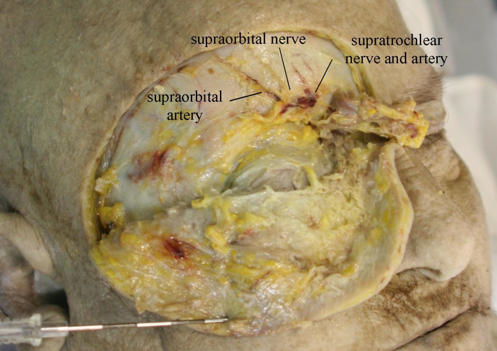
The SON is one of five peripheral branches of the ophthalmic division of the trigeminal nerve (cranial nerve V).  The trigeminal ganglion (TG) gives rise to three divisions, ophthalmic (V1), maxillary (V2), and mandibular (V3).  While the V2 and V3 divisions branch downward, the V1 division branches superior and medially from the TG until it enters the orbit posteriorly via the superior orbital fissure. The nerve then divides into 3, continuing on as the lacrimal nerve, the nasociliary nerve, and the frontal nerve. The frontal nerve then splits into the supraorbital nerve (SON), which exits the orbit with the supraorbital artery via the supraorbital notch (Figure 2), and the supratrochlear nerve (STN), which exits with the supratrochlear artery via the supratrochlear groove or notch (Figure 2). and runs medially in a small groove at the junction of the ocular ridge and the nares.  The SON then branches into a medial and lateral branch over the forehead (Figure 3).

7Ultrasound for evaluation-Fig-2

Figure 2: Supraorbital nerve and artery dissected

7Ultrasound for evaluation-Fig-3

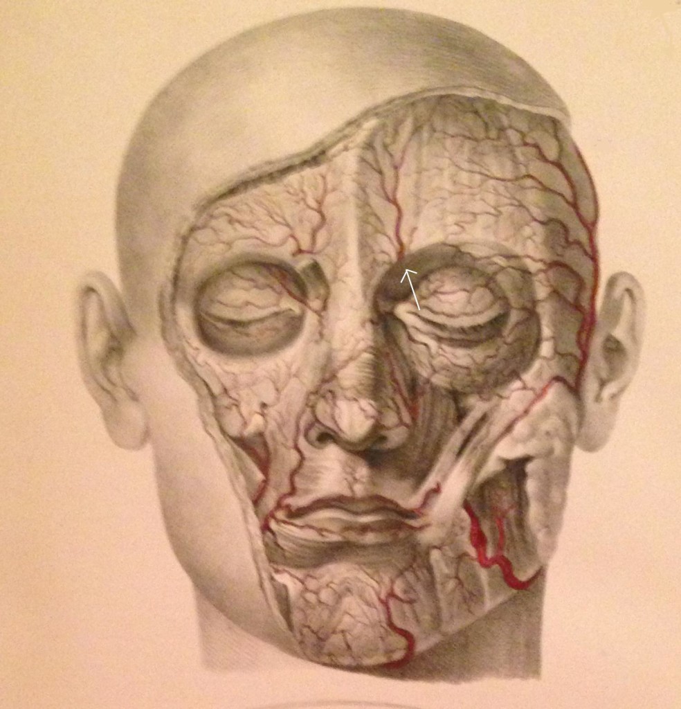
Figure 3: Facial arteries; arrow identifies the supraorbital artery (Image from Quain J, Wilson WJE. The Vessels of the Human Body, 1837)

Separate exits for the medial and lateral SON branches were observed in eight of the 28 nerves (29%) examined by Anderson et al.13  The medial branch continues superiorly up to the vertex of the skull, while the lateral branch moves superior-laterally; both branches supply sensory fibers to the forehead, upper eyelid, and anterior scalp, up to the lambdoidal suture, and both were associated with arteries (often intimately entwined).  Janis et al. found that 73% of the specimens they examined had the SON passing through the corrugator muscle.14

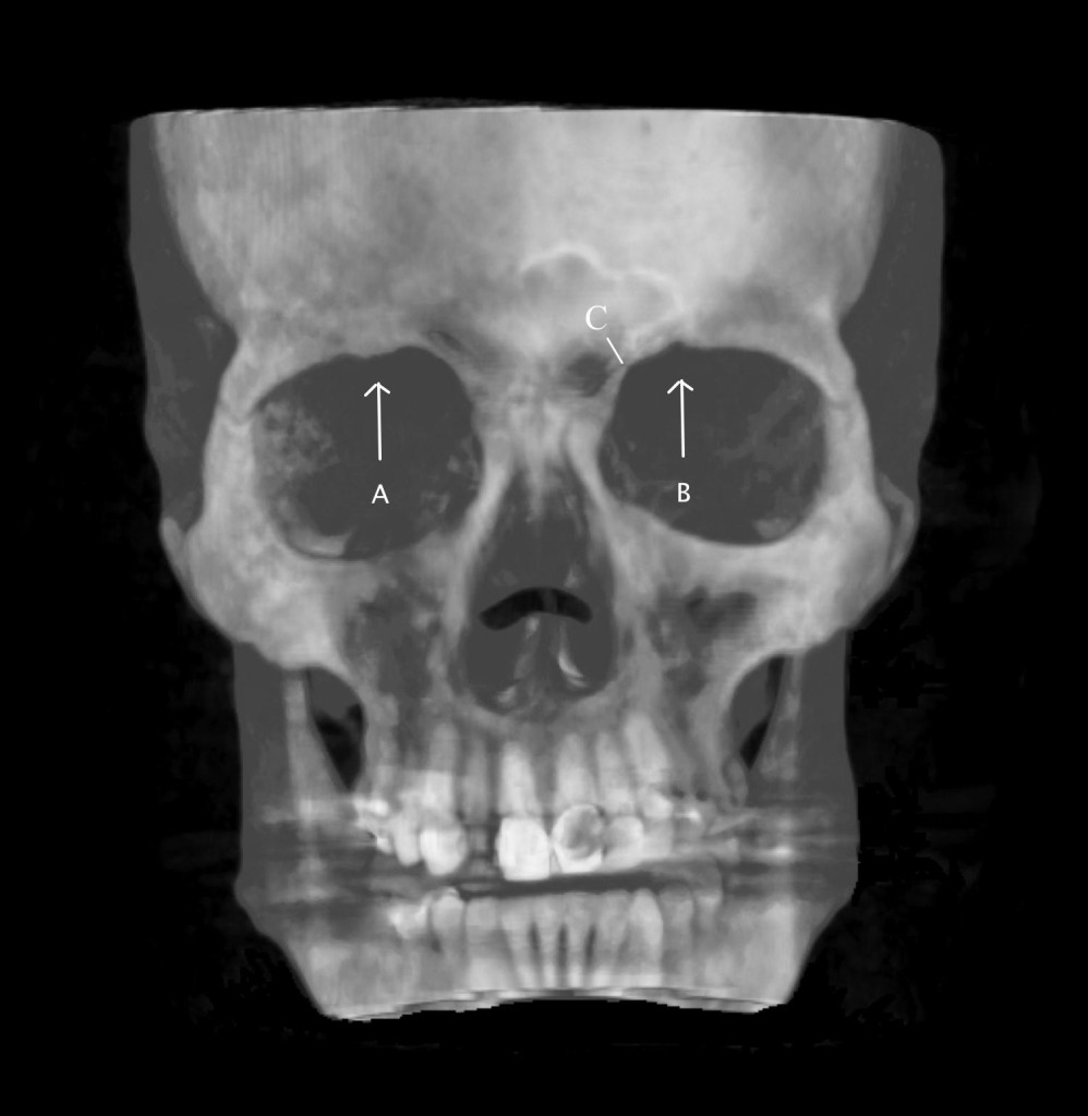
The supraorbital notch may be covered by a small ligament (creating a foramen), which can become thickened or calcified (Figure 4).
7Ultrasound for evaluation-Fig-4

Figure 4: 3D image of the skull.  Arrows show supraorbital notch.  A = obliterated foramen, B = supraorbital notch. C = supratrochlear groove. (Image: Andrea Trescot, MD)

Fallucco and colleagues called this ligament-covered foramen a “miniature carpal tunnel”.15).  In dissections done by Anderson et al.,13 20 of 28 nerves (71%) exited through foramina (with bony or connective tissue bridges) instead of notches.  Fallucco et al.15 dissected 50 SONs and found that 43 of the 50 (86%) had fascial bands across the notch.  These were further divided into “simple” bands (22 of 43 or 51.2%), “partial bony” (13 of 43 or 30.2%), and ‘septums’, which could be horizontal (4 of 43 or 9.3%) or vertical (4 of 43 or 9.3%).  There was a high degree of variability, and the authors noted that the anatomic difference and variable diameters could account for unilateral migraine symptoms.


Entrapment

Entrapment occurs primarily at the supraorbital notch as the nerve leaves the orbit, passing through the supraorbital notch, the orbicularis oculi muscle, the glabellar muscle, and the corrugator muscle, and it can be exacerbated by frowning and squinting (perhaps the reason that some “migraines” respond to use of eyeglasses or botulinum toxin).  Tight fitting goggles16 or an anesthesia mask17 can compress the nerve.  There can also be trauma to the nerve from surgery in the frontal region,17  This headache will often worsen with menses or increased salt intake, perhaps by causing swelling of the nerve in its canal.  In addition, there can be entrapment by scarring17 as well as by thickening or calcification of the supraorbital ligament.


Physical Examination

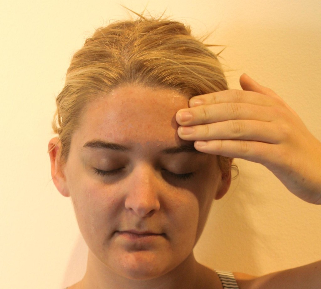
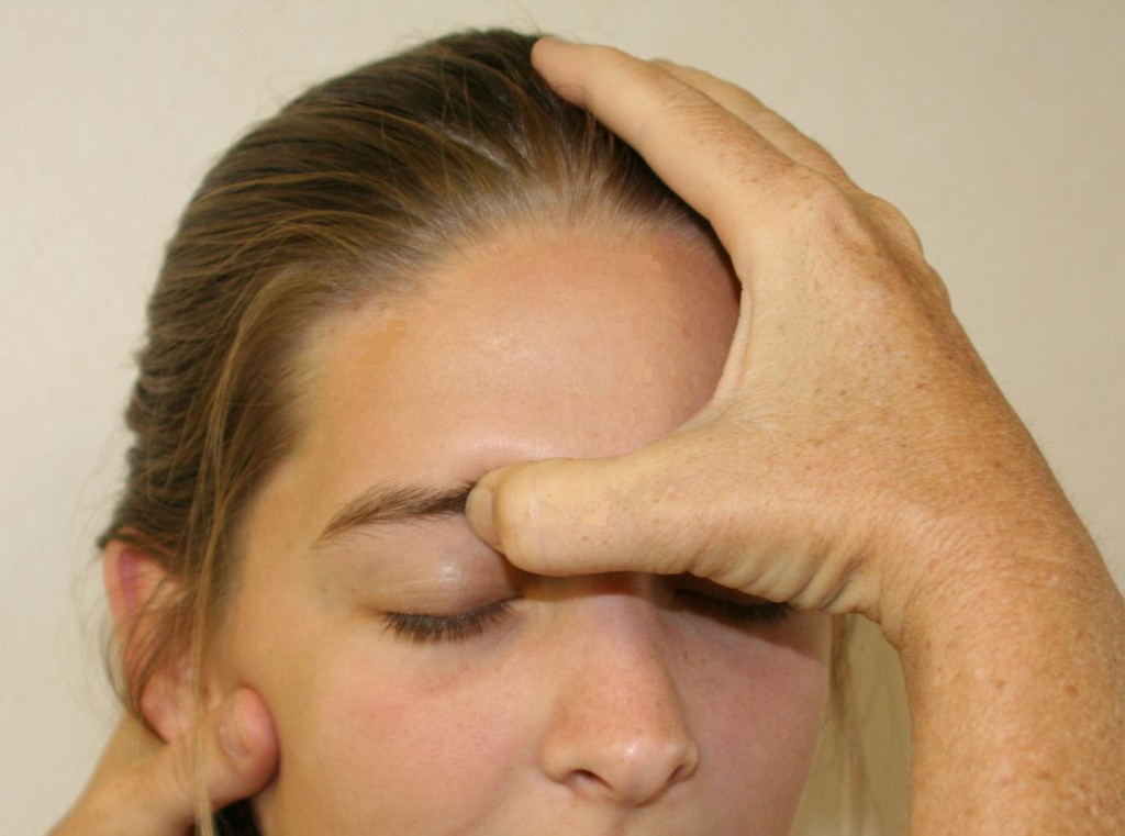
A thorough history should first be obtained to identify any previous trauma or inciting events that could lead to SN.  This is then followed by a visual inspection and physical examination of the forehead, eyebrow, nasal region, and supraorbital foramen.  There will be tenderness to palpation over the supraorbital foramen with possible radiation of symptoms along the nerve distribution of the affected side.  Support the head with the non-examining hand, then palpate over the orbital rim, feeling for the supraorbital notch (Figure 5).  Parasthesias should replicate the pain complaints, and, with careful palpation, the examiner should be able to identify the slim, string-like vertical SON structure.  Move the thumb medially to feel the supratrochlear groove.

7Ultrasound for evaluation-Fig-5

Figure 5: Physical examination of the supraorbital nerve. (Image: Andrea Trescot, MD)

In 2005, Sjaastad and colleagues reported on the supraorbital examination of 1,828 of the inhabitants of Vågå, Norway.12  Only 24 persons (1.3%) did not have a palpable SON; 98% of the individuals had a palpable SON, and 5.4% of the inhabitants (106 of the 1,828 people examined) had increased tenderness over the supraorbital notch.  Twelve of these had a clinical presentation of SN; 10 of the 12 had a history of forehead trauma.

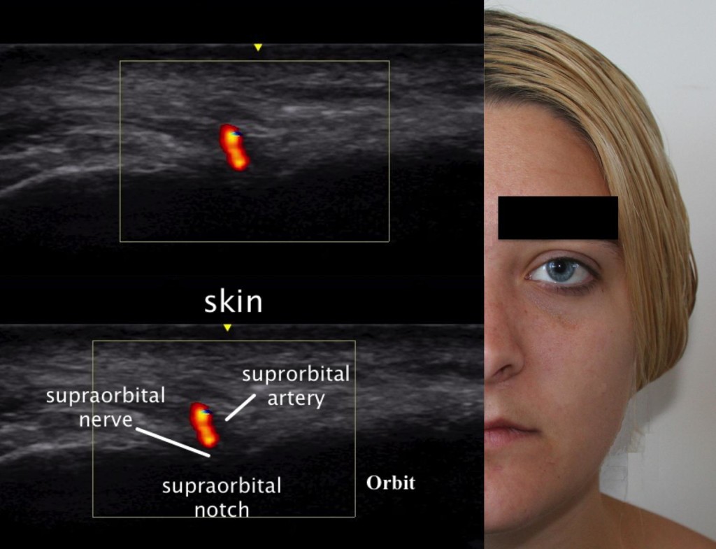
Ultrasound Examination

Using ultrasound (US) to block the supraorbital nerve can be beneficial over the landmark-guided or fluoroscopic approaches.  The linear probe is placed horizontally across the supraorbital notch (Figure 6).  With direct visualization of the supraorbital foramen, proper needle placement can be achieved as well as avoiding inadvertent needle placement into the foramen.  In a recent cadaveric study, Spinner and colleagues18 demonstrated that ultrasound-guided blocks of the supraorbital and infraorbital nerves had greater accuracy than conventional landmark-based techniques.  The results of the study showed that the US accuracy rate was 100% (18 of 18) for the in-plane approach and 94% (17 of 18) for the out-of-plane approach.  Thirty-five injections were considered accurate (97%) with overflow, and one injection was inaccurate.  The study concluded that ultrasound-guided injections had a higher degree of accuracy versus the standard techniques used today.  The US technique would be performed the same as the conventional landmark-guided technique with improved visualization of the target.

7Ultrasound for evaluation-Fig-6

Figure 6:  Ultrasound image of the supraorbital nerve. (Ultrasound Image:  David Spinner, MD; composite created by Andrea Trescot, MD)

AURICULOTEMPORAL NEURALGIA

Temple headaches may be due to entrapment of the auriculotemporal nerve (ATN), a third-division branch of the trigeminal nerve.  Other areas of pain when this nerve is entrapped may include pain in the auricular area and the temporomandibular joint.  This is a common headache site (just visualize all the headache patients rubbing or pressing their temples for relief) shared with migraine headaches, and it may often be misdiagnosed as an intracranial migraine.  There are two distinct clinical presentations of ATN nerve dysfunction, and a variety of names, including auriculotemporal neuralgia, auriculotemporal syndrome, Frey syndrome, Baillarger syndrome, Dupuy syndrome, salivosudoriparous syndrome, and gustatory sweating syndrome.

Clinical Presentation

Entrapment of the ATN is probably the most common of the trigeminal headaches.  Calandre et al.19 evaluated specific areas in the scalp that could be manually palpated to elicit a headache; 42.6% of these “trigger points” were found in the temple region and 33.4% in the suboccipital region.  However, ATN entrapment is rarely diagnosed and represented only 0.4% of the referrals to a tertiary headache outpatient clinic.20

There are two main presentations of irritation of this nerve: Auriculotemporal Neuralgia (ATn) and the rarer Auriculotemporal Syndrome (ATS), both of which can have overlapping presentations, as well as multiple etiologies.

ATn typically presents as paroxysmal attacks of pain in the preauricular and temple regions as well as the retro-orbital region and the teeth.21  The patient with ATn headaches due to ATN entrapment will often awaken at 3-4am with a headache in the auricular area that radiates into the temple (Figure 7) and the retro-orbital as well as the occipital region.

7Ultrasound for evaluation-Fig-7

Figure 7:  Patient’s description of pain from auriculotemporal neuralgia. (Image: Andrea Trescot, MD)

The headache may be unilateral22 or bilateral, and may have associated ear, parotid, and jaw pain or numbness.20  The headache is often throbbing in nature, possibly due to its proximity to the temporal artery.  There is often increased pain with talking, chewing, and menses.23  The intensity of the pain is moderate to severe, and the quality is usually constant, though at times there may be paroxysms of sharp, jabbing, lancinating pain or a throbbing, pounding pain.  The pain may be triggered by trauma to the jaw or temple, as well as by surgery, exacerbated by palpation over the preauricular area or by chewing, and can be associated with severe nausea and vomiting.  The pain characteristics and accompanying nausea and vomiting meet the International Headache Society criteria9 for a “migraine”, though there is no specific listing for ATn.  The early morning headache appears to be related to bruxism or nocturnal jaw clenching.  Because there is a physical connection with the facial nerve (see Anatomy section), there can also be pain in the muscles of facial expression with ATN entrapment.

In 2005, Speciali and Gonçalves 20 evaluated six patients with ATn proven by diagnostic nerve blocks (see Injection techniques section).  All were females with unilateral pain, suffering with symptoms from 1 month to 20 years, and all of the patients had pain with palpation of the preauricular area just above the tragus.  Pain was primarily around the ear, radiating to the head of the mandible and temple in all of the patients.  One patient had knife-like pain triggered by gustatory stimuli (see Frey’s syndrome below).  In 4 patients, the pain radiated to the occipital region, but there was no relief from occipital nerve blocks (see below).  There were complaints of facial tingling or parasthesias in 4 patients, but, notably, temporomandibular joint (TMJ) pathology was absent in all of the patients.  All of the patients had complete or near-complete relief with ATN injections.

Murayama et al.21 described a patient with left ear pain and ipsilateral maxillary second molar pain.  The dental examination was normal, and the tooth and ear pain resolved with a local anesthetic and steroid injection of the ATN, with remission still present 6 months later.

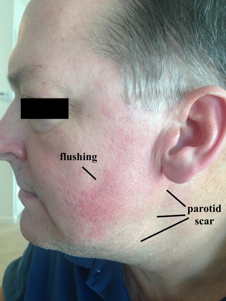
The Auriculotemporal Syndrome, also known as Frey syndrome, was described by Frey in 192324 as a constellation of symptoms including unilateral hyperhidrosis and flushing of the cheek and ear that occurs when eating or drinking anything that stimulates the parotid gland to produce saliva (Figure 8).  Pain is uncommon, but there may be sensory changes in the ATN distribution, and there may be a concomitant trigeminal neuralgia.  Also known as Baillarger syndrome, Dupuy syndrome, salivosudoriparous syndrome, and gustatory sweating syndrome, these symptoms usually occur 2 to 13 months after surgery, open trauma, or infection of the parotid gland, though it has also be described after dislocation of the mandibular condyle.25  The proposed cause is an improper regeneration of the sympathetic and parasympathetic nerves of the parotid gland, and the syndrome occurs in about 5% of surgical patients, even with careful technique.  The severity of symptoms associated with Frey syndrome can range from mild to debilitating.

7Ultrasound for evaluation-Fig-8

Figure 8:  Frey syndrome. Note the well-healed parotid scar and the facial flushing with eating.

Gordon and Fiddian26  reviewed the records of 71 patients who had parotid surgery; 17 patients had “noticeable” Frey syndrome.  Fourteen patients had positive Minors’ test (iodine/starch), with the greater auricular nerve involved in 6 cases, the ATN in 4 cases, and both in 2 cases (with 2 inconclusive tests).  Choi et al.27 evaluated 59 patients after parotidectomy, using subjective symptoms, Minor’s starch iodine test, and thermography, and found a “good” correlation between the 3 indicators.  Pain triggered by taste stimuli can be a significant component of Frey’s syndrome; Scrivani et al.28 described 6 patients with recurrent, episodic shocking facial pain triggered even by the smell of food, which occurred days to weeks after head and neck surgery.

Anatomy

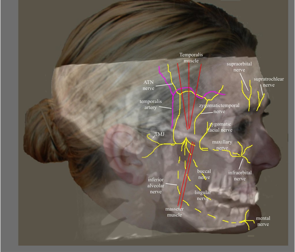
The ATN is a branch of the third division (mandibular) of the trigeminal ganglion.  The mandibular nerve exits the cranium through the foramen ovale and then immediately divides into an anterior and posterior division, providing sensation to the TMJ, tragus, external auditory canal, parotid, base of the auricle, and skin of temporal region (Figure 9).

7Ultrasound for evaluation-Fig-9

Figure 9:  Nerves of the face. (Image: Andrea Trescot, MD)

The anterior division travels between the roof of the infratemporal fossa and the lateral pterygoid muscle (LPM) and is composed of the anterior deep temporal nerve, the posterior deep temporal nerve, and the masseteric nerves.  The posterior division consists of the lingual nerve, inferior alveolar nerve, and ATN, descending medial to the LPM.29  Because the ATN has a long and tortuous course, it is at risk for irritation and entrapment.  The ATN arises by two roots, which travel on either side of the ascending middle meningeal artery and then join together to form a short trunk.  It leaves the foramen ovale, and runs beneath the pterygoideus externus to the medial side of the neck of the mandible.  It then turns upward with the superficial temporal artery, between the external ear and condyle of the mandible, under the parotid gland. After it leaves the gland, it ascends over the zygomatic arch, traveling in front of the temporal mandibular joint (providing the primary enervation of the joint as it goes by) to pierce the temporalis muscle. The ATN then divides into five small branches, including the nerve to the external auditory meatus, which innervates the skin of the meatus and the external tympanic membrane; the parotid branch nerves; the articular branches, which innervate the posterior part of the TMJ; and the superficial temporal nerve, which innervates the skin of the temporal region. The superficial temporal nerve communicates with the facial nerve, the zygomaticotemporal nerve (which is a branch of the maxillary nerve) and zygomatic branches of the facial nerve.

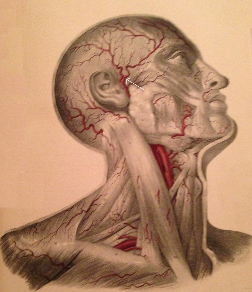
The ATN has been associated with migraine headaches.  Chim et al.30 dissected the ATN in 20 cadavers and found 3 potential compression points along the path of the nerve.  Point 1 and 2 corresponded to fascial bands, with point 1 being 13.1 ± 5.9mm anterior and 5 ± 7mm superior to the superior point of the external auditory meatus and point 2 found at 11.9 ± 6mm anterior and 17.2 ± 10.4mm superior to the most anteriosuperior portion of the external auditory meatus.  The 3rd point represented the interaction of the ATN and the superficial temporal artery (Figure 10), found in 80% of the dissection.  There were 3 types of interactions noted – the artery crossing over the nerve (62.5%), the nerve crossing over the artery (18.8%), or the artery wrapping helically around the nerve (18.8%).

7Ultrasound for evaluation-Fig-10

Figure 10: Temple arteries; arrow identifies the temporal artery (Image from Quain J, Wilson WJE. The Vessels of the Human Body, 1837)

Andersen et al.13 dissected 10 heads and found the superficial ATN branch to be located between 8 and 20mm anterior to the root of the ear helix.  Only 4 of the cadavers had a single branch anterior to the tragus; the rest had multiple branches.  The temporal artery was always found deep to the ATN, and in 3 dissections the ATN connected with the temporal branch of the facial nerve.  There was noted to be a connection between the ATN and lesser occipital nerve in 2 cases, and between the ATN and the greater occipital nerve in 4 cases.

Janis et al.1 dissected 50 cadaver temples and identified that the ATN was always found to lateral to the artery in the upper preauricular temple.  In another 50 cadavers, Janis et al.31 dissected the periorbital and temporal regions, evaluating the path of the zygomaticotemporal branch of the ATN.  In “exactly half” of the specimens, the nerve had no intramuscular path; in the other half of the cadavers, 11 had only a brief course through the muscle, but 14 had a long, tortuous path through the muscle, making entrapment likely.

 Entrapment

Entrapment of the posterior division of the mandibular nerve is not uncommon,20 with one study showing entrapment in 6% of 52 cadavers, primarily at the level of the LPM.29  Another study of 10 cadaver dissections showed myositis, local ischemia, and inflammation of the LPM in patients with premortem complaints of pain in an ATN distribution.32  Spasm of the LPM or local changes within the muscle can entrap the nerve, causing numbness of the external ear and TMJ.29  The ATN can also be entrapped between the medial and lateral pterygoid muscles, causing facial numbness, mandibular pain, or headaches.33

The anatomic relationships between the ATN and the muscles of mastication, the TMJ, and the surrounding vascular structures set up the potential for entrapment, so that ATn can play a role in TMJ pathology, headaches, and external ear pain. Secretomotor fibers within the parotid gland may also be entrapped, leading to impairment of salivation (and therefore triggering Frey syndrome).

Janis et al.1 proposed an entrapment of the ATN by the superficial temporal artery, finding a “direct relationship” between the ATN and the superficial temporal artery in 34% of the cadavers studied, with the ATN wrapped helically around the superficial temporal nerve.  Because of the ATN and facial nerve connection, entrapment of the nerve at this level can cause pain in the muscles of facial expression.29

In addition, the temporal artery entraps the zygomaticotemporal nerve (a branch of the maxillary nerve that exits through the zygomaticotemporal foramen and innervates a small area of the forehead and temporal region), mimicking an ATN.34

 Physical Examination

            Since there are several sites of entrapment, the physical examination must be able to separate each area of pathology.  Examination should include the TMJ (placing the examining fingers over the joint as the patient opens and closes their mouth), the masseter (rolling the examining thumb horizontally as the patient clenches their jaw) and the zygomatic branch of the facial nerve (placing the examining thumb at the anterior condyle of the mandible, just under the zygoma).  The examiner should also palpate of the temporalis muscle (looking for trigger point tenderness) and the superficial temporal artery (to evaluate for temporal arteritis).

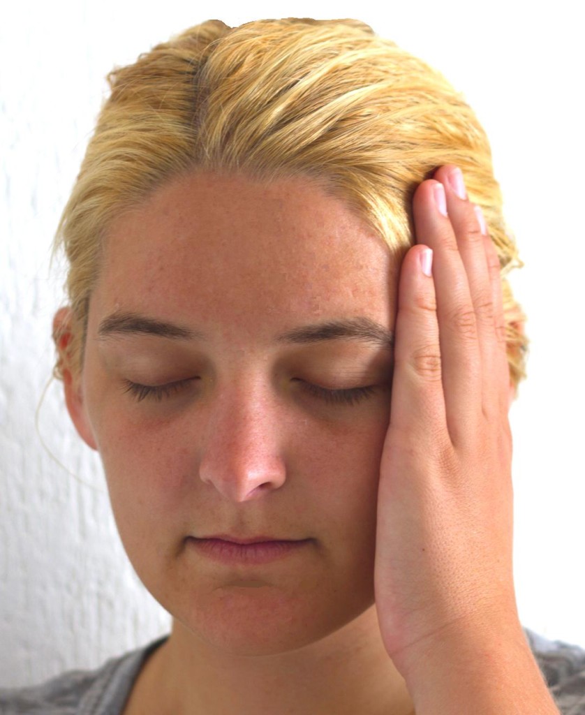
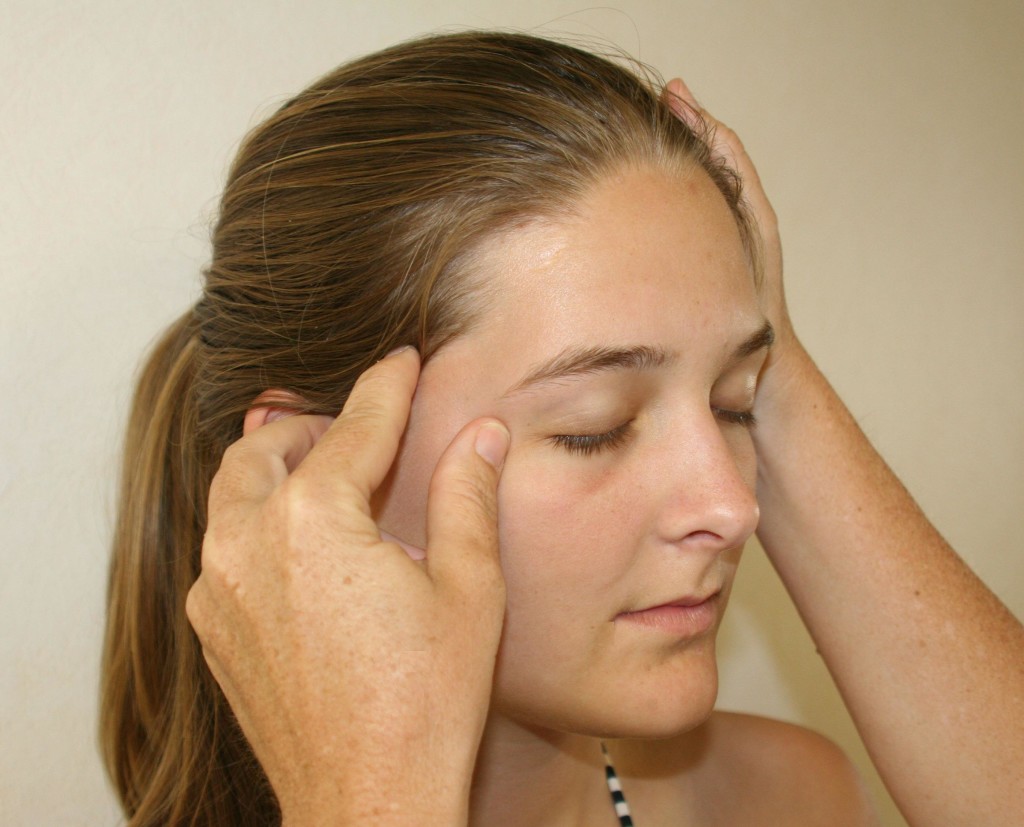
The most clinically common site of entrapment for the ATN is in the temporal fossa; Trescot described finding the ATN by placing the index finger at the apex of an isosceles triangle created by resting (for the right-sided exam) the thumb on the tragus and the middle finger on the canthus (corner of the eye) (Figure 11).23

7Ultrasound for evaluation-Fig-11

Figure 11:  Auriculotemporal nerve examination. (Image: Andrea Trescot, MD)

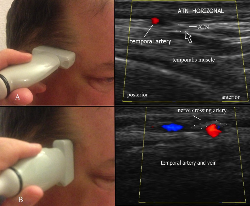
 Ultrasound Guided Injection

In 2007, Shankar and Brethauer35 described an US technique for the ATN injection.  The US probe is placed transversely just above the TMJ, and color Doppler used to identify the superficial temporal artery; the nerve appears as a small hyperlucent bundle (Figure 12A).  The probe is then rotated to perform a longitudinal scan to track the nerve cephalad (Figure12B).  They used a 25g needle from an out-of-plane approach to deliver medication (2cc of 1% lidocaine and 10mg of Depo-Medrol®) around the nerve.

7Ultrasound for evaluation-Fig-12

Fig 12:  Ultrasound evaluation of the auriculotemporal nerve. A = horizontal evaluation; B = vertical evaluation. (Images: Andrea Trescot, MD)

Because of its tortuous path, its proximity to a significant artery (the temporal artery), and its frequent misdiagnosis, US is an excellent tool for the diagnosis and treatment of the ATN

OCCIPITAL NERVE

The greater occipital nerve (GON) entrapment commonly causes occipital and cervicogenic headaches, and can sometimes cause headaches with characteristics similar to migraine headaches.  The anatomic recognition of multiple occipital nerves (the greater occipital, the lesser occipital, and the third occipital nerve), each with different patterns of pain, neurologic origin, and treatment modalities, has been facilitated by the use of precision diagnostic injections.  This article will focus specifically on the GON. There are multiple names used for these nerve entrapments, including “migraine”, “tension headache”, “cervicogenic headaches”,36 and “occipital myalgia-neuralgia syndrome”.37 Sometimes the term “suboccipital injections” has been used to describe greater occipital nerve injections, but, for this article, I use “suboccipital” to mean the C1 nerve root or the high volume occipital decompression. Occipital neuralgia is also known as C2 neuralgia or Arnold’s neuralgia.

Clinical Presentation

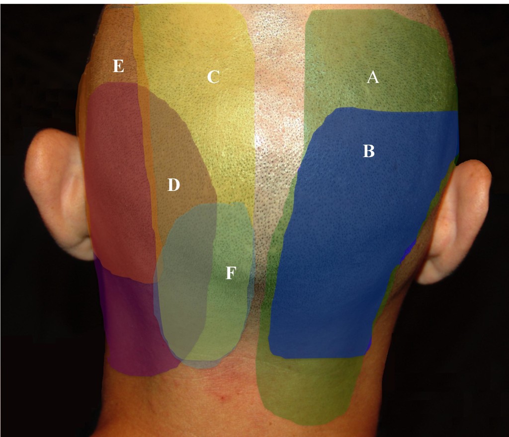
Beruto et al.38 described occipital neuralgia (ON) in 1821 as a sharp, lightening-bolt pain radiating from the occiput to the vertex.  The medical terms occipital neuralgia and cervicogenic headache describe a syndrome of neck and head pain primarily referring to the occiput, as well as the temporal area, forehead, and retro-orbital areas, that may arise some distance away, in the upper cervical spine.  Greater occipital neuralgia characteristically presents as paroxysmal shooting, stabbing pain from the suboccipital region to the vertex (Figure 13).  The cutaneous innervation of the posterior neck and occiput comes primarily from C1, C2, and C3, which provide overlapping dermatomes (Figure 14).  C2 covers the occiput, neck, and submental regions, while the C3 dermatome can span from the clavicle to the mandible to behind and over the ear, pinna, and the angle of the mandible.39  Since the greater occipital nerve is made up of contributions from C1, C2, and C3, there can be a wide range of clinical presentations.

7Ultrasound for evaluation-Fig-13

Figure 13.  Pattern of greater occipital pain. (Image: Andrea Trescot, MD)

7Ultrasound for evaluation-Fig-14

Figure 14.  Pattern of posterior cervical and occipital pain. A = C2/3 facet; B = C3/4 facet; C = greater occipital nerve (GON); D = posterior auricular nerve (PAN); E = lesser occipital nerve (LON); F = third occipital nerve (TON). (Image: Andrea Trescot, MD)

As a subset of cervicogenic headaches (CGH), occipital neuralgia can cause pain and paresthesias to the posterior scalp, the periorbital, temporal and mandibular regions, the external ear and mastoid regions, as well as pain in the neck and shoulders.  The first three cervical spinal nerve segments (C1-C3) that make up the occipital nerves share a relay-station in the brainstem that continues into the upper cervical spinal cord with the trigeminal cell bodies (the cervico-trigeminal complex). The pain of occipital neuralgia and cervicogenic headaches can therefore be referred to structures innervated by the branches of the trigeminal nerve, namely, the forehead, temples, and eyes.

There are three occipital nerves – the greater, the lesser, and the third occipital nerves.  Their patterns of pain are overlapping, and so the reader is encouraged to review information on these nerves as well.

Unilateral ON, perhaps because of the proximity of the occipital artery (see below), can present with throbbing, unilateral headaches associated with photophobia, phonophobia, and nausea; symptoms meet the International Classification of Headache Disorders (ICHD) of a “migraine”.5  There may be a history of occipital pain, often radiating to the face, specifically described as “behind my eye” and “like an ice pick”.  Occipital headaches may start as “tension headaches” in the upper cervical region, but then center at the base of the skull and produce throbbing, unilateral or bilateral pain, accompanied by nausea, photophobia, and phonophobia. Occipital neuralgia has a close relationship with cluster headaches, with several authors40-42 describing the use of occipital blocks to treat cluster headaches. Hemicrania continuum43 and “transformed migraines”44 have also responded to occipital nerve blocks.

Anatomy

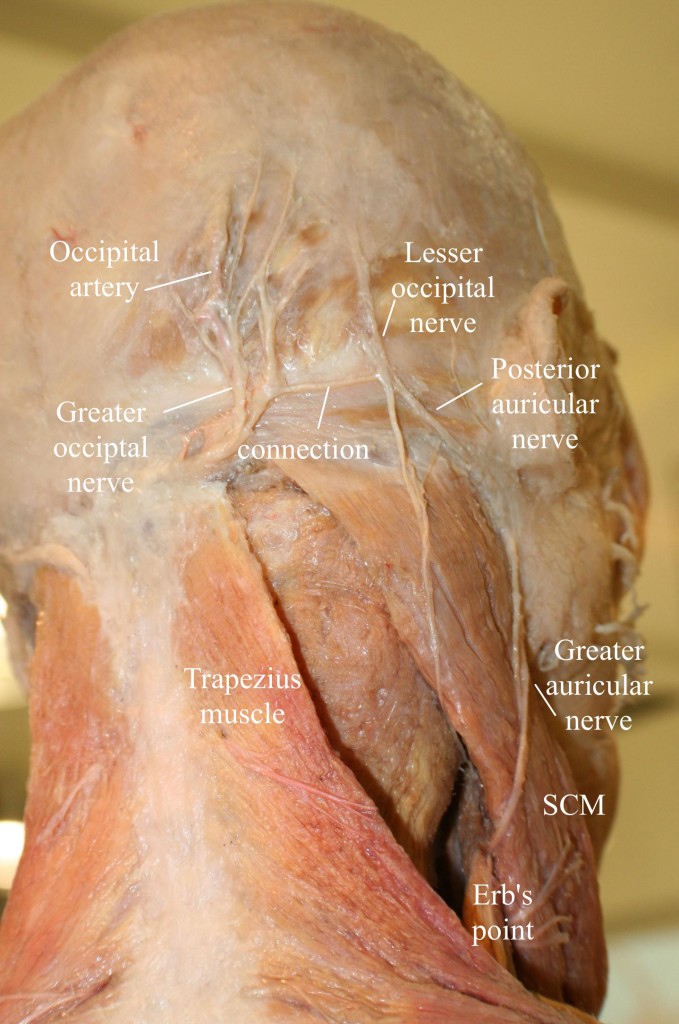
The largest of the three occipital nerves, the GON arises from the posterior ramus of C2 that runs inferiorly between the arch of C1 (atlas) and the lamina of C2 (axis), lateral to the lateral atlantoaxial (AA) joint, deep to the inferior oblique muscle. The GON then curves up over the inferior oblique, between the inferior oblique and the semispinalis capitis muscles (Figure 15).

7Ultrasound for evaluation-Fig-15

Figure 15:  Anatomy of the occipital region, modified from an image from Bodies, The Exhibition, with permission.  Note the connection of the greater and lesser occipital nerves. (Image: Andrea Trescot, MD)

A branch from C3 may join at this point, as the nerve ascends up the neck and over the dorsal surface of the rectus capitis to pierce the semispinalis capitis muscle, deep to the trapezius muscle.  The GON then exits the neck through a muscular sling formed by the aponeurosis of the sternocleidomastoid muscle (SCM) and trapezius muscle at their attachment on the occipital bone (the conjoined tendon), where it is joined laterally by the occipital artery. At this point, the greater occipital nerve is immediately medial to the occipital artery and lateral to the inion or occipital prominence, lying in a palpable groove.  This proximity to the artery may account for the throbbing sensation that often accompanies GON entrapment.

7Ultrasound for evaluation-Fig-16

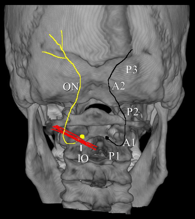
Figure 16.  The path of the occipital nerve. IO = inferior oblique; P1 = Part 1 of the occipital nerve; P2 = Part 2 of the occipital nerve; P3 = Part 3 of the occipital nerve; A1 = site of entrapment of inferior oblique; A2 = site of entrapment by trapezius muscle. (Image: Andrea Trescot, MD)

The GON could therefore be considered to have three parts (Part 1, Part 2, and Part 3) as well as two bends (A1 and A2, where entrapments occur)45 (Figure 16). A cutaneous branch of the suboccipital nerve (the C1 dorsal ramus) will occasionally join the GON as it accompanies the occipital artery. The GON frequently connects with the lesser occipital nerve (Figure 15), which arises from the cervical plexus (formed by the upper four ventral cervical rami). The GON continues to ascend to innervate the skin along the posterior portion of the occiput to the vertex, portions of the ear, and parotid glands.

Shimizu et al.46 looked at the suboccipital region of 24 cadaver heads; the occipital nerve was found to cross over the artery, with the nerve consistently indented by the artery but without histologic evidence of mechanical damage.  Natsis el al.47, based on the dissections of 40 cadavers, noted that GON was noted to get wider as it moved toward the periphery, and, in 3 cases, the nerve split into 2 branches before piercing the trapezius.  The GON and lesser occipital nerves were reunited at the level of the occiput in 80% of the specimens. Becser, Bovim, and Sjaastad48 dissected 10 cadavers and found that twelve of the 20 GONs wrapped around the occipital artery.

Dash et al.49 looked at 19 cadavers and noted that all of the GONs identified were found to pierce the semispinalis capitis.  On the other hand, in the 20 cadavers that Bovim et al.50 dissected (none of whom had a history of headaches), 10 specimens had the occipital nerve piercing the trapezius muscle on one side but not the other.  They also identified 11 nerves with macroscopic signs of possible nerve compression from fibrotic tissue, with 6 compressions accompanied by a “kink” of more than 90°.  Additionally, three of the 40 occipital nerves penetrated the inferior oblique.  They also noted a marked variation of the amount of venous plexus at the level of C2; 50% of the cases had the C2 nerve root surrounded by a marked vascular network.  They noted a significant variation in the path of the occipital nerve, but they also remarked that, for many of the dissections, there was a significant difference noted within the 2 sides of the same individual.

Entrapment

There are several areas of entrapment of the occipital nerve: (A) where the greater occipital nerve emerges from the C2 DRG between the atlas and axis, (B) between the inferior oblique and the semispinalis capitus muscles, (C) where the nerve pierces the semispinalis capitis, and (D) where the nerve exits from the aponeurosis of the trapezius.  Because the GON pierces the nuchal fascia at the base of the skull, it is prone to trauma from flexion/tension injuries, which can lead to entrapment by a spasm of the trapezius muscle.  Repetitive neck contractions secondary to work, recreational activities, and many other activities can cause entrapment and/or scarring of the GON.  The trapezius or sternocleidomastoid muscles can cause entrapment secondary to myofascial spasms.  Head forward positions can entrap the GON at the level of the inferior oblique.  Medical conditions such as osteochondroma (benign tumors of the bone),51 arterial compression of the C2 nerve root by a tortuous vertebral artery, herpes zoster,52 osteoarthritis or rheumatoid arthritis (especially at C1-C2),53,54 or trauma such as blows to the back of the head or posterior fossa surgery, and many others contribute or are the root cause of entrapment or are the contributing factors causing pain.  However, as the ganglion of this nerve interconnects with the trigeminal nerve in the brainstem, pain may be referred to any branch of the trigeminal nerve. The best way to diagnose an occipital nerve entrapment is by physical examination and injection.

Physical Examination

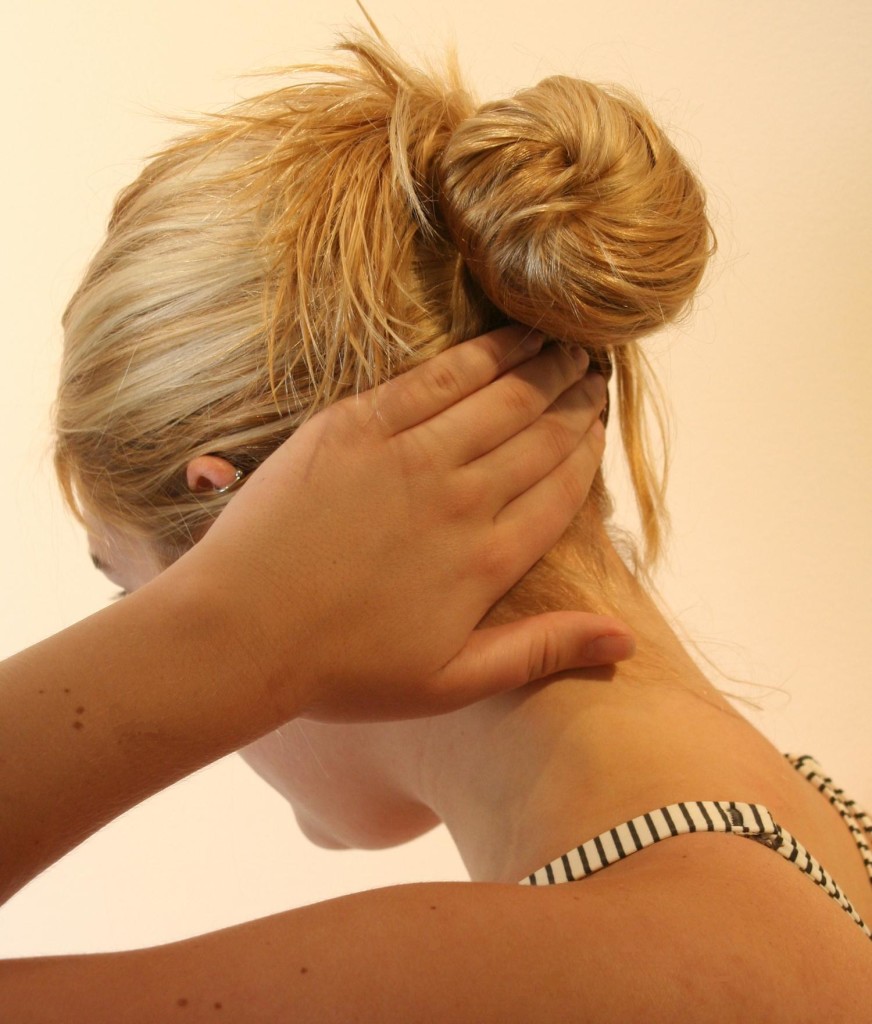
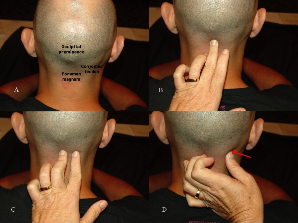
For the physical Examination of the greater occipital nerve, the patient should be positioned sitting with the neck slightly flexed, with the examiner supporting the forehead with the non-examining hand (Figure 17A).  With the examining hand, place the middle finger at the midline base of the head to identify the site of the foramen magnum (Figure 17B).  The index finger is placed at the conjoined tendon attachment (Figure 17C).  The thumb will then palpate the area just lateral to the conjoined tendon, approximately 3cm inferior and 1.5cm lateral to the occipital inion (Figure 17D).  If an area of paresthesia is created, reproducing the patient’s pain and symptomatology, the greater occipital nerve is likely the source.
7Ultrasound for evaluation-Fig-17

Figure 17 A, B, C, and D.  Physical examination of the greater occipital nerve. (Images: Andrea Trescot, MD)

Ultrasound-Guided Technique

There are several sites for injection of the GON under US.  Shim et al. (55) used US-guided injections at the occipital ridge (site 1 on Figure 18) to treat occipital headaches; they measured the distance from the “external occipital prominence” (the inion) to the occipital nerve and artery, and then looked at the results of landmark-guided versus US-guided injections.  They found a greater improvement in pain scores for the US patients at 4 weeks (blind 3.8 ± 0.3 vs. US 2.3 ± 0.2).  Na and colleagues56 did a similar study with 26 patients using flow Doppler versus blind injections at the nuchal ridge.  All of the injections resulted in at least “partial” hypoesthesia; only 30.8% of the blind injections had “complete” anesthesia, while 76.9% of the Doppler-guided injections had “complete” anesthesia. Palamar et al. randomized 23 migraine patients to injections of 1.5cc of local anesthetic (0.5% bupivacaine) or saline under US guidance at the occipital ridge; all actively treated patients noted a significant decrease in headache symptoms.57 Each ON was found to be medial to the occipital artery.

7Ultrasound for evaluation-Fig-18

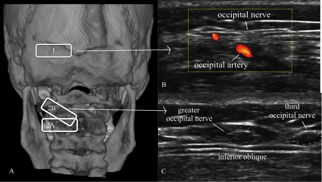
Figure 18.  A = Location of ultrasound transducer: 1 = standard occipital nerve ultrasound site; 2 = US approach with (2A) being the initial probe placement and (2B) being the final probe placement.  B = Distal US occipital nerve and artery. C = Proximal US of greater and third occipital nerve. (Images: Andrea Trescot, MD – modified from Greher et al. [48])

Greher et al.58 described an anatomic study on cadavers using ultrasound to compare the traditional injection site (site 1 on Figure 18) at the nuchal ridge with a more proximal technique (site 2 on Figure 18A).  They identified the bifid spinous process of C2 with the probe in a horizontal position (site 2A on Figure 18A); the probe was then rotated (site 2B on Figure 18A) to visualize the inferior oblique muscle and the GON, which appears as a hypoechoic oval on top of the muscle (Figure 18C). Using 0.1cc injections of dye, they used ultrasound to identify the GON at both sites bilaterally in 10 cadavers. They reported successful injection of the GON distally in 16 of 20 dissections, while all 20 of the proximal GON were stained with dye (i.e., successful).

CONCLUSION

Aggressive treatment of headaches and “migraines” can be gratifying and technically satisfying for the interventional pain physician with the knowledge and skill to provide injection therapy to patients with these pain problems. US imaging and US-directed injections of “epicranial” structures can increase the likelihood of success.

Conflict of interest: None declared by the author

REFERENCES

  1. Janis JE, Hatef DA, Ducic I, Ahmad J, Wong C, Hoxworth RE, et al. Anatomy of the auriculotemporal nerve: variations in its relationship to the superficial temporal artery and implications for the treatment of migraine headaches. Plast Reconstr Surg. 2010;125(5):1422-8. doi: 10.1097/PRS.0b013e3181d4fb05. [PubMed]
  2. Evans RW, Pareja JA. Expert opinion. Supraorbital neuralgia. Headache. 2009;49(2):278-81. [PubMed] doi: 10.1111/j.1526-4610.2008.001328.x.
  3. Guyuron B, Varghai A, Michelow BJ, Thomas T, Davis J. Corrugator supercilii muscle resection and migraine headaches. Plast Reconstr Surg. 2000;106(2):429-34; discussion 435-7. [PubMed]
  4. Gobel H, Heinze A, Heinze-Kuhn K, Jost WH. Evidence-based medicine: botulinum toxin A in migraine and tension-type headache. J Neurol. 2001;248 Suppl 1:34-8. [PubMed]
  5. The International Classification of Headache Disorders, 3rd edition (beta version). Cephalalgia. 2013;33(9):629-808. doi: 10.1177/0333102413485658.[PubMed]
  6. Dalkara T, Zervas NT, Moskowitz MA. From spreading depression to the trigeminovascular system. Neurol Sci. 2006;27 Suppl 2:S86-90. [PubMed]
  7. Beyer TE. Idiopathic supraorbital neuralgia. Laryngoscope. 1949;59(11):1273-5. [PubMed]
  8. Agrawal SM, Kambalimath DH. Trigeminal neuralgia involving supraorbital and infraorbital nerves. Natl J Maxillofac Surg. 2010;1(2):179-82. doi: 10.4103/0975-5950.79226. [PubMed] [Free full text]
  9. The International Classification of Headache Disorders: 2nd edition. Cephalalgia. 2004;24 Suppl 1:9-160. [PubMed]
  10. Trescot AM. Cryoanalgesia in interventional pain management. Pain Physician. 2003;6(3):345-60. [PubMed] [Free full text]
  11. Guerrero AL, Herrero-Velazquez S, Penas ML, Mulero P, Pedraza MI, Cortijo E, et al. Peripheral nerve blocks: a therapeutic alternative for hemicrania continua. Cephalalgia. 2012 Apr;32(6):505-8. doi: 10.1177/0333102412439800. [PubMed] [Free full text]
  12. Sjaastad O, Petersen HC, Bakketeig LS. Supraorbital neuralgia. Vaga study of headache epidemiology. Cephalalgia. 2005;25(4):296-304. [PubMed] [Free full text]
  13. Andersen NB, Bovim G, Sjaastad O. The frontotemporal peripheral nerves. Topographic variations of the supraorbital, supratrochlear and auriculotemporal nerves and their possible clinical significance. Surg Radiol Anat. 2001;23(2):97-104. [PubMed]
  14. Janis JE, Ghavami A, Lemmon JA, Leedy JE, Guyuron B. The anatomy of the corrugator supercilii muscle: part II. Supraorbital nerve branching patterns. Plast Reconstr Surg. 2008 Jan;121(1):233-40. doi: 10.1097/01.prs.0000299260.04932.38. [PubMed]
  15. Fallucco M, Janis JE, Hagan RR. The anatomical morphology of the supraorbital notch: clinical relevance to the surgical treatment of migraine headaches. Plast Reconstr Surg. 2012 Dec;130(6):1227-33. doi: 10.1097/PRS.0b013e31826d9c8d. [PubMed]
  16. O’Brien JC Jr. Swimmer’s headache, or supraorbital neuralgia. Proc (Bayl Univ Med Cent). 2004;17(4):418-9. [PubMed] [Free full text]
  17. Klein DS, Schmidt RE, Jr. Chronic headache resulting from postoperative supraorbital neuralgia. Anesth Analg. 1991;73(4):490-1. [PubMed]
  18. Spinner D, Kirschner JS. Accuracy of ultrasound-guided superficial trigeminal nerve blocks using methylene blue in cadavers. Pain Med. 2012 Nov;13(11):1469-73. doi: 10.1111/j.1526-4637.2012.01480.x. [PubMed]
  19. Calandre EP, Hidalgo J, Garcia-Leiva JM, Rico-Villademoros F. Trigger point evaluation in migraine patients: an indication of peripheral sensitization linked to migraine predisposition? European journal of neurology : the official journal of the European Federation of Neurological Societies. 2006;13(3):244-9. [PubMed]
  20. Speciali JG, Gonçalves DA. Auriculotemporal neuralgia. Curr Pain Head Rep. 2005;9:277-80. [PubMed]
  21. Murayama RA, Stuginski-Barbosa J, Moraes NP, Speciali JG. Toothache referred from auriculotemporal neuralgia: case report. Int Endod J. 2009 Sep;42(9):845-51. doi: 10.1111/j.1365-2591.2009.01599.x. [PubMed]
  22. Damarjian E. Auriculo-temporal neuralgia–an original diagnostic and therapeutic approach. R I Med J. 1970;53(2):100-1. [PubMed]
  23. Trescot AM. Headache: Is it a migraine? Think again. Tech Reg Anesth Pain Manag. 2005;9:68-72.
  24. Frey L. Le syndrome du nerf auriculo-temporal. Rev Neurol (Paris). 1923;2:97-104.
  25. Kamath RA, Bharani S, Prabhakar S. Frey’s Syndrome Consequent to an Unusual Pattern of Temporomandibular Joint Dislocation: Case Report with Review of Its Incidence and Etiology. Craniomaxillofac Trauma Reconstr. 2013 Mar;6(1):1-8. doi: 10.1055/s-0032-1332210. [PubMed] [Free full text]
  26. Gordon AB, Fiddian RV. Frey’s syndrome after parotid surgery. Am J Surg. 1976;132(1):54-8. [PubMed]
  27. Choi HG, Kwon SY, Won JY, Yoo SW, Lee MG, Kim SW, et al. Comparisons of three indicators for Frey’s Syndrome: Subjective symptoms, Minor’s starch iodine test, and infrared thermography. Clinical and experimental otorhinolaryngology. 2013;6(4):249-53.
  28. Scrivani SJ, Keith DA, Kulich R, Mehta N, Maciewicz RJ. Posttraumatic gustatory neuralgia: a clinical model of trigeminal neuropathic pain. J Orofac Pain. 1998;12(4):287-92. [PubMed]
  29. Loughner BA, Larkin LH, Mahan PE. Nerve entrapment in the lateral pterygoid muscle. Oral Surg Oral Med Oral Pathol. 1990;69(3):299-306. [PubMed]
  30. Chim H, Okada HC, Brown MS, Alleyne B, Liu MT, Zwiebel S, et al. The auriculotemporal nerve in etiology of migraine headaches: compression points and anatomical variations. doi: 10.1097/PRS.0b013e3182589dd5. [PubMed]
  31. Janis JE, Hatef DA, Thakar H, Reece EM, McCluskey PD, Schaub TA, et al. The zygomaticotemporal branch of the trigeminal nerve: Part II. Anatomic variations. Plast Reconstr Surg. 2010 Aug;126(2):435-42. doi: 10.1097/PRS.0b013e3181e094d7. [PubMed]
  32. Schmidt BL, Pogrel MA, Hakim-Faal Z. The course of the temporal branch of the facial nerve in the periorbital region. J Oral Maxillofac Surg. 2001;59(2):178-84. [PubMed]
  33. Anil A, Peker T, Turgut HB, Gulekon IN, Liman F. Variations in the anatomy of the inferior alveolar nerve. Br J Oral Maxillofac Surg. 2003;41(4):236-9. [PubMed]
  34. Kemp WJ 3rd, Tubbs RS, Cohen-Gadol AA. The innervation of the scalp: A comprehensive review including anatomy, pathology, and neurosurgical correlates. Surg Neurol Int. 2011;2:178. doi: 10.4103/2152-7806.90699. [PubMed] [Free full text]
  35. Shankar H, Brethauer J. Ultrasound guided steroid injection for auriculo-temporal neuralgia. Anesthesiology. 2007;107:A903. doi: 10.1097/01.anes.0000290617.98058.d9. [Free full text]
  36. Sjaastad O, Saunte C, Hovdahl H, Breivik H, Gronbaek E. “Cervicogenic” headache. An hypothesis. Cephalalgia. 1983;3(4):249-56. [PubMed] [Free full text]
  37. Blume H, Atac M, Golnick J. Neurosurgical treatment of persistent occipital myalgia-neuralgia syndrome. In: Pfaffenrath V, Lundberg P-O, Sjaastad O, editors. Updating in Headache: Springer Berlin Heidelberg; 1985. p. 24-34.
  38. Beruto Y, Lentijo J, Ramus NM. Descodes de med y irugpract. Madrid. 1821;3:145-69.
  39.       Dubuisson D. Treatment of occipital neuralgia by partial posterior rhizotomy at C1-3. J Neurosurg. 1995;82(4):581-6. [PubMed]
  40. Scattoni L, Di Stani F, Villani V, Dugoni D, Mostardini C, Reale C, et al. Great occipital nerve blockade for cluster headache in the emergency department: case report. J Headache Pain. 2006;7(2):98-100.[Free full text]
  41. Peres MF, Stiles MA, Siow HC, Rozen TD, Young WB, Silberstein SD. Greater occipital nerve blockade for cluster headache. Cephalalgia. 2002;22(7):520-2. [PubMed] [Free full text]
  42. Anthony M. Arrest of attacks of cluster headache by local steriod injections in the occipital myalgia-neuralgia syndrome. In: Pfaffenrath V, Lundberg PO, Sjaastad O, editors. Updating in Headache. Berlin: Springer-Verlag; 1985. p. 17-23.
  43. Cortijo E, Guerrero AL, Herrero S, Mulero P, Munoz I, Pedraza MI, et al. Hemicrania continua in a headache clinic: referral source and diagnostic delay in a series of 22 patients. J Headache Pain. 2012;13(7):567-9. [Free full text]
  44. Ashkenazi A, Matro R, Shaw JW, Abbas MA, Silberstein SD. Greater occipital nerve block using local anaesthetics alone or with triamcinolone for transformed migraine: a randomised comparative study. J Neurol Neurosurg Psychiatry. 2008;79(4):415-7. [PubMed] [Free full text]
  45. Gille O, Lavignolle B, Vital JM. Surgical treatment of greater occipital neuralgia by neurolysis of the greater occipital nerve and sectioning of the inferior oblique muscle. Spine (Phila Pa 1976). 2004;29(7):828-32. [PubMed]
  46. Shimizu S, Oka H, Osawa S, Fukushima Y, Utsuki S, Tanaka R, et al. Can proximity of the occipital artery to the greater occipital nerve act as a cause of idiopathic greater occipital neuralgia? An anatomical and histological evaluation of the artery-nerve relationship. Plast Reconstr Surg. 2007;119(7):2029-34; discussion 35-6. [PubMed]
  47. Natsis K, Baraliakos X, Appell HJ, Tsikaras P, Gigis I, Koebke J. The course of the greater occipital nerve in the suboccipital region: a proposal for setting landmarks for local anesthesia in patients with occipital neuralgia. Clin Anat. 2006;19(4):332-6. [PubMed]
  48. Becser N, Bovim G, Sjaastad O. Extracranial nerves in the posterior part of the head. Anatomic variations and their possible clinical significance. Spine (Phila Pa 1976). 1998 Jul 1;23(13):1435-41. [PubMed]
  49. Dash KS, Janis JE, Guyuron B. The lesser and third occipital nerves and migraine headaches. Plast Reconstr Surg. 2005;115(6):1752-8; discussion 9-60. [PubMed]
  50. Bovim G, Bonamico L, Fredriksen TA, Lindboe CF, Stolt-Nielsen A, Sjaastad O. Topographic variations in the peripheral course of the greater occipital nerve. Autopsy study with clinical correlations. Spine (Phila Pa 1976). 1991 Apr;16(4):475-8. [PubMed]
  51. Baer-Henney S, Tatagiba M, Samii M. Osteochondroma of the cervical spine causing occipital nerve neuralgia. Case report. Neurol Res. 2001;23(7):777-9. [PubMed]
  52. Kihara T, Shimohama S. Occipital neuralgia evoked by facial herpes zoster infection. Headache. 2006;46(10):1590-1. [PubMed]
  53. Star MJ, Curd JG, Thorne RP. Atlantoaxial lateral mass osteoarthritis. A frequently overlooked cause of severe occipitocervical pain. Spine (Phila Pa 1976). 1992;17(6 Suppl):S71-6. [PubMed]
  54. Ehni G, Benner B. Occipital neuralgia and the C1-2 arthrosis syndrome. J Neurosurg. 1984;61(5):961-5. [PubMed]
  55. Shim JH, Ko SY, Bang MR, Jeon WJ, Cho SY, Yeom JH, et al. Ultrasound-guided greater occipital nerve block for patients with occipital headache and short term follow up. Korean J Anesthesiol. 2011;61(1):50–4. doi: 10.4097/kjae.2011.61.1.50. [PubMed] [Free full text]
  56. Na SH, Kim TW, Oh SY, Kweon TD, Yoon KB, Yoon DM. Ultrasonic doppler flowmeter-guided occipital nerve block. Korean J Anesthesiol. 2010 Dec; 59(6):394-7. doi: 10.4097/kjae.2010.59.6.394. [PubMed] [Free full text]
  57.       Palamar D, Uluduz D, Saip S, Erden G, Unalan H, Akarirmak U. Ultrasound-guided greater occipital nerve block: an efficient technique in chronic refractory migraine without aura? Pain Physician. 2015;18(2):153-62. [PubMed] [Free full text]
  58. Greher M, Moriggl B, Curatolo M, Kirchmair L, Eichenberger U. Sonographic visualization and ultrasound-guided blockade of the greater occipital nerve: a comparison of two selective techniques confirmed by anatomical dissection. Br J Anaesth. 2010 May;104(5):637-42. doi: 10.1093/bja/aeq052. [PubMed] [Free full text]

Pseudohyperkalemia in infants: A reason to postpone the surgery?

$
0
0

Teena Bansal*, Jatin Lal**
*Assistant Professor; **Professor
Department of Anaesthesiology & Critical Care, Pt. B.D. Sharma University of Health Sciences,
Rohtak-124001, Haryana (India)

Correspondence: Dr Teena Bansal, 19/6 J Medical Campus, PGIMS, Rohtak -124001, Haryana (India); Mobile: +91-9315839374; E-mail: aggarwalteenu@rediffmail.com

Key words: Pseudohyperkalemia; Hyperkalemia; Infants; Hemolysis

Citation:  Bansal T, Lal J. Pseudohyperkalemia in infants: A reason to postpone the surgery? (Correspondence). Anaesth Pain & Intensive Care 2017;21(2):283

 Measurement of serum potassium is not indicated routinely in infants undergoing surgery. It is indicated in some particular conditions, like in patients with vomiting, diarrhoea, ileostomy, colostomy, burns and diuretic therapy. The most common cause of hyperkalemia in infants is pseudohyperkalemia. Sometimes, values of potassium may be falsely elevated and it becomes a dilemma for anesthesiologist.

Laboratory results are the basis of 60-70% of clinical decisions. Potassium is one of the most commonly tested investigation. 32-75% of laboratory errors occur before analysis of the sample i.e. during collection, especially in infants. During the analytical phase, 4-32% of all laboratory errors occur.1

In vitro hemolysis can take place at the time of collection of sample due to a difficult venipuncture, narrow gauge needles and because of extremes of temperature at the time of transport and storage. This hemolysis leads to pseudohyperkalemia as a result of release of potassium from erythrocyte cytosol. This increase in levels of potassium is directly related to plasma Hb concentration. To derive the actual potassium level, a correlation factor of 0.00319 × plasma Hb (mg/dL) has been devised.2

Pseudohyperkalemia in infants should be suspected when the laboratory value of the measured potassium is high but the patient doesn’t manifest signs of hyperkalemia such as weakness, confusion, muscular and respiratory paralysis and abnormal electrocardiogram and surgery need not be postponed in such cases.

Conflict of interest: None

REFERENCES

  1. Asirvatham JR, Moses V, Bjornson L. Errors in potassium measurement: a laboratory perspective for the clinician. N Am J Med Sci 2013;5:255-9 [PubMed] [Free full text] doi: 10.4103/1947-2714.110426.
  2. Dalal BI, Brigden ML. Measurements resulting from hematologic conditions. Am J Clin Pathol 2009;131:195-204 [PubMed] [Free full text] doi: 10.1309/AJCPY9RP5QYTYFWC.

Collaborative research for South Asia

$
0
0

Anuja Unnathie Abayadeera, MD, FRCA, PG Cert MedEd Dundee

Professor in Anesthesiology, University of Colombo, Colombo (Sri Lanka)

Correspondence: Anuja Unnathie Abayadeera, 26, Mallikarama Road, Ratmalana (Sri Lanka);
Telephone: Home 2715651; Mobile: 0777 779868;

E-mail:  anuja@srg.cmb.ac.lkaabayadeera@gmail.com

ABSTRACT

The South Asian region face common challenges in health issues which differ somewhat from those of the developed world.   We lack large scale medical, surgical and anesthetic data which could be compiled to enable us to develop our own guidelines and protocols in keeping with our expertise, finances and resources.   The need is to identify the key issues, train and capacity build researchers, obtain funding and enhance the regulatory agencies. Collaborative research within the region could be the way forward.

Key words: Research; South Asia; Statistics; Epidemiology; Demographic data

Citation: Abayadeera AU. Collaborative research for South Asia. Anaesth Pain & Intensive Care 2017;21(2):125-127

Received: 5 Jun 2017; Reviewed & Corrected: 11 Jun 2017; Accepted: 12 Jun 2017

INTRODUCTION

South Asia,   the most densely populated region, is home to 24.89% of the world’s people.1 On the WHO world health system rankings of 190 countries in 2016, we were placed   76th (Sri Lanka), 88th (Bangladesh), 112th (India), 122nd (Pakistan), 124th (Bhutan), 147th (Maldives), 150th (Nepal), and 173rd (Afghanistan), in comparison to Singapore 6th, UK 18th, Australia 32nd and USA 37th. The indicators used were life expectancy, system performance and inequalities in health, social, economic, system responsiveness and finances. Ethics and social support were included as well as poverty, corruption and the “medical black market”. The WHO agenda for sustainable development by 2030 highlighted equity, human rights, financing research and the use of technology for monitoring and evaluation.  This gives us a broad outline and solid foundation on which to direct our research to assess our weaknesses and share our strengths within the region.

We have similar patterns of an aging population, communicable and non-communicable diseases, surgical disease and trauma. We also have similar social, cultural, economic and political issues.2 There are unmet needs in remote areas and inequitable access to surgical and anesthetic care in urban and rural populations.3

We are aware and have access to health related statistics published by the WHO and medical associations and attempt to follow international guidelines and protocols published by the developed countries. However these may not always suit our patients and resources and there is a dire need for collaboration between our countries to solve our particular problems.

Health research is largely divided into three types. Research in basic sciences aims to understand the pathophysiology of disease and molecular level interactions and interventions. Public health research studies epidemiological data and methodology and approaches to improve health. Clinical research is required to identify causes and outcome of disease states and efficacy of management. Evidence from all three types of research is required to improve the health of individuals and the populations.

In our particular field, we lack large scale anesthetic and surgical outcome related data for the region. Hence, we are unable to formulate guidelines and protocols applicable to our populations. Collaboration across the region, and working together to achieve a common goal, is crucial.2  We need to agree on a shared vision and mission, with influential leadership and team building, to stimulate and coordinate the development of research and dissemination of knowledge, focused on practical guidelines to impact patient safety and cost-benefit.

Areas of study

The areas of study could be outcome data, post-surgical and anesthetic in relation to pre-operative patient characteristics (age, gender, co-morbidities and nutritional status). These would enable formulation of pre-operative optimization guidance for a better surgical and anesthetic outcome. Data on ease of access to surgical and anesthetic care would guide governments in policy matters. The quantity and quality of the available facilities, both physical (operation theatre facilities, anesthetic and monitoring equipment, high dependency and intensive care) and suitably qualified and trained personnel (surgical, anesthetic, nursing etc.) would enable countries to have a regional guidance on standard requirements. It would be useful to share knowledge on surgical procedures and anesthetic techniques used for the various surgical populations and disease states. Further areas that need data are application of anesthetic and surgical safety standards, education and the training provided to the personnel, and availability of funding for research, training, advanced technology and services.

Implementation

Initially, a broad review of the published literature to identify the main challenges and gaps in research and knowledge base should be undertaken. Epidemiological and demographic data taking into account our aging populations, increasing life expectancy, multi-morbidities, and paucity of finances and resources should be prioritized. There is a need to obtain funding, both local and with regional collaborations, and train research personnel and build their capacity. First we need to train the lead researchers and the support personnel. Then we should formulate our research questions, write the proposals and submit for ethical clearance. Collecting the data would be the biggest challenge. The resources available (paper based, web based) would vary within a country and between countries. The best way would be to use an online portal like Google allowing easy collection and compilation, so that analysis could be done at a main center in each country. Finally the regional data should be collated, discussed and published.

Barriers

Estimates of health publications from the South Asian countries are substantially lower than from other countries.2 Health research from the region needs improvement and there is a paucity of large systematic data. Funding for health research is deficient and the proportion from the Gross Domestic Product (GDP) reserved for health and health research is way below that of a high income country. Though National Research agencies in some countries have tried to obtain and provide funding, the implementation has not matched the requirements. The demand for health research by politicians and the public is nonexistent.  Government and research leaders need to think on these aspects for long term benefits. Presence of infrastructure and the human research resource is also at very low level in South Asia. We lack initiative to recruit and train high quality research personnel.

Furthermore, collaboration for research among countries in the region is minimal, although there has been a collaboration with developed countries. The Economist in 2015 reported that out of all collaborative research involving South Asia only 2.2% was at regional level.2

Way forward

Working towards obtaining research funding, to be shared by the governments and industry, is necessary if we are to embark on these projects.  South Asia must invest in capacity building and collaboration between different sectors and the regional countries. Governance of all collaborative research is important. We would need a group of senior cadre to obtain funding, manage finances, ethics oversight, manage the projects, communicate with external resources and ensure monitoring, evaluation and sustainability. Let us have a goal (2025) to produce our own data, protocols and guidelines for better surgical and anesthetic outcomes.

CONCLUSION

The exchange of ideas, exploitation of each other’s strengths and resources, building strong co-worker ties will help to set achievable goals and sustainable choices within the context of ethics and equity. The use of social media for an imaging network and co-authorship and co-funding for multi-center trials will produce a valuable evidence base for ensuring practical guidelines.

Will the SAARC Society of Anesthesiologists4 spearhead this collaboration in research for the region?

Conflict of interest: Nil

REFERENCES

  1. Southern Asia Population [Online]. [cited 2017 May 21]; Available from: URL: worldometers.info/world-population/southern-asia-population.
  2.  Swaminathan S, Qureshi H, Jahan MU, Baskota DK, De Elwis S, Dandona L. Health research priorities and gaps in South Asia. BMJ. 2017 Apr 11;357:j1510. doi: 10.1136/bmj.j1510. [PubMed] [Free full text]
  3.  Nagral S., Hussain M., Nayeem S., Dias R., Enam S. Unmet need for surgery in South Asia. BMJ 2017;357:1-3S [PubMed] [Free full text]
  4. WORLD FEDERATION OF SOCETIES OF ANAESTHESIOLOGISTS [Online]. [cited 2017 May 21]; Available from: URL:http://www.wfsahq.org/

Dexmedetomidine in cardiac anesthesia

$
0
0

Fazal Hameed Khan, FCPS, EDIC

Professor of Anesthesiology, Aga Khan University Hospital (AKUH), Stadium Road, Karachi 74800, (Pakistan)

Correspondence: Professor Fazal Hameed Khan, Consultant Anesthesiologist, The Aga Khan University Hospital, Stadium Road, Karachi 74800, (Pakistan); Phone: +92 301 8223899; Email: fazal.hkhan@aku.edu

ABSTRACT

There is an ever increasing number of invasive cardiovascular procedures performed annually all over the world. Most of these procedures are known to have a high complication rate; the most common cause of these complications being the surgical stress response with resultant impaired myocardial oxygen supply and demand ratio. The use of dexmedetomidine in cardiac anesthesia has shown promising results in decreasing the complication rates in these patients. It is shorter acting, highly selective alpha-2 adrenoceptor agonist and has analgesic, sedative, anxiolytic and sympatholytic properties. Published studies and meta-analyses have demonstrated beneficial role of perioperative use of dexmedetomidine in cardiac anesthesia. It is relatively a new drug and in order to further confirm its beneficial effects during cardiac anesthesia, more well designed, clinical trials are needed to make evidence based recommendations for its use in specific circumstances and establish its permanent place in cardiac anesthesia.

Key words: Dexmedetomidine; Anesthesia; Cardiac surgery

Citation: Khan FH. Dexmedetomidine in cardiac anesthesia. Anaesth Pain & Intensive Care 2017;21(2):128-130

Received: 17 June 2017, Reviewed & Accepted: 17 June 2017

There has been a global increase in the invasive cardiovascular procedures in the recent years. It is estimated that approximately 7 million such procedures are performed annually.1 The Society of Thoracic Surgeons reports major complications following valve and coronary artery bypass surgical procedures to be as high as 30.1%.2 The most common factor contributing to these major complications is the surgical stress response causing imbalance between myocardial oxygen supply and demand.3 Advancement in surgical techniques, anesthesia drugs, better understanding of cardiopulmonary bypass and improved post-operative care have remarkably improved outcomes following cardiac surgery.

Dexmedetomidine is considered as a pure alpha-2 agonist as it has a high selectivity for alpha-2 receptors. It is 1600 times more specific for alpha-2 compared to alpha-1 receptors. It produces analgesia, anxiolysis and reduction of systemic norepinephrine release. It affects the regulation of wakefulness and nociceptive transmission by acting on the locus ceruleus in the brain stem and causes analgesia by acting on the dorsal horn of the spinal cord.4

It has an α half-life of six minutes which makes it suitable for use as an intravenous agent. It was initially approved by US Food and Drug Administration (FDA) for 24 hours use as a sedative for adult mechanically ventilated patients. FDA subsequently gave approval in 2008 for it to be used in adult patients undergoing monitored anesthesia care. In 2008, it got the FDA approval for use in monitored anesthesia care in adults. Later on, it got the approval for use as a mild sedative in adult ICU patients in Europe.5

The drug has recently gained wide spread recognition with improved availability, and is currently being used as a drug of preference in the intensive care unit (ICU), neuro- anesthesia and cardiac anesthesia.6 Meta-analysis conducted on the use of dexmedetomidine in non-cardiac surgery patients has shown that it increases the incidence of bradycardia, shortens the duration of mechanical ventilation and decreases the length of stay in ICU.7

The use of dexmedetomidine in cardiac anesthesia is on the rise as several recently published studies have demonstrated cardiovascular stability in cardiac surgery due to its use. The properties of being a good analgesic, sedative, anxiolytic and sympatholytic agent made it a useful agent to be used with routine anesthetic intravenous induction drugs. It attenuates the hypertensive response to endo tracheal intubation in cardiac surgery patients,8 and produces stable hemodynamics at the time of incision and sternotomy, considerably reducing the requirement of other intravenous anesthetic agents.9 It was once believed that perioperative use of dexmedetomidine provides cardiac protection; however, this was not supported by a study conducted by Tosun Z et al.10

Very few meta-analyses are available in the literature addressing the use of dexmedetomidine in cardiac surgery patients. One such study demonstrated a reduction in the incidence of delirium, ventricular tachycardia and duration of mechanical ventilation in cardiac surgery patients by the use of dexmedetomidine.11 Another meta-analysis12 evaluated the outcome of cardiac surgery patients with the use of dexmedetomidine. The authors showed that dexmedetomidine use reduces the risk of atrial fibrillation, ventricular tachycardia and postoperative delirium. It caused hypotension and bradycardia specifically in adult patients and these side effects were of concern to the cardiac anesthesiologist. These side effects have been described and evaluated in many studies conducted on adult patients.13 However, these known side effects of dexmedetomidine i.e. hypotension and bradycardia are now being explored peri-operatively in cardiac surgery patients as a treatment for blunting the pressor responses and tachyarrhythmia’s. In other words once considered harmful to the cardiac patients, these effects are being considered beneficial.

Dexmedetomidine is also found to benefit patients with pulmonary hypertension undergoing mitral valve replacement. It does this by attenuating the rise in systemic and pulmonary vascular resistances following sternotomy, decreasing fentanyl requirements and not allowing the mean arterial and pulmonary artery pressures to rise in these patients.14

It lacks cholinergic effects and causes improved sleep architecture.15 These effects are considered to be of benefit in the prevention of emergence delirium. It is currently being considered as the drug of choice for the prevention of delirium in the critically ill patients and is included in the 2012 guidelines for providing analgesia and sedation in the ICU. In spite of some favorable studies suggesting it as a drug of choice for preventing delirium, the available literature does not support this in cardiac surgery patients.

The sedative, anxiolytic and minimal hemodynamic effects of the drug in the peri-operative period have resulted in its increased use in cardiac surgery. Cardiac anesthesiologists are now using it frequently based on the available evidence as an agent to suppress the hemodynamic response, as a therapeutic option for tachyarrhythmia, as sedation during mechanical ventilation and to decrease delirium especially in old age patients. However, it being a relatively new drug, in order to further confirm its beneficial effects during cardiac anesthesia, more well designed clinical trials are needed to explore its claimed benefits and make evidence based recommendations for its use in specific circumstances and establish its permanent place in cardiac anesthesia.

Conflict of interest: Nil

REFERENCES

  1. Patel MR, Jneid H, Derdeyn CP, Klein LW, Levine GN, Lookstein RA, et al. Arteriotomy closure devices for cardiovascular procedures: a scientific statement from the American Heart Association. Circulation. 2010;122:1882–1893. [PubMed] [Free full text] doi: 10.1161/CIR.0b013e3181f9b345.
  2. Shahian DM, O’Brien SM, Filardo G, Ferraris VA, Haan CK, Rich JB, Normand SL, DeLong ER, Shewan CM, Dokholyan RS, Peterson ED, Edwards FH, Anderson RP. Society of Thoracic Surgeons Quality Measurement Task Force. The Society of Thoracic Surgeons 2008 Cardiac Surgery Risk Models: Part 3-Valve Plus Coronary Artery Bypass Grafting Surgery. Ann Thorac Surg. 2009; 88:S43–S62. [PubMed] doi: 10.1016/j.athoracsur.2009.05.055.
  3. Halter JB, Pflug AE, Porte DJ. Mechanism of plasma catecholamine increases during surgical stress in man. J of Clin Endocr and Metab. 1977;45:936–944. [PubMed] DOI:1210/jcem-45-5-936
  4. Afonso J, Reis F. Dexmedetomidine: current role in anesthesia and intensive care. Rev Bras Anestesiol. 2012;62:118-133 [PubMed] [Free full text] doi: 10.1016/S0034-7094(12)70110-1.
  5. Kamibayashi T, Maze M. Clinical uses of alpha-2-adrenergic agonists. Anesthesiology. 2000;93(5):1345–9. [PubMed] [Free full text]
  6. Ishikawa S, Kugawa S, Neya K, Suzuki Y, Kawasaki A,Hayama T, et al. Hemodynamic effects of dexmedetomidine in patients after cardiac surgery. Minerva Chir 2006;61:215‑
  7. Park JB, Bang SH, Chee HK, Kim JS, Lee SA, Shin JK. Efficacy and safety of dexmedetomidine for postoperative delirium in adult cardiac surgery on cardiopulmonary bypass. Korean J Thorac Cardiovasc Surg. 2014;47: 249–254. [PubMed] [Free full text] doi: 10.5090/kjtcs.2014.47.3.249.
  8. Menda F, Koner O, Sayin M, Ture H, Imer P, Aykac B. Dexmedetomidine as an adjunct to anesthetic induction to attenuate hemodynamic response to endotracheal intubation in patients undergoing fast-track CABG. Ann Cardiac Anaesth. 2010;13(1):16–21. [PubMed] [Free full text] doi: 10.4103/0971-9784.58829.
  9. Kabukcu HK, Sahin N, Temel Y, Titiz TA. Hemodynamics in coronary artery bypass surgery: effects of intraoperative dexmedetomidine administration. Anaesthesist. 2011;60(5):427–31. [PubMed] doi: 10.1007/s00101-010-1842-3.
  10. Tosun Z, Baktir M, Kahraman HC, Baskol G, Guler G, Boyaci A. Does dexmedetomidine provide cardioprotection in coronary artery bypass grafting with cardiopulmonary bypass? A pilot study. J Cardiothorac Vasc Anesth. 2013;27(4):710–5. [PubMed] [Free full text] doi: 10.1053/j.jvca.2012.12.013.
  11. Lin YY, He B, Chen J,d Wang ZN. Can dexmedetomidine be a safe and efficacious sedative agent in post-cardiac surgery patients? A meta-analysis. Crit Care 16: R169, 2012. [PubMed] [Free full text] doi: 10.1186/cc11646.
  12. Geng J, Qian J, Cheng H, Ji F, Liu H (2016) The Influence of Perioperative Dexmedetomidine on Patients Undergoing Cardiac Surgery: A Meta-analysis. PLoS ONE 11(4): e0152829. [PubMed] [Free full text] doi:10.1371/journal.pone.0152829
  13. Bharati S, Pal A, Biswas C, Biswas R. Incidence of cardiac arrest increases with the indiscriminate use of dexmedetomidine: a case series and review of published case reports. Acta Anaesthesiol Taiwan. 2011;49(4):165–7. [PubMed] doi: 10.1016/j.aat.2011.11.010.
  14. But AK, Ozgul U, Erdil F, Gulhas N, Toprak HI, Durmus M, Ersoy MO. The effects of preoperative dexmedetomidine infusion on hemodynamics in patients with pulmonary hypertension undergoing mitral valve replacement surgery. Acta Anaesthesiol Scand 2006;50:1207-1212. [PubMed] [Free full text] DOI: 10.1111/j.1399-6576.2006.01136.x
  15. Nelson LE, Lu J, Guo T, Saper CB, Franks NP, Maze M. The alpha2-adrenoceptor agonist dexmedetomidine converges on an endogenous sleep-promoting pathway to exert its sedative effects. Anesthesiology. 2003;98(2):428–36. [PubMed] [Free full text] doi:0000542-200302000-00024

Calcific tendonitis and reflex sympathetic dystrophy in a patient with bilateral frozen shoulder syndrome

$
0
0

Anastasios Petrou1, Alexandros Mavrodontidis2, Georgios Karfakis3, Petros Tzimas1, Antonia Liarmakopoulou4, Georgios Papadopoulos5

1Assistant professor; 5Professor

Department of Anesthesiology & Intensive Care, Faculty of Medicine, School of Health Sciences, University of Ioannina, Ioannina, Hellas

2Staff Orthopedic, Department of Orhtopaedics, University Hospital of Ioannina, Ioannina, Hellas

3Resident in Anesthesiology; 4Staff Anesthetist

Department of Anesthesiology, University Hospital of Ioannina, Ioannina, Hellas.

Correspondence: Anastasios Petrou, Department of Anesthesiology, Faculty of Medicine, School of Health Sciences, University of Ioannina, PO BOX: 1186, 45100, Ioannina, Hellas; Tel: +302651009419; Cell: +30697278149; E-mail: apetrou@cc.uoi.gr

ABSTRACT

Calcific tendonitis is a common painful syndrome of the shoulder region that affects mainly women of 40 to 60 years of age. It usually remains asympatomatic at the early stages and in some patients, but produces severe and sometimes protracted pain during the resolution phase. Complex regional pain syndrome (CRPS) and the “frozen shoulder” syndrome are the main entities that need to be considered in the differential diagnosis of the syndrome.  Although there are specific criteria to diagnose any of these painful syndromes but occasionally some of these may co-exist and make diagnosis and appropriate treatment quite a challenging task.

We present a case with bilateral calcific tendonitis of the shoulders, complicated with causalgia and reflex sympathetic dystrophy (RSD) syndrome that make the two arms of the CRPS. After failure of the conservative treatment (e.g., non-steroidal anti-inflammatory medications, opioids, physiotherapy, intra-articular steroids) to treat both pain and causalgia, we applied repetitive trials of electroacupuncture together with auricular acupuncture and one trial of intravenous regional anesthesia. The patient gradually responded to treatment and regained normal, painless mobility of the shoulders. She returned to normal life activities after five years of debilitating pain in both of her shoulders.

We believe that electroacupuncture deserves further clinical research in painful musculoskeletal disorders like calcific tendonitis.

Key words: Electroacupuncture; Auricular acupuncture; Complex regional pain syndrome; Reflex sympathetic dystrophy; Calcific tendonitis; Intravenous regional anesthesia

Citation: Petrou A, Mavrodontidis A, Karfakis G, Tzimas P, Liarmakopoulou A, Papadopoulos G. Calcific tendonitis and reflex sympathetic dystrophy in a patient with bilateral frozen shoulder syndrome. Anaesth Pain & Intensive Care 2017;21(2):255-259

Received: 24 Apr 2017, Reviewed: 29 Apr 2017, Accepted: 28 Jun 2017

INTRODUCTION

Calcific tendonitis of the shoulder is a common, painful disorder that arises out of deposition of calcium into the rotator cuff tendons, affecting mainly the supraspinatus tendon and rarely the infraspinatus and the subscapularis tendons.1-4 It has a mean prevalence of 2.7% in asymptomatic patients and mainly affects women in 4th to 6th decades of age and people with sedentary life.5,6

Calcific tendonitis may present with painful but diverse pathological profiles such as adhesive capsulitis, rupture of the rotator cuff tendons, osteolysis of the humeral head curvature and ossifying tendonitis. When symptomatic, after an initial obscure and mostly painless stage, the disease produces severe pain that affects the shoulder area and leads to restricted arm movements.5,7 It usually does not radiate to the elbow joint or the hand. Adhesive capsulitis, also known as frozen shoulder syndrome, is quite often accompanied by protracted periods of pain and immobility.5

CRPS type 1, consist of the so called RSD entity that is thought to derive from excessive sympathetic reaction of a joint and the peri-articular tissues to traumatic or other triggers, like prolonged immobilization. CRPS is characterized by prolonged or excessive pain and changes in skin color, temperature, and/or swelling in the affected area. Type 2 CRPS refers to causalgia syndrome that is derived from partial nerve injury. Typically type 1 CRPS is confirmed not to be associated with nerve injury as is the case for type 2 CRPS 8.

We present a case of a middle aged woman with bilateral frozen shoulder syndrome and calcific tendonitis complicated with long standing RSD of the arm that was treated with acupuncture and intravenous regional anesthesia (IVRA).

CASE REPORT

A 50 years old woman presented in our pain clinic with bilateral frozen shoulder and calcific tendonitis. The diagnosis was set by an orthopedic surgeon based on the painful symptoms that started three years earlier in the right shoulder and two months back in the left shoulder.

At presentation in the pain clinic, the patient was quite anxious and un-willing to provide a detailed history or to co-operate fully in the initial examination. She reported a Visual Analogue Scale (VAS) score of 7 out of 10 in both shoulders. The pain also radiated to the elbows and was intolerable (VAS: 10) during typical arm movements. It was accompanied by intense causalgia (reported again at VAS 10) and prevented her from a peaceful sleep at night during the last months. Consequently the patient was much depressed and reported a score of 18 on the Beck Depression Inventory (BDI) 9.

Even the slightest movement of the arms or digital palpation of the shoulder and scapula produced severe pain. All the corresponding muscles of the upper arms were atrophic while both hands were cold and cyanotic. On digital palpation, the examiner identified multiple trigger points at the scapular and arm muscles.

Arm movements were severely decreased. Abduction (right and left arm respectively) was measured at 60o and 80o, frontal elevation at 70o and 80o while internal rotation could just reach the thighs for both arms.

In the past, the patient received three trials of intra-articular cortisone injections on each shoulder that resulted to insignificant improvement. Non-steroidal anti-inflammatory medications, weak opioids and physiotherapy also failed to produce good outcomes.

We radiologically confirmed the diagnosis of calcific tendonitis together with a clinical confirmation of CRPS of indiscriminate type, as the patient had causalgia together with RSD.

The patient consented to the application of electroacupuncture therapy. We followed a standard electroacupuncture protocol with needling at the GB34, LI4, TW5 , LI15,  TW14, GB21, Chien Chen, SI9, SI12, S37, S38 acupuncture points, bilaterally (Figures 1-4). The electrical current was applied for 60 seconds at each point (2Hz – 180 mA) and the trial was repeated every 4 days.10

8Calcific tendonitis and reflex

Figure 1: Acupuncture points at the shoulders (back)

8Calcific tendonitis and reflexF-2

Figure 2: Acupuncture points at the shoulders (front)

8Calcific tendonitis and reflexF-3

Figure 3: Acupuncture points at the hands (back)


  8Calcific tendonitis and reflexF-4

Figure 4: Acupuncture points at the legs (lateral & frontal)


In addition to the above electroacupuncture protocol we applied auricular acupuncture for CPRS treatment at the Shenmen, Sympathetic I & II (Figure 5) with semi-transparent, semi-permanent needles on every other electroacupuncture session (i.e., every 8 days).

Finally trigger points were identified and treated with electroacupuncture (as above) in combination with injections of 1 ml of inj lidocaine 2% on each point. We also advised the patient to attempt pendular movements of the arms as far as possible.

8Calcific tendonitis and reflexF-5

Figure 5: Auricular acupuncture points

After 10 electroacupuncture sessions the patient eventually reported satisfactory reduction of the pain (VAS: 3, both at rest and on movements) at the shoulder region together with a reduction of the causalgia feeling to a VAS of 5.

On clinical examination, the hands gradually regained normal warmth and color and the arm movements reached to 120o and 130o in abduction (right and left arm respectively), 130o and 140o in frontal elevation and in the internal rotation the patient could reach the L5 spine on her back.

As she continued to feel quite stressed and demanded more plausible results, we proposed a stellate ganglion blockade that she declined. So, we discussed the option of regional intravenous anesthesia treatment that she accepted. The trials included the infusion of 20 ml of inj lidocaine 0.2% together with 125 mg of methylprednisolone (in 20 ml), making a total volume of 40 ml.11-13

We noticed that the patient implicitly raised her right arm to 170o of abduction after the inflation of the fist cuff to 220 mmHg. After two IVRA trials the patient reported complete abolition of causalgia in both shoulders and restored abduction and frontal elevation to 180o.

She remains pain free and scored 5 at BDI on repetitive testing even 18 months after her treatment.

DISCUSSION

Adhesive capsulitis, as a complication of calcific tendonitis5,14-16 is thought to result from protracted immobilization of the shoulder joint, due to pain from the calcium deposits. Some of these cases become painless as the range of movement of the shoulder joint deteriorates significantly with time.17 It can also result from systemic diseases like coronary artery disease, hyper- or hypothyroidism, diabetes mellitus and cancer.1823  It is still unclear if it is a result of or a trigger of inflammation of the surrounding tissues.

It seems that a considerable number of patients with frozen shoulder syndrome present significant decreases in bone density in the affected joint and it has been proposed that this could be an important consideration for therapeutic intervention.24

In the presented case, the diagnosis of CRPS was based on the extreme sensitivity of the shoulder region, the deterioration of free movement of the joint, the reported causalgia and the radiologic findings of calcific tendonitis and adhesive capsulitis.

Frozen shoulder syndrome and RSD have a few common radiologic features. They both present calcific deposits in radiologic evaluation and enhanced radionuclide deposition at the affected tissues.24

Since 1979, the World Health Organization has endorsed acupuncture as a potentially useful mode of treating musculoskeletal pain.  Electroacupuncture is usually welcomed by the patients, because it carries very low risk and in combination with exercise, contributes significantly to rehabilitation and restoration of normal functionality of the shoulder, as in the currently presented case.25,26

We applied a combination of auricular and somatic acupuncture to treat CRPS. This intervention resulted in approximately 50% decrease in pain intensity and significant improvement in the range of abduction at the right arm, but it was still unsatisfactory to the patient. IVRA with lidocaine and methylprednisolone was chosen to be the final therapy for this complex pain with good results.

There are only scarce evidence in the literature on the effectiveness of IVRA on CRPS. Poplawski Z et al. have published a series of 16 patients with post-traumatic algodystrophy of the extremities that were followed for 5 years (in mean). The application of IVRA with local anesthetics and corticosteroids in 28 affected hands or feet, resulted in significant improvement in 75% of cases.11 They concluded that the most decisive factor for a successful treatment was the elapsed time between the start of symptoms and the application of therapy to be less than six months.

Zyluk A et al. reported similar results with the application of IVRA in combination with physiotherapy in a smaller series of 36 patients that were followed for 12 months.12

CONCLUSION

So, IVRA is considered a simple, safe and cost-effective technique that offers similar results with other, more established analgesic techniques. As the mechanism of action is not completely understood in the case of CRPS we believe that this technique deserves further investigation in chronic pain syndromes.

Conflict of Interest: None of the author have any conflict of interest by any means with the content of the present case report.

Authors’ Contribution

AP, PT, AL: literature search, drafting, review and final preparation of manuscript

AM, GK, GP: literature search, drafting and final review of manuscript

REFERENCES

  1. Balke M, Bielefeld R, Schmidt C, Dedy N, Liem D. Calcifying tendinitis of the shoulder: midterm results after arthroscopic treatment. Am J Sports Med. 2012;40:657-661. doi: 10.1177/0363546511430202. [PubMed] [Free full text]
  2. Cho NS, Lee BG, Rhee YG. Radiologic course of the calcific deposits in calcific tendinitis of the shoulder: does the initial radiologic aspect affect the final results? J Shoulder Elbow Surg. 2010;19:267-272. J Shoulder Elbow Surg. 2010 Mar;19(2):267-72. doi: 10.1016/j.jse.2009.07.008. [PubMed]
  3. Depalma AF, Kruper JS. Long-term study of shoulder joints afflicted with and treated for calcific tendinitis. Clin Orthop. 1961;20:61-72. [PubMed]
  4. Lippmann RK. Observations concerning the calcific cuff deposit. Clin Orthop. 1961;20:49-60. [PubMed]
  5. Merolla G, Dave AC, Paladini P, Campi F, Porcellini G. Ossifying tendinitis of the rotator cuff after arthroscopic excision of calcium deposits: report of two cases and literature review. J Orthop Traumatol. 2015;16:67-73. [Free full text]
  6. Bosworth BM. Calcium deposits in the shoulder and subacromial bursitis: a survey of 12,122 shoulders. JAMA. 1941;116:2477-2482.
  7. Bayam L, Ahmad MA, Naqui SZ, Chouhan A, Funk L. Pain mapping for common shoulder disorders. Am J Orthop (Belle Mead NJ). 2011;40:353-358. [PubMed]
  8. Galer BS, Schwartz L, Allen RJ. Complex regional pain syndromes – Type I: reflex sympathetic dystrophy, and type II: causalgia. In: Loeser JD, Butler SH, Chapman CR, Turk DC, editors. Bonica’s Management of Pain. Third ed. Philadelphia: Lippincott, Williams & Wilkins; 2001. p. 388-411.
  9. Beck AT, Ward CH, Mendelson M, Mock J, Erbaugh J. An inventory for measuring depression. Arch Gen Psychiatry. 1961;4:561-571. [PubMed] [Free full text]
  10. Richards D. Simple Health Maintenance. 2nd ed. St Georges, SA: Superior Health Products PTY Ltd; 1991. 172 p.
  11. Poplawski ZJ, Wiley AM, Murray JF. Post-traumatic dystrophy of the extremities. J Bone Joint Surg Am. 1983;65:642-655. [PubMed]
  12. Zyluk A. Results of the treatment of posttraumatic reflex sympathetic dystrophy of the upper extremity with regional intravenous blocks of methylprednisolone and lidocaine. Acta Orthop Belg. 1998;64:452-456.
  13. Soucacos PN, Diznitsas LA, Beris AE, Malizos KN, Xenakis TA, Papadopoulos GS. Clinical criteria and treatment of segmental versus upper extremity reflex sympathetic dystrophy. Acta Orthop Belg. 1998;64:314-321. [PubMed]
  14. Rokito AS, Loebenberg MI. Frozen shoulder and calcific tendonitis. Curr Opin Orthop. 1999;10:294-304. doi: 10.1097/00001433-199908000-00008. [Free full text]
  15. Neviaser TJ. Adhesive capsulitis. Orthop Clin North Am. 1987;18:439-443. [PubMed]
  16. Chen SK, Chou PH, Lue YJ, Lu YM. Treatment for frozen shoulder combined with calcific tendinitis of the supraspinatus. Kaohsiung J Med Sci. 2008;24:78-84. doi: 10.1016/S1607-551X(08)70101-3. [PubMed] [Free full text]
  17. Noel E. Treatment of calcific tendinitis and adhesive capsulitis of the shoulder. Rev Rhum Engl Ed. 1997;64:619-628. [PubMed]
  18. Minter WT, 3rd. The shoulder-hand syndrome in coronary disease. J Med Assoc Ga. 1967;56:45-49. [PubMed]
  19. Wohlgethan JR. Frozen shoulder in hyperthyroidism. Arthritis Rheum. 1987;30:936-939. [PubMed]
  20. Cheville AL, Tchou J. Barriers to rehabilitation following surgery for primary breast cancer. J Surg Oncol. 2007;95:409-418. [PubMed]
  21. Bowman CA, Jeffcoate WJ, Pattrick M, Doherty M. Bilateral adhesive capsulitis, oligoarthritis and proximal myopathy as presentation of hypothyroidism. Br J Rheumatol. 1988;27:62-64. [PubMed]
  22. Ogilvie-Harris DJ, Myerthall S. The diabetic frozen shoulder: arthroscopic release. Arthroscopy. 1997;13:1-8. [PubMed]
  23. Arkkila PE, Kantola IM, Viikari JS, Ronnemaa T. Shoulder capsulitis in type I and II diabetic patients: association with diabetic complications and related diseases. Ann Rheum Dis. 1996;55:907-914. [PubMed] [Free full text]
  24. Muller LP, Muller LA, Happ J, Kerschbaumer F. Frozen shoulder: a sympathetic dystrophy? Arch Orthop Trauma Surg. 2000;120:84-87. [PubMed]
  25. Lathia AT, Jung SM, Chen LX. Efficacy of acupuncture as a treatment for chronic shoulder pain. J Altern Complement Med. 2009;15:613-618. doi: 10.1089/acm.2008.0272. [PubMed] [Free full text]
  26. Cheing GL, So EM, Chao CY. Effectiveness of electroacupuncture and interferential eloctrotherapy in the management of frozen shoulder. J Rehabil Med. 2008;40:166-170. [PubMed] [Free full text]

Ultrasound is here to stay!

$
0
0

Tariq H. Khan, MBBS, MCPS, FCPS

Consultant Anesthesiologist & Pain Specialist

Correspondence: Tariq H. Khan, 60-A, Nazim-ud-Din Road, F-8/4, Islamabad 44000 (Pakistan); Cell: +92 321 5149 709; E-mail: apicare@yahoo.com

ABSTRACT

Only in a few decades, ultrasonography has revolutionized the diagnostic approach in many of the medical specialties. Although the obstetricians were the first ones to use it to the advantage of the patients, many other medical and surgical specialties followed them. Anesthesiologists were not very late in this race, and they soon studied and found its multiple uses in the practice of anesthesiology, interventional pain management, intensive care, trauma and resuscitation. Huge cost on the ultrasound machines, administrative inertias and lack of adequate training facilities have been the main obstacles in adopting this modality to its full potential in non-developed countries. It’s the need of the time that cheaper but adequate versions of the machines are developed and due stress is laid on the professional training in its use at all postgraduate training courses.

Ultrasound is here to stay!

Key words: Ultrasound; Ultrasonography; Imaging; Radiodiagnostics; Interventional pain management; Maternal mortality

Citation: Khan TH. Ultrasound is here to stay! Anaesth Pain & Intensive Care 2017;21(2):131-133

Received: 28 Jun 2017, Reviewed & accepted: 15 Jul 2017

Medical ultrasound is commonly known as ultrasonography (USG) or as diagnostic sonography. It uses the application of ultrasound for diagnostic imaging. USG means graphically recording the results of an ultrasound (US) examination. It is used to visualize internal body tissues e.g. tendons, muscles, joints, blood vessels, nerves, bones and visceral organs.

The advent of USG was based on the principle used to detect industrial flaws in ships. Not surprisingly, it was first used in clinical practice by an obstetrician Ian Donald, who developed the first prototype system with an engineer Tom Brown. Obstetricians quickly picked up the new modality to grasp the advantages to the benefit to the mothers and neonates, thus helping save the most maternal and fetal lives than any other modality. General surgeons were the next, who requested USG examination of abdomen and its contents to confirm or rule out the diagnosis. It remained the domain of the radiologists till very recent times. Only a few decades back we witnessed radiologists struggling with newly acquired ultrasound machines, which had been added to their armament with the rapid advancement in diagnostic radiology, but with which technology, my old friends had no formal training in their college years. The training mostly comprised of self-learning on live humans, by hit and trial methods and through instructional manuals and videos. It lead to many a misadventures, as the surgeons and other colleagues started to rely heavily on the results of ultrasonic examination for definitive diagnoses of complex diseases. Slowly the level of expertise in the corps of radiology improved, so the number of machines in the hospitals also increased to a point that no radiology department was considered complete without ultrasound specialists or the machines.1 The postgraduate training in radiology was also exponentially improved, so that all radiologists were now also considered as ultrasonologists. The level of expertise, however, varied as it is in all other specialties.

It was roughly at this turbulent period when the advantages of the presence of an ultrasound machine in the operating rooms were recognized. Many a times a situation would arise, when the services of a good radiologist and his portable US machine had to be summoned to operating room suite to help take some crucial decisions. The number of such visits were ever-increasing and the anesthesiologists were quick to grasp the situation to their advantage. Why not to get hold of few machines for the operating rooms? Precisely this was the era in which a new generation of radiologists – interventional radiologists was born. The machines were now better developed, enough to show us the fine vessels and the nerves with precision to be targeted therapeutically. It was soon proved that US guided central venous cannulation was fast, easier and safer. The peripheral arteries and veins were the next to be cannulated with the help of US.2

Regional anesthesia techniques, including nerve roots, peripheral nerves or nerve plexuses were largely attempted under landmark guidance. The technique required prolonged experience, a high degree of precision, and focus to the detail, to be successful. Still, the results remained variable, and frequent failures led to this very important branch of anesthesia slide into disrepute. The advent of US ushered a new era of safety and precision in the field of regional anesthesia.3,4

The neural structures are usually located superficially in children, so that higher frequency US probes can be used for better resolution. Moreover, the spine interspaces and intervertebral foramina allow the ultrasonic beam to penetrate through, to visualize deeper structures.5,6

So, the US has particularly a very important use in regional techniques in pediatric patients. It also increases the safety factor manyfold in this population of the patients.5,6

A renewed interest in nerve blocks gave birth to the concept of a balanced anesthesia, in which the operator tries to derive maximum benefits of he each modality, while minimizing the associated side effects and / or complications. The rapid progress in this visual guidance modality has been the main driving force behind the birth of modern interventional pain management. Earlier, C-arm fluoroscopy and injections of radio opaque dye were used to visualize the intended target. It left much to chance and exposed the patient, as well as the staff, to very high doses of radiation. The equipment was bulky and costly and required the services of a trained radiographer. Visualization with MRI had even more problems than the fluoroscope technique, as the patient, the staff and the procedure trolley had to be moved to the MRI suite. USG solved these problems and eliminated the radiation hazard to a large extent, although some practitioners still prefer to use it along with USG and dye injections to achieve the precision and accuracy about the site to deposit the drug. Moreover, fluoroscopes cannot be used in obstetric patients. This reminds us that US was first used for clinical purposes in 1956 in Glasgow, after an obstetrician Ian Donald and engineer Tom Brown developed the first prototype systems based on an instrument used to detect industrial flaws in ships.1

The old generation of anesthesiologists was slow to get hold of the changing trends, but the newly qualified ones were bold enough to learn this new skill and bring it to good use. The machines were costly and the inertia of the administrators at the reigns of the hospitals was too much to overcome. This was the single large factor which materially slowed up the development of this fine diagnostic modality in our country, and put hurdles in keeping pace with the advanced countries. But this dark period is hopefully over, and the anesthesiologists of most of the developed countries have learnt and apply the skills of US not only in vascular access and regional nerve techniques but also in complex areas, like spinal or epidural analgesia and the diagnosis and treatment of musculoskeletal disorders.7

With more progress the application of the same principle in the shape of echocardiography revolutionized the medical branch of cardiology. It is now used to diagnose heart disease, whether ischemic or valvular, at very early stages. Rather, the technique has now been in practice to diagnose fetal cardiac and other abnormalities during intrauterine life and has helped in intrauterine surgical corrective procedures. Perioperative echocardiography has helped timely diagnosis of potentially fatal cardiac dysfunction and quick interventions to save so many lives. This indication of US has been slow to be picked up by our anesthesiologists, but the time has come that learning US and echocardiography skills becomes essential part of anesthesiology training.8

In the very recent past US has found many new applications in the OR and ICU setting. It has been shown to be useful in preanesthesia clinics in diagnosis of airway abnormalities, either congenital or acquired, and potential difficult intubation which may alert an anesthesiologist to take adequate precautions or to modify his plan of anesthesia. Similarly, it has been advocated for its accuracy in confirmation of correct placement of endotracheal or gastric tubes.9-12 Surgeons may use it for visualizing vocal cord movements to diagnose recurrent nerve damage after a thyroidectomy.13

Anaesthesia, Pain & Intensive Care has been on the fore-front to introduce and propagate US in Pakistan and surrounding countries. Uptill now, the journal has published more than thirty papers on the use of US.14-17 The first title picture based upon the use of US in transversus abdominis plane block was published in August 2012, in connection with a special editorial on the same topic.18 Later on a special issue on US was published in September 2015, which comprised of more than thirty full length articles and editorials on this particular theme (http://www.apicareonline.com/table-of-contents/20150701/).

It is sad that majority of hospitals in non-developed countries lack this useful diagnostic and therapeutic tool. Only some of the bigger hospitals enjoy free availability of US. The anesthesia societies need to make administrators at all levels recognize its importance and stress upon early procurement of all operating room complexes and ICU’s. The societies have also the responsibility to get US included in the list of essential equipment by medical practice regulating authorities for ORC and ICU’s. On the sidelines, US training must be a part of every postgraduate training curriculum for the specialties of anesthesiology, pain management, intensive care and resuscitation.

Conflict of interest: None

REFERENCES

  1. Prashant K. Gupta, Kumkum Gupta,1 Amit Nandan D. Dwivedi, and Manish Jain. Potential role of ultrasound in anesthesia and intensive care. Anesth Essays Res. 2011 Jan-Jun;5(1):11–19. [PubMed] doi:  4103/0259-1162.84172
  2. Hind D, Calvert N, McWilliams R, Davidson A, Paisley S, Beverley C, et al. Ultrasonic locating devices for central venous cannulation: Meta -analysis. BMJ. 2003;327:361. [Free full text] [PubMed]
  3. Maecken T, Grau T. Ultrasound imaging in vascular access. Crit Care Med. 2007;35:S178–85. [PubMed]
  4. Kapral S, Krafft P, Gosch M, Fleischmann D, Weinstabl C. Ultrasound imaging for stellate ganglion block: direct visualization of puncture site and local anaesthetic spread.A pilot study. Reg Anesth. 1995;20:323–8. [PubMed]
  5. Rapp H, Grau T. Ultrasound guided regional anaesthesia in paediatric patients. Tech Reg Anesth Pain Manag. 2004;8:179–98.
  6. Marhofer P, Bosenberg A, Sitzwohl C, Willschke H, Wanzel O, Kapral S. Pilot study of neuraxial imaging by ultrasound in infants and children. Paediatr Anaesth. 2005;15:948–52. [PubMed]
  7. Furness G, Reilly MP, Kuchi S. An evolution of ultrasound imaging for identification of lumbar vertebral level. Anaesthesia. 2002;57:277–83. [PubMed]
  8. Rubin M. Cardiac ultrasonography. Emerg Med Clin North Am. 1997;15:745–62. [PubMed]
  9. Bohme G. Clinical contribution to ultrasound diagnosis of the larynx (echolaryngography) Laryngorhinootologie. 1989;68:510–5. [PubMed]
  10. Werner SL, Smith CE, Goldstein JR, Jones RA, Cydulka RK. Pilot study to evaluate the accuracy of ultrasonography in confirming endotracheal tube placement. Ann Emerg Med. 2007;49:75–80. [PubMed]
  11. Tsui BC, Hui CM. Sublingual airway ultrasound imaging. Can J Anesth. 2008;55:790–1. [PubMed]
  12. Bertram S, Emshoff R, Norer B. Ultrasonographic anatomy of the anterior neck: Implications for tracheostomy. J oral Maxillofac Surg. 1995;53:1420–4. [PubMed]
  13. Friedman EM. Role of ultrasound in the assessment of vocal cord function. Ann Otol Rhinol Laryngol. 1997;106:199–209. [PubMed]
  14. Contractor HU, Shah VA, Gajjar VA. Ultrasound guided superficial cervical plexus and interscalene brachial plexus block for clavicular surgery. Anaesth Pain & Intensive Care 2016;20(4):447-450 [Free full text]
  15. Shah VA, Bajaj M. Ultrasound guided rectus sheath block in management of pain in laparoscopic tubal ligation. Anaesth Pain & Intensive Care 2016;20(4):436-438 [Free full text]
  16. Gupta KK, Attri JP, Singh A. Ultrasound guided brachial plexus block. Anaesth Pain & Intensive Care 2016;20(2):187-192 [Free full text]
  17. Kupeli I, Ozcan S, Bedir Z, Karakurt. Epidural anesthesia and ultrasound-guided femoral nerve block in a patient with femur shaft fracture receiving combined anticoagulant treatment. Anaesth Pain & Intensive Care 2016;20(1):65-67. [Free full text]
  18. Martin DP, Tobias JD. Transversus abdominis blockade: Ready for use in the pediatric population? (Editorial). Anaesth Pain & Intensive Care 2012;16(2):115-18 (Free full text)

A prospective randomized controlled double blind study of the effects of intravenous dexmedetomidine on subarachnoid block with hyperbaric bupivacaine for elective inguinal hernia repair in adult male patients

$
0
0

Arjuna Bhagavatha1, Deepa Kattishettar2, Aruna Tegginamatha3

1Junior Resident; 2Assistant Professor; 3Associate Professor

Department of Anesthesiology, Mysore Medical College & Research Institute, South Western Railway Office, Near Mysore Railway Station, Irwin Road, Medar Block, Yadavagiri, Mysuru, Karnataka 570001, (India)

Correspondence: Dr Deepa Kattishettar, #1613, 6th Cross, 6th Main, Vijayanagar 2nd Stage, Mysuru 570017, Karnataka, (India); Phone: 09611104343; E-mail: deepapalax@gmail.com

ABSTRACT

Background and objectives: Intravenous (IV) dexmedetomidine is used as an adjuvant to general anesthesia due to its excellent analgesic and sedation properties. These properties may be useful to prolong the duration of sensory and motor block with spinal anesthesia. Hence, this study was designed to assess the effects of IV dexmedetomidine on the onset, duration and the hemodynamic characteristics of subarachnoid block (SAB) with hyperbaric bupivacaine 0.5% for elective inguinal hernia repair in adult male patients.

Methodology: This prospective randomized double blind study was conducted in 60 adult male patients belonging to American Society of Anesthesiologists (ASA) class I and II undergoing elective inguinal hernia repair. The enrolled patients were divided into 2 groups (n=30) to receive either 0.5 µg/kg dexmedetomidine intravenous bolus over 10 min (Group D) or saline infusion (Group C) prior to subarachnoid block with 0.5% hyperbaric bupivacaine 12.5 mg. Parameters assessed were the time of onset, highest level of sensory block, time to two segment sensory regression, total duration of analgesia, onset and duration of motor block, in addition to hemodynamic parameters at various intervals.

Results: Faster onset time of sensory block (1.29 ± 0.26 min vs. 3.38 ± 0.62 min), prolonged two segment sensory regression time (105.47 ± 8.71min vs. 71.3 ± 9.4 min) and prolonged duration of sensory block (249.5 ± 36.32 min vs. 154.17 ± 13.46 min) were observed in Group D compared to Group C. Similarly, rapid onset (2.92 ± 0.73 min vs. 6.52 ± 0.91 min) and prolonged duration of motor block (192 ± 28.44 min vs135.43 ± 12.64 min) were noted in Group D. Mean sedation score in Group D was 3.83 ± 0.379 vs. 1.83 ± 0.379 in Group C. Time of request for rescue analgesia was significantly prolonged in Group D compared to Group C. Hemodynamic parameters and incidence of side effects were similar in both the groups.

Conclusion: Premedication with single dose of intravenous dexmedetomidine 0.5 µg/kg prior to subarachnoid blockade with 0.5% hyperbaric bupivacaine hastens the onset and increases duration of sensory and motor block, with maintenance of stable hemodynamics and arousable sedation in infra umbilical surgeries in adult male patients.

Key words: Intravenous; Dexmedetomidine; Bupivacaine; Spinal anesthesia

Citation: Bhagavatha A, Kattishettar D, Tegginamatha A. A prospective randomized controlled double blind study of the effects of intravenous dexmedetomidine on subarachnoid block with hyperbaric bupivacaine for elective inguinal hernia repair in adult male patients. Anaesth Pain & Intensive Care 2017;21(2):134-140

Received: 14 Jun 2016, Reviewed: 26 Jun 2016, Corrected: 30 Dec 2016, Accepted: 09 Feb 2017

INTRODUCTION

Spinal anesthesia (SA) is a commonly used regional anesthesia technique in lower abdominal surgeries as it is economical and easy to perform. The intrathecal local anesthetic 0.5% hyperbaric bupivacaine with dextrose is appropriate for surgeries lasting for 2 to 2.5 hours.1 Intrathecal hyperbaric bupivacaine alone is not sufficient to produce postoperative analgesia and hence some adjuvant may have to be added along with local anesthetic. Many adjuvants have been tried to prolong the duration of analgesia and to enhance the spinal anesthetic efficacy. Adjuvants used during SA reduce the dose of local anesthetic agents and their side effects.2,3 Many adjuvants such as opioids (fentanyl), ketamine, neostigmine and alpha-2 agonists like clonidine and dexmedetomidine prolong bupivacaine SA.4,5 One of the drawbacks of using spinal adjuvants when mixed with local anesthetics is the change in pH and baricity of the drug. Drugs have also been used through intravenous routes to enhance the quality of spinal block and prolong the duration of postoperative analgesia.

Dexmedetomidine is a highly selective alpha-2 adrenoceptor agonist, with α21 ratio eight to ten times higher than that of clonidine is an attractive option as an adjuvant during SA.6 Although systemically administered clonidine has been used before bupivacaine SA, not many studies have been done using dexmedetomidine for the same purpose.

We hypothesized that intravenous bolus of dexmedetomidine would prolong the duration of SAB with 0.5% hyperbaric bupivacaine. The aim of our study was to assess the effects of 0.5 µg/ kg of IV dexmedetomidine on the onset and duration of sensory and motor blocks and the effects on hemodynamic characteristics, sedation and adverse effects, when given 10 min before SA with 2.5 ml of 0.5% hyperbaric bupivacaine for elective inguinal hernia repair surgery.

METHODOLOGY

After obtaining approval from institutional ethical committee, male patients aged between 18-55 years, American Society of Anesthesiology (ASA) class I and II, scheduled for elective inguinal hernia repair conducted at our hospital from July 2014 to June 2015, were enrolled for this randomized, controlled double blind clinical study. Written informed consent was taken from every patient. Patients with body mass index (BMI) > 28 kg/m2, patients having any absolute contraindications for SA like raised intracranial pressure, severe hypovolemia, bleeding diathesis, local infection and patients with severe comorbid diseases e.g., diabetes, hypertension, cardiovascular diseases, psychiatric and neurologic diseases, were excluded from the study.

The study population of 60 patients were randomly divided into two groups of 30 each using computer generated randomization table. All the patients received oral alprazolam 0.5 mg and ranitidine hydrochloride 150 mg the night before surgery. Patients were kept nil by mouth overnight. On arrival to the operating room the patients were connected to multiparameter monitor (NIBP, SpO2, and ECG) and the baseline vital parameters were recorded. An IV line with 18G cannula was secured and all patients were preloaded with 10 ml/kg of ringer lactate solution. Group D received IV dexmedetomidine 0.5 µg/kg bolus in 10 ml of normal saline over 10 min before SA. Similarly, Group C received 10 ml of normal saline over 10 min before SA. Study drug was prepared by a senior anesthesiologist not involved in the study. All spinal blockades were performed by the same anesthesiologist who was also an observer. Thus, the patient and the observer were blinded to the study. Ten minutes after the end of the study drug infusion the patients were placed in the flexed lateral position with operative side downwards. The operating table was kept flat and SA was administered in the L3–L4 subarachnoid space through the midline approach with 12.5 mg of hyperbaric bupivacaine. The patients were turned to supine posture immediately and supplemental oxygen was given.

The following parameters were noted in all patients: Onset of sensory and motor blockade, maximum level of sensory and motor blockade attained and the time taken for the same, maximum level of sensory block achieved, time for two segment sensory regression and regression to S1 dermatome, total duration of analgesia, total duration of sensory and motor block were noted. Sensory block was tested using a blunt tipped 27 gauge hypodermic needle in the midaxillary line at every 30 sec for the first 2 min, every minute till the maximum level of block was achieved every 10 min till the end of surgery and thereafter every 30 min until sensory block was resolved. Quality of motor block was assessed by modified Bromage scale7 (0 = no paralysis; 1 = unable to raise extended leg; 2 = unable to flex knee; 3 = unable to flex ankle). Level of sedation was noted using Ramsay Sedation Score8 (1 – Anxious or agitated; 2 – Cooperative and tranquil; 3 – drowsy but responsive to command; 4 – Asleep but responsive to glabellar tap; 5 – Asleep but sluggish response to tactile stimulation; 6 – Asleep and no response). The score was re-assessed every 10 min after drug administration for up to 180 min and every 15 min thereafter till the patient was awake. Excessive sedation was defined as score > 4 out of 6.

Hemodynamic monitoring was done at 1, 3, 7 and 9 min after completion of the study drug and after the subarachnoid block every 1 min for the first 5 min, every 5 min for the next 15 min, every 10 min for the next 30 min and once in 15 min till the end of surgery followed by hourly monitoring in the postoperative period.

Hypotension was defined as more than 20% reduction in the mean arterial pressure below the baseline value and it was treated with fluid boluses and increments of inj mephentermine 3 mg IV. Bradycardia was defined as heart rate less than 50 beats per min and was managed with inj atropine 0.6 mg IV. Total duration of surgery was noted.

Onset of sensory block was defined as the time taken from the completion of intrathecal bupivacaine till the time the patient stopped feeling pin prick at T10 level and onset of motor block when patient attained complete loss of motor power (Bromage 3). Recovery time was defined as the time taken from the maximum level of sensory block attained till the time the sensation regressed by 2 segments.

Patients were monitored during the postoperative period for analgesia and side effects like shivering, sedation, postoperative nausea and vomiting and treated accordingly. Postoperative pain assessment was done using Visual Analogue Scale (0 – 10 cm line). Duration of analgesia was defined as time taken from the completion of sub arachnoid block till the patient complained of pain at the surgical site and inj diclofenac 75 mg intramuscular was given as a rescue analgesic if score was ≥ 4.

Statistical analysis: Data are presented as Mean ± SD or number of patients (percentage) per category. p < 0.05 was considered statistically significant. Sampling was purposive sampling, done using the formula S=z2pq/d2 where z is constant, p is prevalence, q is (1-p) and d is significance level. In this study, considering hospital prevalence of 4% and confidence interval of 95% z was 1.96 and d was 0.05 and applying this formula S= sample size was 60 patients. The data obtained was statistically analyzed using Student’s t test and chi square test using SPSS version 22.

RESULTS

Subarachnoid block was successful in all the patients and all 60 patients completed the study. Demographic parameters with regard to age, height, weight, duration of surgery were comparable between the groups (Table 1).

Table 1: Demographic data

Parameters Group D Group C p value
Mean age (y) 40.60 ± 7.90 40.07 ± 6.82 NS
Mean weight (kg) 65.03 ± 5.83 61.80 ± 4.90 NS

Values are in mean ± SD. NS- Non significant.

Onset of sensory block was significantly faster in Group D (1.29 ± 0.26 min) when compared to Group C (3.38 ± 0.62 min) (p < 0.001). The median maximum level of sensory block was T8 (T6-T10) in Group D compared to T10 (T8-T12) in Group C. The time required for two segment sensory regression was also significantly prolonged in Group D (105.47 ± 8.71 min) when compared to Group C (71.3 ± 9.41 min) (p < 0.001). Mean time for the onset of motor block was significantly shorter in Group D (6.9 ± 0.885 min) in comparison to Group C (13.05 ± 2.08 min) (p < 0.001). The mean duration of motor block was significantly longer in Group D (192 ± 28.44 min) when compared to Group C (135.43 ± 12.64 min) (p < 0.001).The total duration of analgesia was significantly prolonged in Group D (177.67 ± 25.86 min) when compared to Group C (114.2 ± 13.46 min) (Table 2).

The mean Ramsay Sedation Score (RSS) was higher in Group D (3.83 ± 0.329) when compared to Group C (1.83 ± 0.407) which was statistically significant (p < 0.001) (Table 2).

We observed that the trend of mean heart rate in Group D appears to be lower than that of Group C, but there was no statistically significant difference among the groups. The mean heart rates between the groups were maintained between 60 – 85 beats / min indicating the hemodynamic stability in the dexmedetomidine Group at the given dosage. The trend of MAP between the groups showed no significant difference between the groups. But, Group D had lower MAP 3 min after bolus injection of dexmedetomidine up to 20 min after SA when compared to Group C.

The incidence of side effects like hypotension, bradycardia and shivering were not statistically significant between the groups (Table 3)

Table 2: Sensory and Motor blockade characteristics

Parameters Group D Group C p value
Mean duration of surgery (min) 82 ± 9.05 73 ± 14.12 NS
Mean time of onset of sensory analgesia at T10 (min) 1.29 ± 0.26 3.38 ± 0.62  < 0.001
Mean time to achieve highest level of sensory block (min) 4.48 ± 0.55 7.10 ± 1.03  < 0.001
Median maximum level of sensory blockade T8(T6-T10) T10(T8-T12) NS
Mean time for two segment sensory regression (min) 105.47 ± 8.71 71.3 ± 9.41  < 0.001
Mean time for sensory regression to S1(min) 249.50 ± 36.32 154.7 ± 13.46  < 0.001
Mean time of onset of Grade I motor block (min) 2.92 ± 0.73 6.52 ± 0.91  < 0.001
Mean time of onset of Grade IV motor block (min) 6.9 ± 0.885 13.05 ± 2.08  < 0.001
Mean duration of motor block (min) 192 ± 28.44 135.43 ± 12.64  < 0.001
Mean total duration of analgesia (min) 177.67 ± 25.86 114.2 ± 13.46  < 0.001
Sedation score (by Modified Ramsay Sedation Score) 3.83 ± .329 1.83 ± .407  < 0.001

Values are in mean ± SD. NS- Non significant.

 Table 3: Adverse effects

Parameters Group D Group C P value
Hypotension 4 (11.33) 0 (0) NS
Bradycardia 2 (6.66) 0 (0) NS
Shivering 0 3 (10) NS

Values are in numbers and percentage. NS- Non significant.

Figure 1: Comparison of intraoperative mean heart rate (HR) in bpm per min between Group D and Group C
A prospective randomized . Fig 1

Figure 2: Comparison of intraoperative mean arterial pressure (MAP) in mmHg between Group D and Group C A prospective randomized . Fig 2

Figure 3: Comparison of Ramsay sedation score (RSS) between Group D and Group C.
A prospective randomized . Fig 3

The mean Ramsay Sedation Score (RSS) was higher in Group D when compared to Group C which was statistically significant (p < 0.001).

DISCUSSION

Several techniques and drug regimens have been used from time to time to alleviate the anxiety component and to prolong the postoperative pain relief during regional anesthesia.9,10

Alpha-2 adrenoceptor agonists like clonidine and dexmedetomidine have been used for the same purpose with promising results. The latter is a highly selective α2 adrenoceptor agonist with α2: α1 binding ratio of 1620:1 compared to 220:1 for clonidine.1

Dose range of intravenous dexmedetomidine 0.1 to 10 µg/kg has been commonly used but higher doses have been found to produce significant incidence of bradycardia and hypotension.11,12 An evaluation of the analgesic effects of different doses of intravenous dexmedetomidine (0.25, 0.5, and 1 µg/kg) on ischemic pain in healthy volunteers demonstrated moderate analgesia with a ceiling effect at 0.5 µg/kg.13 Hence, dexmedetomidine 0.5 µg/kg was administered slowly over a period of 10 min in our study.

In our study, injection of dexmedetomidine infusion 0.5 µg/kg bolus prior to subarachnoid block has been found to hasten the onset of sensory block in the study group in comparison to control group (1.29 ± 0.26 min vs. 3.38 ± 0.62 min). The time for peak sensory level was found to be shorter in Group D compared to Group C (4.48 ± 0.549 min vs. 7.1 ± 1.039 min). Similar observations were made by Chandrashekharappa et al.14 who noted significantly shorter onset of sensory block and time for maximum level of sensory block, when dexmedetomidine 0.5 µg/kg was used for IV premedication when compared to the control group. Reddy et al.15 also observed a faster onset of sensory block and time for peak sensory level in dexmedetomidine and clonidine groups. Our study is also comparable to a study by Harsoor et al.16 where dexmedetomidine 0.5 µg/kg was used for IV premedication before subarachnoid block but, maintenance infusion of dexmedetomidine at the rate of 0.5 µg/kg/h was used throughout surgery. They also noted a faster onset of sensory block and shorter time for maximum level of sensory block. The authors suggested that direct analgesic, supraspinal analgesic and/or vasoconstrictive actions of dexmedetomidine may be the mechanism involved.17 The effect seems to be mediated through both presynaptic and postsynaptic α2 receptors.18 In addition, dexmedetomidine infusion may result in increased activation of α2 receptors at the spinal cord resulting in inhibition of nociceptive impulse transmission.16 This mechanism may explain the supraspinal action of intravenous dexmedetomidine in prolongation of subarachnoid block.

The time for two segment regression was prolonged in Group D (105.47 ± 8.71 min) when compared to Group C (71.3 ± 9.41 min). There was a significant prolongation in mean time for two dermatomal regression of sensory blockade in studies by Kaya et al19, Tekin et al.20and Hong et al.21 Similar results were reported by some other researchers.14,15,16,22,23,24 Single dose of intravenous dexmedetomidine was given by Chandrashekharappa et al.14 and Kaya et al.19 All other studies had groups receiving both loading and maintenance doses of intravenous dexmedetomidine.

The mean duration of sensory blockade in our study i.e., time for sensory regression to S1 dermatome is significantly prolonged in Group D (249.5 ± 36.32 min) when compared to Group C (154.17 ± 13.46 min). The mean duration of sensory blockade was significantly prolonged in studies by Al Mustafa et al.,25 and Whizar-Lugo et al.26 In many of the mentioned studies mean duration for sensory regression to S1 dermatome was comparable to our study.14,15,16,23 The total duration of analgesia was significantly prolonged in Group D when compared to the Group C in our study. The time for the first rescue analgesic was also prolonged in other studies.14,21,26

In this present study, the mean time for onset of motor blockade was significantly shorter in Group D (2.92 ± 0.73 min) when compared to Group C (6.52 ± 0.91 min) (p < 0.001). This result is consistent with Reddy et al.15 who noted the time of onset of motor block was reduced by dexmedetomidine (3.64 ± 0.75 min) but not by clonidine (4.21 ± 1.49 min) when compared with placebo (4.57 ± 0.83 min) and Chandrashekharappa et al.14 also noted significant shorter onset of motor blockade in dexmedetomidine group. However, in studies by Kaya et al.19 the mean time for onset of motor blockade was comparable in dexmedetomidine and control groups which was not statistically significant.

In our study, the onset of bradycardia and hypotension was slow and transient. This could be explained by the fact that we used a lower dose of dexmedetomidine infusion at a slow rate. Hemodynamic response following dexmedetomidine infusion depends upon the dose and speed of infusion.16 A sequence of transient increase in blood pressure with reflex bradycardia, followed by hypotension is seen with higher dose and rapid infusion.27,28 Other studies have noted bradycardia as a side effect, with incidences varying from 30–40%. Sometimes requiring treatment with atropine following the use of a bolus dose of 1 µg/kg of dexmedetomidine and maintenance infusion higher than 0.4 µg/kg.22,23 An insignificant decrease in heart rate and blood pressure in patients receiving dexmedetomidine was noted by Harsoor et al.16 and Al Mustafa et al.25

Most of the patients in our study had arousable sedation. The sedation score using modified Ramsay Sedation Score was higher in the Group D compared to Group C. Respiratory depression assessed by fall in saturation was not noted in Group D and SpO2 was maintained well in all of the patients. Harsoor et al.16 and Kaya et al.19 also made similar observations in their studies. Dexmedetomidine induced intra-operative sedation eliminates the need of additional sedatives, thus providing optimal conditions for the surgeon and the patient.29

LIMITATIONS

The limitations of this study were; only male patients were included since the incidence of inguinal hernia is more common in male population. Another limitation was only inguinal hernia surgeries were chosen for this study. However, the results of our study may be applicable to all infraumbilical surgeries in either gender and further studies need to be conducted to assess the same.

CONCLUSION

 We conclude that dexmedetomidine 0.5 µg/kg bolus infusion prior to subarachnoid block with hyperbaric bupivacaine quickens the onset of sensory and motor block, prolongs the duration of sensory and motor block and thus postoperative pain relief with minimal changes in hemodynamic profile. It also results in arousable sedation without respiratory depression.

Conflict of interest: Nil declared by the authors

Authors’ contribution:

ABKR: Conduct of study, literature search, statistical analysis

DK & AT: Conduct of study, manuscript editing

REFERENCES

  1. Miller RD, Erikson LI, Fleisher LA, Wiener-Kronish GP, Young WI. Spinal epidural and caudal anesthesia. Miller’s Anaesthesia. . Philadelphia PA: Elsevier Churchill Livingstone. 2010;7:1624.
  2. Chilvers CR, Goodwin A, Vaghadia H, Mitchell GW. Selective spinal anesthesia for outpatient laparoscopy. V: Pharmacoeconomic comparison vs general anesthesia. Can J Anaesth. 2001;48:279–83 [PubMed]
  3. Liu SS, McDonald SB. Current issues in spinal anesthesia. Anesthesiology. 2001;94:888–906. [PubMed]
  4. MikkoPitkänen. Spinal (Subarachnoid) blockade. In: Cousin MJ, Bridenbaugh PO, Carr DB, Horlocker TT, editors, Neural Blockade in Clinical Anaesthesia and Management of Pain. 4th ed. Philadelphia: Lippincot Williams and Wilkins; 2009. pp.213-38
  5. Rhee K, Kang K, Kim J, Jeon Y. Intravenous clonidine prolongs bupivacaine spinal anesthesia. Acta Anaesthesiol Scand. 2003;47:1001–5 [PubMed]
  6. Venn RM, Bradshaw CJ, Spencer R. Preliminary UK experience of dexmedetomidine, a novel agent for postoperative sedation in the intensive care unit. Anaesthesia. 1999;54:1136-142 [PubMed]
  7. Bromage PR, Burfoot MF, Crowell DE, Pettigrew RT. Quality of epidural blockade. I. influence of physical factors. Br J Anaesth 1964;36:34252. [PubMed]
  8. Ramsay MA, Savege TM, Simpson BR, Goodwin R. Controlled sedation with alphaxalone–alphadolone. Br Med J 1974;2:656-9. [PubMed]
  9. Hohener D, Blumenthal S, Borgeat A. Sedation and regional anaesthesia in the adult patient. Br J Anaesth.2008;100:8–16. [PubMed]
  10. Helgeson LE. Sedation during regional anaesthesia: Inhalational versus intravenous. Curr Opin Anaesthesiol. 2005;18:534–9. [PubMed]
  11. Feld JM, Hoffman WE, Stechert MM, Hoffman IW, Ananda RC. Fentanyl or dexmedetomidine combined with desflurane for bariatric surgery. J Clin Anesth. 2006;18:24–8 [PubMed]
  12. Ramsey MA, Saha D, Hebeler RF. Tracheal resection in the morbidly obese patient: The role of dexmedetomidine. J Clin Anesth. 2006;18:452–4. [PubMed]
  13. Jaakola ML, Salonen M, Lehtinen R, Scheinin H. The analgesic action of dexmedetomidine-a novel α2-adrenoceptor agonist-in healthy volunteers. Pain. 1991;46:281–5. [PubMed]
  14. Chandrashekharappa K, Ravindra C G, Kumara A B, Kiran M. Intravenous Dexmedetomidine Premedication on spinal Anaesthesia with Hyperbaric Bupivacaine in Patients Undergoing Total Abdominal Hysterectomies. IJHSR. (2015); 5(8): 155- 61. [Online] [Free full text]
  15. Reddy VS, Shaik NA, Donthu B, Reddy Sannala VK, Jangam V. Intravenous dexmedetomidine versus clonidine for prolongation of bupivacaine spinal anesthesia and analgesia: A randomized double-blind study. J Anaesthesiol Clin Pharmacol. 2013 Jul-Sep; 29(3): 342–7. [PubMed]
  16. Harsoor SS, Rani DD, Yalamuru B, Sudheesh K, Nethra SS. Effect of supplementation of low dose intravenous dexmedetomidine on characteristics of spinal anaesthesia with hyperbaric bupivacaine. Indian J Anaesth 2013;57:265-9. [PubMed] [Free full text] DOI:4103/0019-5049.115616
  17. Ebert TJ, Hall JE, Barney JA, Uhrich TD, Colinco MD. The effects of increasing plasma concentrations of dexmedetomidine in humans. Anesthesiology 2000; 382–94 [Pubmed].
  18. Coursin DB, Maccioli GA. Dexmedetomidine. Curr Opin Crit Care2001;7:2216. [PubMed]
  19. Kaya FN, Yavascaoglu B, Turker G, Yildirim A, Gurbet A, Mogol EB, et al. Intravenous dexmedetomidine, but not midazolam, prolongs bupivacaine spinal anesthesia. Can J Anaes 2010;57:39-45. [PubMed]
  20. Tekin M, Kati I, Tomak Y, Kisli E. Effect of dexmedetomidine IV on the duration of spinal anesthesia with Prilocaine: a double-blind, prospective study in adult surgical patients. Curr Ther Res Clin Exp. 2007;68:313-24. [PubMed]
  21. Hong JY, Kim WO, Yoon Y, Choi Y, Kim SH, Kil HK. Effects of intravenous dexmedetomidine on low-dose bupivacaine spinal anaesthesia in elderly patients. Acta Anaesthesiol Scand 2012;56:382-7. [PubMed]
  22. Elcıcek K, Tekin M, Kati I. The effects of intravenous dexmedetomidine on spinal hyperbaric ropivacaineanesthesia. J Anesth 2010;24:544-8. [PubMed]
  23. Gupta K, Tiwari V, Gupta PK, Pandey MN, Agarwal S, Arora A. Prolongation of subarachnoid block by intravenous dexmedetomidine for sub umbilical surgical procedures: A prospective control study. Anesth Essays Res. 2014 May-Aug; 8(2): 175–8. [PubMed]
  24. Kiran Kumar S, Anand kumar C, Murthy SGK. A Comparative Prospective, Randomised, Double Blind Study of the Effect of IV Dexmedetomidine on Subarachnoid Block Versus 0.9 % Normal Saline as Control. Sch J App Med Sci 2014; 2(5A):1517-23. [Full Link]
  25. Al-Mustafa MM, Badran IZ, Abu Ali HM, Al-Barazangi BA, Massad IM, Al-Ghanem SM. Intravenous dexmedetomidine prolongs bupivacine spinal analgesia. Middle East J Anaesthesiol; 2009. [PubMed]
  26. Whizar-Lugo V, Gómez-Ramírez IA, Cisneros-Corral R, Martínez-Gallegos N. Intravenous dexmedetomidine vs. intravenous clonidine to prolong bupivacaine spinal anaesthesia. A double blind study. Anestesiaen Mexico 2007;19:143-6. [Free full text]
  27. Mason KP, Zurakowski D, Zgleszewski S, Prescilla R. Fontaine PJ, Dinardo JA. Incidence and predictors of hypertension during high – dose dexmedetomidine sedation for paediatric MRI. Paediatr Anaesth 2010;20:516 [PubMed]
  28. Sudheesh K, Harsoor S. Dexmedetomidine in anaesthesia practice: A wonder drug? Indian J Anaesth 2011;55:323-4. [Link]
  29. Hall JE, Uhrich TD, Barney JA, Arain SR, Ebert TJ. Sedative, amnestic, and analgesic properties of small – dose dexmedetomidine infusions. Anesth Analg 2000;90:699-705.[PubMed]

Comparison of insertion characteristics between LMA- Supreme and Laryngeal tube with suction in patients undergoing short duration surgery: a prospective analytical cohort study

$
0
0

Shashank Chitmulwar, MD1, Charulata Deshpande, MD, DA2

1Resident; 2Professor

Department of Anesthesiology, Topiwala National Medical College & BYL Nair Charitable Hospital, Mumbai, Maharashtra, (India)

Correspondence: Dr. Charulata Deshpande, 2/24, Haji Ali Govt. Colony, K. Khadye Marg, Mumbai-400 034, Maharashtra, (India); Phone: +91-022-23542972, Mobile: 09322655611, 09820345856; E-mail: desh56@hotmail.com

ABSTRACT

Background & Objective: A number of supraglottic airway devices (SADs) are being used for airway management and newer devices are being introduced. This study compared insertion characteristics of two SADs namely LMA Supreme (LMAS) and Laryngeal tube with suction (LTS) in short duration surgery, including insertion success rate, ease of insertion, time taken to insert and the number of attempts required to secure the airway.

Methodology: This prospective analytical cohort study compared the insertion characteristics for two devices in short duration surgeries. Sixty six ASA Class I and II patients were divided into two groups of 33 each.

Insertion characteristics of the two airway devices were assessed for insertion success rate, ease of insertion, number of insertion attempts, time required for successful insertion and leak pressure. Ease and time for insertion of Ryle’s tube, hemodynamic and respiratory parameters during and immediately post insertion as well as postoperative airway morbidity (sore throat, dysphagia and hoarseness) was also compared.

Statistical analysis: Data was analyzed with SPSS statistical software. Statistical significance: p value < 0.05 was considered to be statistically significant.

Results: Both LMAS and LTS secured effective airway in less than 30 sec. LTS was inserted in first attempt in 69.7% patients compared to 84.8% in LMAS group. LTS was easy to insert with no resistance in 42.4% patients and LMAS in 69.7% patients. Mean time for establishment of an effective airway was 24.06 ± 2.54 sec and 20.39 ± 2.19 sec with LTS and LMAS respectively while for Ryle’s tube (RT) insertion it was 18.70 ± 2.40 and 17.27 ± 2.30 sec. LMAS was associated with lower leak pressure and thus lower incidence of laryngotracheal complications.

Conclusion: Both LMAS and LTS are useful alternatives to endotracheal intubation and provide effective and safe airway within 30sec. Success rate of insertion at first attempt is higher with LMAS. The LMAS was easier and faster to insert than the LTS and RT insertion was easier and faster through LMAS. The airway leak pressure was higher with LTS. Both devices are associated with minimal hemodynamic response. Incidence of post-operative airway morbidity was less with LMAS. LMAS was found to be a reliable and better airway management option for patients undergoing short surgical procedures under general anesthesia.

Key words: Supraglottic airway devices; Supreme LMA; Laryngeal Tube with suction; Leak pressure

Citation: Chitmulwar S, Deshpande C. Comparison of insertion characteristics between LMA- Supreme and Laryngeal tube with suction in patients undergoing short duration surgery: a prospective analytical cohort study. Anaesth Pain & Intensive Care 2017;21(2):187-193

Received: 5 Dec 2016, Reviewed: 25 Oct, 22 Nov 2016, Corrected: 12 Mar 2017, Accepted: 19 Apr 2017

INTRODUCTION

A number of supraglottic airway devices (SADs) have been introduced in airway management, and offer a simple and effective alternative to endotracheal intubation.1-3

There has been a rapid rise in day care surgery throughout the world. In our institute the percentage of day care surgeries is around 15-20%. With increase in the popularity of the day care surgeries, SADs have become more popular among anesthesiologists as they are less invasive and can be used in a spontaneously breathing patient without the use of a muscle relaxant. This leads to shorter stay in post-anesthesia care unit and early discharge from hospital.4-6

LMA Supreme (LMAS) and Laryngeal Tube with Suction (LTS) are second generation SAD that provide gas tight seal and have a gastric drainage facility thus overcoming the disadvantage of the risk of aspiration with the use of classic LMA. LMAS is widely accepted in the airway management during short duration elective surgical procedures.5,6 We wanted to evaluate and compare the insertion success rate, insertion characteristics as well as the quality of airway secured using LTS in patients presenting for short duration surgeries at our hospital. We compared these findings with LMAS.

METHODOLOGY

This study was conducted at a tertiary health care center during the period July 2013 to February 2015.

After obtaining permission from the institutional ethics committee and written informed consent 66 ASA grade I & II patients of both sexes, between 18-60 years of age, with BMI of < 30 kg/m2 scheduled for elective surgeries lasting for less than 2 h, were recruited in this study.

The exclusion criteria was: Patients with less than 2.5cm mouth opening, abnormality of neck or cervical spine disease, history of hoarseness, gastro-oesophageal reflux disease , active or a recent history of upper respiratory infection , obstructive sleep apnea and increased risk of aspiration.

We tested the hypothesis that these devices did not differ in insertion characteristics when used to achieve airway without use of muscle relaxant.

The primary objective of the study was to assess and compare LMAS and LTS with respect to insertion success rate. The secondary objectives were to also compare ease of insertion of the device, time taken for the insertion of the device, number of attempts required for securing the airway, leak pressure, ease of insertion of gastric tube, time taken for insertion of gastric tube and any post-operative laryngo-tracheal complications like post-operative sore throat, dysphagia, and hoarseness.

Sample size was calculated on the basis of the anticipated difference in success rate of insertion between the two devices, we used published data of success rate for insertion from a previous study2. In this study, the insertion success rate was 70% for LTS and 95% for LMA Supreme. Assuming the power of study 80% and significance level of 0.05, we calculated a sample size of 33 patients per group with a total of 66 patients using following formula
Comparison of insertion-Figure-1
Although use of both LMAS and LTS is a standard practice in our hospital as an alternative to endotracheal intubation, the availability of these two devices depends on presence of department funds for purchase of these devices and the availability of ethylene oxide gas sterilization facility for LTS. Thus the allocation of a patient to a particular group depended on which device was available on the day of surgery. A convenience sampling method was therefore used to select and classify the patients depending upon which device was available for use. Group 1 had LMAS of appropriate size inserted and Group 2 had LTS of appropriate size inserted.

All patients were assessed the day before surgery, and the device to be inserted was explained to the patient All patients were investigated as per the institutional protocol with complete blood count, chest X-ray, random blood sugar and blood urea, serum creatinine, serum electrolyte, serum bilirubin liver enzymes and ECG in all patients above 40 years of age.

On the day of surgery, An appropriate sized SAD to be inserted was prepared. We used the appropriate size of airway device as per manufacturer’s recommendation. All insertions were made by an anesthesiologist with minimum two year experience and who had inserted each SAD at least 50 times. Only two attempts were allowed for insertion of the device. If there was failure to secure airway in two attempts, the patient was intubated with appropriate sized endotracheal tube. We also observed for any incidence of desaturation, coughing, bronchospasm or vomiting.

In the operating room the intravenous access was secured. Standard monitoring was with pulse oximeter, non-invasive blood pressure monitor, Cardioscope™ for ECG monitoring, capnometer and cuff pressure monitor.

The same standard balanced anesthesia technique and standard monitoring was used for both groups. All the SADs were inserted by the same qualified anesthesiologist without using a muscle relaxant. All patients were premedicated with IV glycopyrrolate 0.004 mg/kg, IV midazolam 0.03 mg/kg, IV ondansetron 0.08mg/kg and IV fentanyl 2 mcg/kg. After pre-oxygenation, anesthesia was induced with IV propofol 2.5-3 mg/kg. Adequate depth of anesthesia was confirmed when there was no motor response to jaw thrust.3

Patients were given the sniffing position to allow introduction of SADs. Semi sniffing position was given for insertion of LTS.

LMAS size 3 was used for small adult weighing 30-50KG, four for medium adult weighing 50-70 kg and five for large adult weighing 70-100 kg

LTS size 3 was used for small adult with height < 150 cm, 4 for medium adult with height of 155-180 cm 50-70 kg and 5 for large adult with height of  > 180 cm

The efficacy of the device to achieve a secure airway was assessed by the following parameters.

Success rate at insertion was assessed as percentage of patients with successful insertion in first and second attempts.

Ease of insertion of the device was assessed by using a subjective scale4,5 of 1─4 [1-Very easy (no resistance), 2-Easy (mild resistance), 3-Difficult (moderate resistance), 4-Very difficult (inability to insert the device)]. Time taken for insertion of the selected device was counted as the time from the moment the face mask was removed until the first capnography upstroke on the capnometer after insertion of the device. Number of insertion attempts required to secure the airway was documented.

The leak pressure around the cuff was graded as follows: The intra-cuff pressure was measured using a hand held aneroid Portex manometer and the cuff pressure was maintained at 60 cmH2O. The air volume used to inflate the cuff was adjusted (either increased or decreased) to achieve the intra-cuff pressure of 60 cmH2O. The pressure limit of the anesthesia circuit was set to 40 cmH2O, the adjustable pressure limiting valve was closed and airway pressure was increased steadily with a continuous flow of oxygen (3 l/min). Leakage was defined as air escape audible with a stethoscope placed on the larynx, and leak pressure was defined as the airway pressure at which leakage was first detected. It was graded as 1- < 20 cmH2O, 2 as 20-30 cmH2O, 3 if > 30 cmH2O.

Ease of insertion of gastric tube was graded on 1-3 scale: 1- Easy, 2- Difficult, 3- Unable to pass.5 Time taken for insertion for gastric tube was counted from the start of the gastric tube insertion to the confirmatory insufflation of air into stomach heard on auscultation over epigastrium

The safety of the device was assessed by hemodynamic parameters (heart rate and mean arterial pressure) at baseline, at insertion, and in immediate post insertion period. Respiratory parameters i.e. oxygen saturation and end tidal carbon dioxide (EtCO2) were measured after insertion and throughout surgery. Any incidence of coughing, laryngospasm, desaturation and vomiting was also noted.

Incidence of postoperative complications was noted: Sore throat, dysphagia and hoarseness at one hour and 24 hours, grading of sore throat was done on a sore throat scale i.e. 0 – no sore throat, 1 – mild sore throat (complaint of sore throat only on asking), 2 – moderate sore throat (complaint of sore throat on his own), 3 – severe sore throat (associated with throat pain).

The patient was excluded from the study if number of attempts of insertion of the SAD was more than 2 or there was lack of square wave capnograph tracing on connection of circuit or inadequate tidal volume or excessive gastric insufflation and was considered as failed case. In these patients endotracheal tube was used to secure the airway.

RESULTS

A total of 66 patients were studied with 33 patients in each group. Analysis of results between the groups was done using Chi-square test and Fischer’s exact test. For statistical significance p value < 0.05 was considered to be statistical significant.

Both groups were comparable with regards to age, sex, weight, and ASA grade and baseline hemodynamic parameters with no statistically significant difference between the two groups (Table 1).

Table 1: Comparison of ASA grade, Demographic & Haemodynamic Parameters

Variables Group Statistics p-value
Mean SD Median IQR Chi-Square Fisher’s Exact
Age (yrs) LTS 35.82 10.46 34.00 17.50 Unpaired t test 0.169
LMAS 32.33 9.90 32.00 17.00 Difference is not significant
Weight kg LTS 55.58 7.56 56.00 14.50 Unpaired t test 0.133
LMAS 52.88 6.81 52.00 11.00 Difference is not significant
ASA Grade LTS ASA I: 20 ASAII: 13 0.108 0.180
LMAS ASA I: 26 ASA II: 7 Difference is not significant
Sex LTS 12 Females 21 Males 0.215 0.321
LMAS 17 Females 16 Males
Preop Pulse Rate LTS 77.52 12.71 76.00 21.00 Unpaired t test 0.247
LMAS 74.03 11.51 70.00 15.50 Difference is not significant
Preop MAP LTS 76.30 12.58 7.81 78.00 Unpaired t test 0.466
LMAS 77.93 10.46 76.00 15.50 Difference is not significant

Comparison of Number of Attempts at Insertion and Insertion Success Rate: LTS was inserted in first attempt in 69.7% patients compared to 84.8% in LMAS group. This difference in the success rate at first attempt was statistically significant with a p value of 0.01.

Second attempt was required in 30.3% patients from LTS group compared to 15.2% patients from

LMAS group. The difference in the number of attempts was statistically insignificant with a p

Value of 0.142. The overall success rate was 100% in both groups.

Comparison of ease of insertion: In LTS group, the device was very easy to insert with no resistance in 42.4% patients, easy with mild resistance in 36.4% patients and difficult with moderate resistance in 21.2% patients.

In LMAS group the device was very easy to insert with no resistance in 69.7% patients, easy with mild resistance in 30.3% patients and difficult with moderate resistance in 0 % patients. This difference was statistically significant with a p value of 0.009.

Comparison of Leak Airway Pressure: In LTS group, the airway leak pressure was less than 20 cmH2O in 45.5% and 20-30 cmH2O in 54.5% patients. In LMAS group, the airway leak pressure was less than 20 cmH2O in 81.8% and 20-30 cmH2O in 18.2%. This difference was statistically significant with a p value of 0.002.

Comparison of Ease of RT insertion:

In LMAS group, RT was easy to pass in 87.9% patients and difficult in 12.1% patients. In LTS group, RT was easy to pass in 81.8% patients and difficult in 18.2% patients. The difference was statistically not significant with a p value of 0.492.

Comparison of Time for Effective Airway and Ryle’s Tube Insertion:

TFEA (time for effective airway- time from the moment the face mask was removed until the first capnography upstroke after insertion) and TFRT (time for RT- from the start of gastric tube insertion to confirmatory insufflation of air into stomach heard on auscultation over epigastrium) were monitored. Mean time for airway device insertion was 24.06 ± 2.54 sec and 20.39 ± 2.19 sec in LTS and LMAS groups respectively while time for RT insertion was 18.70 ± 2.40 sec and 17.27 ± 2.30 sec in LTS and LMAS group respectively. The difference in both these parameters was statistically significant with p value of 0.000 for TFEA and 0.017 for TFRT.

Comparison of Post-operative Sore Throat at One Hour:

Sore throat at 1 hour – No sore throat was noted in 93.9% cases in LMAS and 69.7% in LTS group. Mild sore throat was noted in only 6.1% cases in LMAS and in 24.2% cases in LTS group. Moderate sore throat was noted in 0% cases in LMAS and 6.1% cases in LTS group. Thus 28.3% patients from LTS group had some degree of sore throat while only 6.1% patients had sore throat in LMAS group. Severe sore throat was not found in any of the patients from either group. This difference in the incidence of mild sore throat at 1 hour (24% for LTS and 6% for LMAS) was found to be statistically significant using Pearson Chi – Square test (P=0.034).

Comparison of Post-Operative Sore Throat at 24 Hour:

No sore throat was present in 97% cases in LMAS and 87.9% in LTS group while mild sore throat was present in 3% case in LMAS and in 12.1% in LTS group. Moderate and severe sore throat was not found in any cases 24 hour after surgery in either of the group. This difference was not found to be statistically significant with a p value of 0.163 using Pearson Chi – Square test.

Comparison of Post-Operative Dysphagia:

We evaluated and graded dysphagia as nil, mild and severe 2. Dysphagia was not noted in any patient in LMAS group while mild dysphagia was noted in 5 patients (15.2%) in LTS group and this difference was found to be statistically significant (p=0.02). Hoarseness was present in one patient in each group (3%) and it was statistically insignificant (p=1.000).

Heart rate, MAP, SPO2 and ETCO2 was noted and compared at following times in both groups: preoperative, pre-induction, at insertion, 5, 10, and 15 min after insertion. No statistical significant difference was observed between the two groups at any time with a p value of > 0.05 for all outcomes at all periods.

DISCUSSION

SADs have become popular in airway management as a missing link between the facemask and the endotracheal tube in terms of both anatomical position and degree of invasiveness. These devices are often the first to be used in securing airway in emergency situations and in difficult to ventilate and intubate patients, both in-hospital and out-of-hospital1. Their less invasive nature and the ability to introduce them without muscle relaxation makes them an attractive option to secure airway in short duration and day care surgeries as the recovery if faster.2-5

This prospective cohort study compared Supreme LMA and LTS with respect to the insertion characteristics and achievement of a secure airway. In this study the airway could be secured in all patients with in two attempts and the overall insertion success rate was 100%. The insertion success rate at first attempt was statistically and clinically higher with LMAS. However the difference in the number attempts was not statistically significant.

In a randomized study, Russo et al compared I gel, LMAS and LTS in 120 patients. The first time insertion success rate was 72% in LMAS and 53% in LTS2. Beleña, et al studied the efficacy of Supreme LMA in 140 female patients undergoing gynecological laparoscopic surgeries. In 123 patients, insertion was successful in the first attempt (87.8%), in 16 patients in the second attempt (11.4%), and in one patient in the third attempt (0.7%).6 Cook, et al evaluated LMAS in 100 non-paralyzed patients in terms of insertion characteristics.7 In 90% patients it was successful in the first attempt and in 10% of patients in second attempt.

All the above mentioned studies indicate that the success rate at first insertion attempt with LMAS is clinically higher. Results from our study also show that LMAS has higher insertion success rate at first attempt as compared to LTS.

Henlin et al8 found LMAS very easy to insert in 61.4% patients, easy to insert in 30.7% patients and difficult to insert in none of the patients. In LTS group, it was very easy in only 16.3% patients, easy in 43.9% patients, and difficult in 13.3% patients. The results of our study are similar to the results of these investigators.

Leak airway pressure was determined as a function of cuff pressure for the SAD with inflatable cuffs (LMAS and the LTS). A higher oropharyngeal leak pressure is an indicator of efficacy and safety of cuff seal when using SADs. Our study showed that LTS had better sealing pressure and has better efficacy of the seal and fit with the anatomy of supraglottic region.

Henlin et al also showed that airway leak pressure was higher in the LTS which was statistically significant.8

Ease of insertion of a drainage tube is an indicator of proper positioning of an SAD. Ryle’s tube can be inserted with ease only if the SAD sits on the tip of upper esophagus in case LMAS and if the esophageal cuff is properly positioned in the upper esophagus in case of LTS. In our study, the RT insertion was faster through LMAS

The time required for achieving the effective airway (TFEA) showed that it took longer to achieve secure airway with LTS as compared to LMAS. Russo et al found insertion time in LMAS group as 11 ± 9 sec and 14 ± 10 sec in LTS group (p = 0.173) which was statistically insignificant2. However this study does not mention the criteria chosen for measuring the time interval as well as who did the insertions. Thus it is difficult to compare our results with the results of this study.

Henlin et al8, defined insertion time as the time from SAD preparation (removal of SAD from the package, lubrication etc.) to confirmation of effective ventilation with a visible ETCO2 tracking on the monitor. The study found the average insertion time for LMAS group as 70.4 ± 32.5 sec and for LTS group was 107.3 ± 67.9 sec respectively and this difference was statistically significant. Our study too found that it takes longer to achieve effective airway with LTS as compared to LMAS. Thus LMAS is better than LTS with respect to Time needed to secure effective airway.

Cook et al7 found median insertion time of LMAS to be 18 ± 8 sec3which is similar to the time taken for successful LMAS insertion from our study.

Thus LMAS is easier to insert, takes shorter time for securing airway and requires fewer attempts at successful insertion than LTS. As LMAS is preformed to closely mimic the anatomy of the upper airway it has more success rate even in the hands of inexperienced anesthesiologist.

A statistically higher incidence of sore throat was found at one hour with LTS in the immediate postoperative period (p=0.034). This could be attributed to higher leak pressure in LTS group as compared to LMAS group. The leak pressure is an important factor which predicts postoperative airway morbidity. Incidence and severity of postoperative sore throat is known to be markedly reduced with lower cuff pressure and leak pressures.

A statistically higher incidence of mild dysphagia was noted in LTS group. There was no difference in the incidence of hoarseness between the two groups. This shows that LTS leads to higher incidence of post-operative airway morbidity as compared to LMAS.

The hemodynamic parameters such as pulse rate and mean arterial pressure and SpO2 as well as EtCO2 were compared and no statistical significant difference was observed between the two groups during the entire observation period with respect to these parameters.

Gupta et al compared I-gel and supreme LMA, and observed no significant difference in mean heart rate (beat/min) and arterial pressure (mmHg) between the 2 groups (p-value > 0.05).9

Dahaba et al also assessed the hemodynamic response to insertion of Proseal laryngeal mask airway (PLMA) and LTS in 60 patients4 and concluded that the LTS produces a greater and more sustained hemodynamic response than does the PLMA (p < 0.005). In our study we did not find any statistically or clinically significant difference in the hemodynamic parameters in both the groups.

LIMITATIONS

The nature of the study did not allow blinding regarding the airway inserted; this could have led to researcher bias. We also did not use a fibreoptic bronchoscope to visualize the larynx. We do not routinely do a fibreoptic bronchoscopy as it is a time consuming procedure and it reduces the patient turnover in our department dealing with heavy patient load.

CONCLUSION

In this study we found that both LMAS and LTS are easy and effective alternatives to endotracheal intubation. Both the SADs secured effective airway in less than 30 seconds.

The insertion success rate at first attempt was higher with    and LMAS was easier to insert as compared to LTS. Time for effective airway was shorter with the use of LMAS. The time for Ryle’s tube insertion was also shorter through LMAS suggesting better anatomical positioning LTS was associated with higher leak pressure and a higher incidence of laryngo-tracheal complications.

Conflict of Interest: None declared by the authors

Author Contributions:

SC-Conduction of study

CND-concept and manuscript editing

REFERENCES

  1. Ramaiah R., Das D., Bhananker SM, Joffe A.M. Extraglottic airway devices: A review. Int J of Critical illness and Injury science, Jan- March 2014; 4(1):77-87. doi: 10.4103/2229-5151.128019. [PubMed] [Free full text]
  2. Russo SG, Cremer S, Galli T, Eich C, Bräuer A, Crozier TA, et al. Randomized comparison of the I-gel, the LMASupreme, and the Laryngeal Tube Suction-D using clinical and fiberoptic assessments in elective patients. BMC Anesthesiology 2012, 12(18) :2-9. [Free full text]
  3. Drage MP, Nunez J, Vaughan RS, Asai T. Jaw thrusting as a clinical test to assess the adequate depth of anaesthesia for insertion of the laryngeal mask. Anaesthesia, 1997;51 (12): 1167-70. [PubMed] [Free full text]
  4. Dahaba AA, Prax N, Gaube W, Gries M, Rehak PH, Metzler H. Haemodynamic and catecholamine stress responses to the Laryngeal Tube-Suction Airway and the Proseal Laryngeal Mask Airway. Anaesthesia, April 2006, 61(4): 330–334. [PubMed] [Free full text]
  5. Richez B, Saltel L, Banchereau F, Torrielli R, Cros AM. A new single use supraglottic airway device with a noninflatable cuff and an esophageal vent: an observational study of the i-gel. Anesth Analg 2008; vol 106:1137-9. doi: 10.1213/ane.0b013e318164f062. [PubMed]
  6. Beleña JM, Núñez M, Gracia JL, Pérez JL, & Yuste J. The Laryngeal Mask Airway Supreme: safety and efficacy during gynaecological laparoscopic surgery. SAJAA. 2012; 18(3): 143-147. [Free full text]
  7. Cook TM, Gatward JJ, Handel J, Hardy R, Thompson C, Srivastava R, Clarke PA. Evaluation of the LMA Supreme in 100 non-paralysed patients. Anesthesia, May 2009; 64 (5) :555-62. doi: 10.1111/j.1365-2044.2008.05824.x. [PubMed] [Free full text]
  8. Tomas H, Sotak M, Kovaricek P, Tyll T, Balcarek L, Michalek P. Comparison of Five 2nd-Generation Supraglottic Airway Devices for Airway Management Performed by Novice Military Operators, BioMed Res Int. 2015;201898. [Free full text]
  9. Gupta V, Mehta N, Gupta S, Mahotra K. Comparative evaluation of supraglottic airway devices I-gel and Supreme LMA in patients undergoing surgery under general anesthesia. Ind J Clin Anaesth. 2015;2(2):86-91. [Free full text]

Article 1

$
0
0

Richa Saroa, MD
Associate Professor, Department of Anesthesiology & Intensive Care, Block D, Level V, Government Medical College, Chandigarh (India)

Portable, Calibration-Free Spirometer


Trends & Technology

nDD Medical technologies have unveiled a spirometer that does not need calibration as well as maintenance with a rechargeable battery. A color touchscreen is used to select settings and to review the readings, which are also interpreted to the ATS/ERS guidelines. The spirometer can connect to in-clinic EMRs to upload its readings via a Bluetooth connection to a paired PC or via a USB cable.

Source: Easy one Air Spirometer, nDD Medical Technologies, Switzerland

www.nddmed.com

Drip Assist Infusion Rate Monitor


Shiftlabs have introduced a device that automates the calculation of IV infusion drip rates and total infusion volumes while also being able to alert a nurse when a drip rate changes or stops. It has a  benefit of  accurate medication dosing as well as infusion time and cost savings. It allows the clinician to set the rate of a gravity IV drip while the technology automatically monitors drip rate and volume to ensure safe dosing. The device does not require calibration, needs minimal training, and operates using one AA battery, making it incredibly portable
Trends & Technology2                        .

Source:  Drip Assist, Shiftlabs

www.shiftlabs.com

On-Demand Portable iNO

Third Pole aims at developing next generation life-­saving therapies capable of serving new cardio-pulmonary markets. Their initial product leverages an iridium spark electrode, calcium chloride scavenger and filter  to deliver continuous inhaled nitric oxide (NO) generated from readily available ambient air. The technology could be integrated with standard ventilators, inhalers, or implantable devices, enabling the use of iNO for pulmonary hypertension and other diseases in ways never before seen.

Trends & Technology3

Source: Third Pole, Boston, US

www.pole3.com

Forehead Pulse Oximeter

Masimo has  introduced the TFA-1 disposable forehead sensor that lets clinicians get pulse oximetry readings from the forehead instead of the fingertip thus avoiding the local inconsistencies. The device uses  proprietary SET Measure-through Motion and Low Perfusion technology to provide accurate readings. It can measure oxygen saturation (SpO2), pulse rate (PR), perfusion index (PI), and PVI, changes in the perfusion index that occur due to breathing.

Trends & Technology4
Source: Masimo,CA

www.masimo.com

Miniature Sensor Measures Velocity of Blood Flow Below Skin
Trends & Technology5

Kyosera Corporation Japan, has developed a tiny optical sensor for measuring blood flow within subcutaneous tissue. The sensor measures 1.6 mm by 3.2 mm and only 1 mm in height and  can be integrated into various devices, including smartphones and wearable activity trackers. Within the sensor is a laser that shines light onto the skin, and a photodiode that converts light returning from the skin into an electrical signal. By detecting and measuring the Doppler shift of the returning light compared to what the laser emits, the device can extrapolate how fast red blood cells are moving. The sensor only works on certain parts of the body that includes the ear lobe, fingers, and the forehead. Readings from such a device may help assess how injured tissue is healing, produce evidence of dehydration, and detect altitude sickness.

Source: Kyocera Corporation, Japan

global.kyocera.com

Sedation Mask

Accutron, part of Crosstex International, unveiled its new AXESS nitrous oxide/oxygen nasal sedation mask that is designed to optimize comfort, minimize anxiety particularly in children, reduce opportunities for displacement, and allow for easy access to the mouth for dental and orthodontic procedures. It’s lightweight and stays out of the way of the eyes and  the nostrils as it doesn’t have any protruding nipples within its interior. It works with a reusable scavenging circuit that sucks up unused gas and recycles it automatically without blowing toward the clinical staff. The scavenging circuit can be processed after each use using steam sterilization. It’s available in three sizes, the smallest being for kids that has the option of having bubble gum or mint scent.
Trends & Technology6

Source: Axess low profile nasal mask, Accutron, NY

www.accutron-inc.com

Canadian Anesthesiologists Society Annual Meeting

$
0
0

Title: Canadian Anesthesiologists Society Annual Meeting
Location: Niagara Falls, Canada
Link out: Click here
Description: With the theme of ‘Competence By Design – The Future of Education and Assessment in Anesthesiology – From Residency to Retirement’, the conference is committed to feature medical education, patient care, and programming to feature number of vital practice areas, research innovations, and the newest techniques from notable speakers and educators.
Start Date: June 23, 2017
Start Time: 9:00 am
End Date: June 26, 2017
End Time: 5:00 pm

Urgent aortic valve replacement in severe aortic stenosis with severe left ventricular dysfunction and severe pulmonary hypertension: a perioperative multidisciplinary management approach

$
0
0

Mohammad Irfan Akhtar, FCPS1, Sobia Butt, MBBS2, Syed Shahabuddin, FCPS3, Mohammad Hamid, DABA4

1Associate Professor; 2Resident level-III, 4Associate Professor

Department of Anesthesiology, Aga Khan University Hospital, Stadium Road, Karachi (Pakistan)

3Assistant Professor, Department of Surgery, Aga Khan University, Hospital, Stadium Road, Karachi.

Correspondence: Dr Mohammad Irfan Akhtar, FCPS, Associate Professor, Department of Anesthesiology, Aga Khan University Hospital, Stadium Road, Karachi (Pakistan); E-mail: mohammad.irfan@aku.edu.

ABSTRACT

Severe aortic stenosis (AS) with reduced left ventricular systolic function and pulmonary artery hypertension (PH) is associated with poor outcome if remained untreated We report a case report of a 62 years old male patient weighing 69 kg had progressive dyspnea for 5 years and was diagnosed cardiac patient, and was scheduled for an urgent aortic valve replacement. He had severely reduced left ventricular (LV) function and severe pulmonary hypertension. The patient was put on bypass with special emphasis on myocardial protection. Tissue valve was placed and patient was successfully put off cardiopulmonary bypass on high inotrope score, which was tapered after some time. The patient was shifted to CICU after chest closure and was extubated on fast track mode. The patient was followed up for three months showing improvement in symptoms and LV function

The objective of reporting the case is to highlight the role of multidisciplinary integrated approach in the perioperative period for best patient outcome.

Key words: Severe Aortic stenosis; Aortic valve replacement; Pulmonary Hypertension

Citation: Akhtar MI, Butt S, Shahabuddin S, Hamid M. Urgent aortic valve replacement in severe aortic stenosis with severe left ventricular dysfunction and severe pulmonary hypertension: a perioperative multidisciplinary management approach. Anaesth Pain & Intensive Care 2017;21(3):366-369

Received – 22 August 2017; Reviewed – 24 August 2017; Corrected – 28 August 2017; Accepted – 15 Septemvber 2017

INTRODUCTION

Severe aortic stenosis (AS) with reduced left ventricular systolic function and pulmonary artery hypertension (PH) is associated with poor outcome if remain untreated.1 In severe but symptomatic AS, risk of sudden death is 1%. Eventually, symptoms of angina, exertional syncope, or heart failure occur and the prognosis becomes poor with average survival being 2 years, a 50% incidence of sudden death and a monthly mortality of 2%.2

Aortic valve replacement is required to modify the natural history; however, in the presence of severe pulmonary hypertension it is associated with an increased mortality.3,4 Perioperative management of these patients is always a great challenge for the anesthesiologist especially at the time of induction of anesthesia. We report a case in which the aortic valve area was severely reduced to 0.3 cm2 with severely reduced LV function and severe pulmonary hypertension to highlight the role of multidisciplinary approach in the perioperative period for the best patient outcome.

CASE REPORT

A 62 years old male patient, 69 kg and height 172 cm, had progressive dyspnea for the last five years. Initially dyspnea came on moderate exertion but for the last few months, it appeared even on mild exertion (NYHA–III) and was associated with diaphoresis and palpitations. Occasionally he had had orthopnea too. He was admitted in a peripheral hospital and was diagnosed as a case of severe AS and referred to our hospital. He was scheduled for an urgent aortic valve replacement.

The patient underwent a thorough preoperative assessment and optimization. ECG showed normal sinus rhythm with RBBB. He had deranged LFTS with an increased SGPT (668 units/L). He also required upper and lower GI endoscopies for anemia. Chest x-rays were suggestive of bilateral effusion that was drained.

Echo report showed aortic valve area of 0.3 cm2, LV EF 25%, severe HT and mild RV dysfunction. Aortic valve area by PISA was 0.36 cm2, peak pressure gradient of 110 mmHg and mean pressure gradient of 75 mmHg. Estimated pulmonary artery systolic pressure was 60 mmHg.

Considering his advanced age and need for the aortic valve replacement, surgical disease in the coronary arteries was ruled out by cardiac catheterization prior to surgery.

Pre-induction invasive monitoring lines were inserted under local anesthesia after counseling the patient with USG guidance in addition to the routine pre-induction radial arterial line. Defibrillating combo pads were placed proactively to manage lethal ventricular arrhythmias, as CPR is usually ineffective in severe AS in cases of cardiac arrest.

Cardiac surgeon and the perfusionist in their capacity were vigilant at induction to manage hemodynamic compromise at induction. The patient was induced and intubated with fentanyl 250 μg, etomidate 12 mg and atracurium 40 mg with vigilant hemodynamic monitoring by all team members.

TOE probe was inserted after induction to monitor heart function continuously. BIS monitoring was done to monitor depth of anesthesia and to prevent awareness. BIS was kept at 40-60. Patient’s systemic pressures remained stable despite high PA pressures.

Pulmonary artery catheter (PAC) was inserted after pericardial opening to avoid any lethal arrhythmias. Baseline PA pressures were 81/38 with systemic pressures of 99/65 and CVP was 17.

Chest was opened and the patient was put on bypass. MAP was kept around 70 mmHg during bypass with maintenance of adequate urine output and other necessary metabolic parameters. Temperature was dropped down to 32º C. Cardioplegic solution was delivered initially through aortic root to arrest the heart and then after opening the aortic root, it was delivered through right and left coronary ostia. Aortic root opened and on inspection the valve was heavily calcified with bicuspid morphology. Tissue valve was inserted after opening the aortic root. Total bypass time and cross clamp time were 130 min and 100 min respectively.

At rewarming, the patient was given 16 meq of magnesium sulphate and loaded with 150 mg of amiodarone to prophylactically address reperfusion ventricular arrhythmias. Ventilation was started after endobronchial suctioning. After optimizing the metabolic and oxygenation  /ventilation status and activation of AV sequential pacing at 90 with DOO mode due to sinus bradycardia, the patient was started to be off the bypass with infusions of epinephrine 0.1 μ/kg/min,  nor-epinephrine 0.05 μ/kg/min and dobutamine 5 μ/kg/min. The systemic pressures remained stable with systolic pressures above 100 and PAs initially 2/3rd systemic, but eventually the PAs came down to less than half of the systemic pressure. Immediately post-valve replacement reversibility of PH showed that it was secondary to tightly stenosed aortic valve. TOE showed no intra-cardiac air (left atrium and left ventricle), an appropriate aortic valve placement and function with no leak.

The patient’s chest was closed after appropriate hemostasis and shifted to cardiac intensive care unit (CICU). The patient was extubated after 6 h in the CICU with optimized hemodynamics, chest tube output, metabolic and extubation parameters. He remained stable post-extubation.

On third postoperative day, he was shifted to special care unit. His symptoms improved remarkably. He was discharged home on 6th postoperative day after an uneventful hospital course on beta-blockers, ACE inhibitor and diuretic.

He was followed up in the clinic within 2 weeks and found to be doing well. Indirect telephonic follow up at 3 months revealed improved functional status.

DISCUSSION

Calcific aortic stenosis (AS) has become one of the most frequent types of valvular heart disease (VHD) among elderly patients. Prevalence of aortic valve disease (AVD) increases with age and the incidence of calcific AS is on the rise as the general age of the population increases. Severe AS with reduced LV function has high operative mortality.5,6 Presence of pulmonary artery hypertension makes it further challenging.  According to a study severe PH in patients with severe AS is associated with increased rates of in-hospital adverse events and decreased 5-year survival after Surgical aortic valve replacement (AVR).7  SVRis the only effective corrective treatment, prolongs survival, and greatly improves symptoms.  LV dysfunction is due to afterload mismatch plus diastolic dysfunction due to concentric hypertrophy as seen in severe AS. Aortic valve replacement results in improvement in symptoms and survival. In patients undergoing AVR with reduced LV function mortality is around 10-25%. Severe AS is defined as an aortic valve area (AVA) of 1 cm2 and or indexed AVA 0.6cm2 /m2 and a mean trans-valvular gradient (40 mm/ Hg) based on Doppler echocardiography. In our patient the valve area was 0.3 cm2, that is less than critical stenosis, along with reduced LV function and severe PAH.

Providing safe effective anesthesia for these procedures is through understanding the pathophysiology of AS, knowing details of echocardiographic findings and of the processes and potential complications of these complex procedures in high-risk patients. In our reported case, the high operative risk was attributed to severe LV dysfunction with severe pulmonary hypertension in addition to severely reduced aortic valve area.

During intraoperative management, critical periods including induction of anesthesia, sternotomy, aortic cannulation, and institution and withdrawal of cardiopulmonary bypass should be proactively dealt with multi-disciplinary effort. Induction of anesthesia must be done with drugs having stable pharmacodynamics and with slow dose titration as the drugs have got prolong circulation time and onset of action due to fixed low cardiac output state. Maintenance of SVR is very crucial as blood pressures are maintained by normal to high afterload. Cardioplegia of a severely hypertrophied LV can be challenging particularly with aortic valve incompetence or coexisting coronary artery disease but can be aided by direct coronary ostial or retrograde coronary sinusplegia. In the majority of patients with adequate LV function and after correction of afterload mismatch by valve replacement, weaning from bypass is uneventful. When intraoperative complications occur, they frequently relate to poor ventricular function, air embolism, and bleeding. Ventricular epicardial pacing wires reduce the risks of immediate and delayed complete heart block as done in our case.

The potential benefit of aortic valve replacement like relief of symptoms, improved quality of life and prolongation of survival outweighs extraordinary risk in patients with LV dysfunction and pulmonary hypertension. Our patient presented with congestive heart failure, supposed to be worst symptom and associated with dismal outcome in terms of survival. Considering a very high mortality we took all the considerations in account and made our perioperative plan as a team of anesthetist, surgeon and perfusionist. The surgical team did its job by taking care of myocardial protection as in these patients due to left ventricular hypertrophy, adequate cardioplegia delivery is challenging. Similarly appropriate sizing of valve is important to have minimal gradient postoperatively. Perfusionist played their role as an important team member contributing equally to the best surgical outcome by ensuring the cardiac electrical quiescence and maintenance of optimal pump flows, perfusion pressures and metabolic parameters.

The three interventional options for severe symptomatic AS include surgical AVR, TAVI, or balloon valvuloplasty of the aortic valve. The decision to offer intervention is dependent upon risk–benefit ratio assessment. Surgical AVR remains the gold-standard intervention for severe AS. Despite a poor prognosis without intervention, at least one-third of patients with severe symptomatic AS are not surgically intervened because of high probability of perioperative risk.8,9

The anesthetic management of this specific group of patients remains challenging as far as induction of anesthesia, pre and post CPB strategies are concerned. The presence of LV dysfunction and Pulmonary HTN requires extra vigilance and team effort to achieve desirable surgical outcome.

Aortic valve intervention improves survival and improves symptoms in patients with severe AS. History and echocardiography are indicated when intervention is mandatory. Surgical valve replacement can be undertaken with very low morbidity and mortality in the majority of patients. Perioperative management in patients with severe AS compounded by severe LV dysfunction and severe PH is extremely challenging, requiring thorough perioperative multidisciplinary preparation to address the associated complications like hemodynamic collapse at induction, lethal arrhythmias plus difficult weaning coming off the bypass.

Conflict of interest: None declared by the authors

Authors’ Contribution:

MIAK + SB – Manuscript writing, literature search

SS – Manuscript editing

MH – Manuscript review

REFERENCES

  1. Connolly HM, Oh JK, Schaff HV, Roger VL, Osborn SL, Hodge DO, et al. Severe aortic stenosis with low transvalvular gradient and severe left ventricular dysfunction:result of aortic valve replacement in 52 patients. Circulation. 2000 Apr 25;101(16):1940-6. [PubMed] [Free full text]
  2. Bonow RO, Carabello BA, Chatterjee K, de Leon AC Jr, Faxon DP, Freed MD, et al. 2008 Focused update incorporated into the ACC/AHA 2006 guidelines for the management of patients with valvular heart disease: a report of the American College of Cardiology/American Heart Association Task Force on Practice Guidelines (Writing Committee to Revise the 1998 Guidelines for the Management of Patients With Valvular Heart Disease): endorsed by the Society of Cardiovascular Anesthesiologists, Society for Cardiovascular Angiography and Interventions, and Society of Thoracic Surgeons. Circulation. 2008 Oct 7;118(15):e523-661. doi: 10.1161/CIRCULATIONAHA.108.190748. [PubMed] [Free full text]
  3. Levy F, Laurent M, Monin JL, Maillet JM, Pasquet A, Le Tourneau T, et al. Aortic valve replacement for low-flow/low-gradient aortic stenosis: operative risk stratification and long-term outcome: a European multicenter study. J Am Coll Cardiol. 2008 Apr 15;51(15):1466-72. doi: 10.1016/j.jacc.2007.10.067. [PubMed] [Free full text]
  4. Melby SJ, Moon MR, Lindman BR, Bailey MS, Hill LL, Damiano RJ. Impact of pulmonary hypertension on outcomes after aortic valve replacement for aortic valve stenosis. J Thorac Cardiovasc Surg. 2011 Jun;141(6):1424-30. doi: 10.1016/j.jtcvs.2011.02.028. [PubMed] [Free full text]
  5. Vaquette, B, Corbineau H, Laurent M, Lelong B, Langanay T, de Place C, et al. Valve replacement in patients with critical aortic stenosis and depressed left ventricular function: predictors of operative risk, left ventricular function recovery, and long term outcome. Heart10 (2005): 1324-1329. [PubMed] [Free full text]
  6. Naicker A, Brown S, Ponnusamy S. Outcomes following aortic valve replacement for isolated aortic stenosis with left ventricular dysfunction. SA Heart. 2016;13(4):290-6.
  7. Zlotnick DM, Ouellette ML, Malenka DJ, Desimone JP, Leavitt BJ, Helm RE, et al. Effect of preoperative pulmonary hypertension on outcomes in patients with severe aortic stenosis following surgical aortic valve replacement. The American journal of cardiology. 2013: 112 (10);1635-1640.
  8. Vahanian A, Baumgartner H, Bax J, Butchart E, Dion R, Filippatos G, et al. Guidelines on the management of valvular heart disease: The Task Force on the Management of Valvular Heart Disease of the European Society of Cardiology. Eur Heart J 2007; 28: 230–68. [PubMed] [Free full text]
  9. Holmes DR, Mack MJ. Transcatheter valve therapy: a professional society overview from the American College of Cardiology Foundation and the Society of Thoracic Surgeons. Ann Thorac Surg. 2011 Jul;92(1):380-9. doi: 10.1016/j.athoracsur.2011.05.067. [PubMed] [Free full text]

Acute kidney injury and posterior reversible encephalopathy syndrome- a case report

$
0
0

Alka Chandra, MD, DNB, MBA (Hospital Management)1, Anshumali Gupta,DNB2, Divya Arora, DA3, Sanjeev Singh Chauhan, MD, DM, DNB4

1Senior Specialist; 2Assistant Professor, 3Resident,

Department Of Anaesthesia & Critical Care, North Delhi Municipal Corporation (NDMC) Medical College & Hindu Rao Hospital, DR. J.S. Kkaranwal Memorial Road, Malka Ganj, New Delhi, Delhi 110007, (India)

4Neurologist, Department of Neurology, North Delhi Municipal Corporation (NDMC) Medical College & Hindu Rao Hospital, DR. J.S. Kkaranwal Memorial Road, Malka Ganj, New Delhi, Delhi 110007, (India)

Correspondence: Dr Alka Chandra, 802, South Delhi Apt, Sector 4, Dwarka, New Delhi (India); Phone: +9560044454; E-mail: dralkadelhi@yahoo.co.in

ABSTRACT

A 30 year old postpartum female presented to us with clinical features of headache, vomiting, hypertension, blurring of vision and altered sensorium. On investigating she had anemia and deranged kidney function tests. The computerised tomography (CT) scan head findings were suggestive of posterior reversible encephalopathy syndrome (PRES), which was confirmed by magnetic resonance imaging (MRI) of the brain. The patient responded to symptomatic treatment and was discharged with normal MRI of head and normal kidney function test. We would like to highlight this neuro-radiological condition, which if treated promptly, has a good outcome.

Key words: Hypertension; Kidney function tests; Posterior reversible encephalopathy syndrome; PRES
Citation: Chandra A, Gupta A, Arora D, Chauhan SS. Acute kidney injury and posterior reversible encephalopathy syndrome- a case report. Anaesth Pain & Intensive Care 2017;21(3)377-379
Received – 20 Aug 2017; Reviewed – 10 Sep 2017; Corrected & Accepted – 15 Sep 2017

INTRODUCTION

Posterior reversible encephalopathy syndrome (PRES) is characterized by symmetrical bilateral sub cortical-cortical hyper intensities in T2 weighted images. The parieto-occipital lobes are more often involved. The regional heterogeneity of the arterial sympathetic innervation which is maximal in the anterior circulation is the reason for occipital lobes and other posterior brain regions to be at higher risk for hydrostatic edema.1

It is a clinical and neuro-radiological entity characterized by a disorder of cerebrovascular autoregulation whose neuro-radiological and clinical alterations are usually completely reversible.2 Although an ischemic evolution is possible , prompt treatment is necessary to prevent irreversible brain damage.3 Inspite of being increasingly recognized neurological disorder, its pathogenesis  remains poorly understood. Through this case report we have tried to discuss the details of available diagnostic and management tools.

CASE REPORT

A 30 years old woman, with intrauterine death (IUD) and antepartum haemorrhage (APH) but with no other systemic disease, was referred to our tertiary care hospital after vaginal delivery in a peripheral hospital. In the postpartum period she had nausea, vomiting, headache, blurring of vision, altered sensorium, high blood pressure of 170/100 mmHg and a  pulse rate  of 92 beats/min. Empiric antibiotics were started but urine and blood cultures showed negative results at a later date. Serum hemoglobin level was 7 g/dl, hematocrit of 26.9%. Injectable iron was started. Her kidney function tests showed blood urea 80 mg/dl and creatinine 4.5mg/dl. Anticipating need for dialysis the viral markers were sent which were negative. Aggressive hydration was done and then patient was sent for CT scan of head, which showed hypodense lesions involving occipital region (Figure 1) suggesting edema. MRI brain confirmed the diagnosis of PRES as shown in Figure 2.

5Acute kidney injury and posterior-fig-1

Figure 1: CT Scan showing hypodense lesions involving occipital regions

5Acute kidney injury and posterior-fig-2

Figure 2: T2-Weighted MRI (fluid attenuated inversion recovery) showing vasogenic edema in right occipital lobe consistent with PRES

The patient was put on regular antihypertensives and injection mannitol for vasogenic edema. The vitals were gradually controlled and kidney functions also returned back to normal within 5 days period. The MRI brain repeated after 2 weeks showed complete resolution of neuro-radiological features of PRES.

DISCUSSION

The lesions of PRES are best visualized with MRI T2 weighted images at the height of symptoms. It characteristically shows diffuse hyper intensity selectively involving the parieto-occipital white mater. Occasionally the lesion may involve the grey mater. However, computed tomography can also be used satisfactorily to detect hypodense lesions of posterior leukoencephalopathy.4

The differential diagnosis of PRES is a broad one. Venous thrombosis or subdural or intracerebral or subarachnoid hemorrhages can also present with headache, seizures, reduced consciousness and focal neurological signs. It is important to consider the diagnosis of posterior circulation stroke because its treatment (urgent thrombolysis) and prognosis, both differ from those in PRES. Basilar artery thrombosis can present with progressive neurological deficits and can result in hemiparesis, coma and locked in syndrome.5

In our case, the patient had acute kidney injury due to antepartum haemorrhage, which was the cause for hypertension; and not due to preeclampsia or eclampsia which are commonly thought causes for hypertension in pregnancy, as the urinary protein or oedema were not present and there was no history of convulsions or hypertension. Immediate CT brain ruled out venous sinus thrombosis and cerebral hemorrhage. Although not 100% sensitive, CT may also demonstrate venous snus thrombosis, arterial ischemia or thrombosis.6

In PRES, the cause of acute hypertension are commonly acute kidney injury or eclampsia but can be seen in autonomic disturbances like Guillain-Barre Syndrome.7,8  Venous sinus thrombosis can be rapidly diagnosed by CT or MR venography. However MR venography could not be done because of its unavailability.

Our patient responded very well to antihypertensives and diuretics along with antibiotic coverage. The symptoms reverted within 5 days, MRI repeated after 2 weeks showed no residual findings.

The cause of PRES remains controversial but the most popular theory is that severe hypertension causes interruption of brain autoregulation. Uncontrolled hypertension leads to hyperperfusion and cerebral vessel damage, resulting in interstitial extravasation of proteins and fluids, causing vasogenic edema. An alternative theory is that PRES is a result of a systemic inflammatory state causing endothelial dysfunction. When blood pressure is high the vasoconstriction that occurs during autoregulation could exacerbate such a preexisting inflammatory endothelial dysfunction causing hypoxia and subsequent vasogenic edema.9 Rapid withdrawal of the trigger hastens the recovery, thus avoiding complications. Corticosteroids should theoretically improve vasogenic edema but there is no evidence of their use in PRES. No clinical trials have evaluated the management of PRES.6

Many studies have mentioned hemorrhage as major cause of pregnancy related acute kidney injury with PPH on the lead. Pregnancy is responsible for 15-20% AKI in developing countries. Our patient had APH which was probably the cause of AKI.10

CONCLUSION

It is important for the intensivist to be cognizant of this entity, as a delay in the diagnosis and treatment can result in permanent damage of the affected brain tissues. Early recognition of this brain disorder may eliminate the need for extensive, invasive investigations. Prompt treatment without delay carries a favourable prognosis without unnecessary repeated neuroimaging.

Conflict of interest: Nil
Author contribution: AC, DA – Concept, literature search, manuscript preparation

AG – Data acquisition, analysis

SSC – Manuscript review

REFERENCES

  1. AYH,Buonanno FS, Schrter PW, Le DA, Wang B, Gonzalez RG, Koroshetz WJ. Posterior Leukoencephalopathy without severe hypertension: utility of diffusion weighted MRI. Neurology 1998;51(5):1369-76 [PubMed]
  2. Pugliese S, Finocchi V, Borgia ML,  Nania C, Della Vella B, Pierallini A, Bozzao A.  Intracranial hypotension and PRES: case report. J Headache Pain 2010;11(5):437-40. [PubMed] [Free full text] doi: 10.1007/s10194-010-0226-z
  3. Hinchoy J, Chaver C , Appignani B, Breen J, Pao L, Wang A, et al. A reversible posterior leukoencephalopathy syndrome. N Engl J Med 1996;334:494-500 [PubMed] [Free full text]
  4. Akhvlediani R, Mogylevsky A, Contant S. Clinical report of posterior leukoencephalopathy syndrome. Georgian Med News 2008;154:22-6 [PubMed]
  5. Hobson EV, Craven I, Blank SC. posterior reversible encephalopathy syndrome: a truly treatable neurologic illness. Perit Dial int 2012;32:590-4 [PubMed] [Free full text] doi: 10.3747/pdi.2012.00152.
  6. Roth C, Ferbert A. The posterior reversible encephalopathy syndrome- what’s certain, what’s new? Pract Neurol 2011;11:136-44. [PubMed] [Free full text] doi: 10.1136/practneurol-2011-000010.
  7. Barikatte G, Gaber T, Eshieff NU. Posterior reversible encephalopathy syndrome as a complication of Guillain- Barre Syndrome. J Clin Neuro Sci.2010;17-924-6. [PubMed] doi: 10.1016/j.jocn.2009.11.009.
  8. Jorge S, Lopes JA, Dc Almeida E, Martins PM. Posterior reversible encephalopathy syndrome and chronic kidney disease. Nefrologia 2007;27:650. [PubMed] [Free full text]
  9. Bartynski W. Posterior reversible encephalopathy syndrome, Part 2: controversies surrounding pathophysiology of vasogenic edema. AJNR Am J Neuroradiol 2008;29:1043-9. [PubMed] [Free full text] doi: 10.3174/ajnr.A0929
  10. Nafar MS, Shah AR, Warin IA, Reshi AR, Banday KA, Bhat MA, et al. Pregnancy related acute kidney injury: A single centre experience from the Kashmir valley. Indian J Nephrol 2008;18:159-61. [PubMed] [Free full text]  doi: 10.4103/0971-4065.45291.

Peripheral nerve stimulator (PNS) guided adductor canal block: A novel approach to regional analgesia technique

$
0
0

Shiv Kumar Singh1, Ritesh Roy2, Gaurav Agarwal2, Chandrasekhar Pradhan2

1Consultant Anesthesiologist, Royal Liverpool University Hospitals, Prescot Street, Liverpool, UK L7 8XP.
2Consultant Anesthesiologist, CARE Hospitals, Bhubaneswar, Odisha, India.

Correspondence: Ritesh Roy, Consultant Anesthesiologist, Plot No. 582, Jagannath Complex, Flat No. 102, Sahid Nagar, Bhubnaeswar-751007, Odisha, (India); E-mail: drriteshroy@yahoo.com; Phone: 9437101886; Mobile: 91 9437101886

ABSTRACT

Adductor canal block (ACB) till now has been administered mostly by anesthesiologists who have access to ultrasound machines. It can be done blindly but the success rate is poor and variable. In this article, we describe peripheral nerve stimulation (PNS) guided ACB. Use of PNS will not only widen the acceptance of this block but also improve the success rate of analgesia for surgeries around the knee. We also describe the anatomical basis of this block and our experince with PNS guided ACB.
Key words: Adductor canal; Adductor canal block; Ultrasound; Peripheral nerve stimulation;
Citation: Singh SK, Roy R, Agarwal G, Pradhan C. Peripheral nerve stimulator (PNS) guided adductor canal block: A novel approach to regional analgesia technique. Anaesth Pain & Intensive Care 2017;21(3):340-343
Received – 4 May 2017; Reviewed – 17 May, 26 Jun, 5 Jul 2017; Corrected – 20 Jul 2017; Accepted – 5 Sep 2017

INTRODUCTION

Adductor canal block (ACB) has recently gained popularity in day case surgery and as part of enhanced recovery for procedures around the knee. This popularity is due to the fact that ACB does not produce motor blockade and hence allows for early ambulation and discharge from the hospital. Van der Wal and colleagues first described the approach to adductor canal way back in 1993; they described it as “subsartorial approach” to saphenous nerve.1 In 2009, Horn et al described the anatomical basis to the ultrasound-guided approach for saphenous nerve blockade2 and in the same year, Manickam et al described the feasibility and efficacy of ultrasound-guided block of the saphenous nerve in the adductor canal.3 Lund et al in 2011 described the use of continuous adductor canal block for post-op analgesia for knee surgeries.4

APPLIED ANATOMY OF THE ADDUCTOR CANAL

The adductor canal was first described by John Hunter in 1786. Adductor canal is a fibromuscular canal that contains the superficial femoral vessels and saphenous nerve along with a variable amount of fibrous tissue.5 This aponeurotic tunnel is located in the middle third of the thigh and runs from the apex of the femoral triangle (Scarpa’s triangle) to an opening in adductor magnus through which the femoral vessels reach the popliteal fossa. The adductor canal is conical or pyramidal in shape and triangular in cross-section. The boundaries are represented anterolaterally by vastus medialis (VM), posteriorly by adductor longus (AL) and adductor magnus (AM) muscles and anteromedially by a strong aponeurosis that extends between the adductors across the vessels to vastus medialis, the vasto-adductor membrane (VAM). The sartorius muscle along with its fascia lies anteriorly. The adductor canal contains branches from the femoral nerve, namely, Saphenous nerve (SN) (sensory), and the nerve to vastus medialis (NVM) (muscular branch). In the distal part of the canal, branches from the obturator nerve can also be found (Figure 1 and 2) The SN and NVM contribute to the innervations of the anteromedial knee joint and are the important targets of adductor canal block. It is these muscular branches in the distal part of the adductor canal that can be stimulated using PNS and local anesthetic (LA) can be deposited for peri-operative analgesia.6

1Peripheral nerve stimulator (PNS)-Fig1

Figure 1: The contents of the adductor canal; the vessels and the nerves

1Peripheral nerve stimulator (PNS)-Fig2

Figure 2: The boundaries of the adductor canal and the contents

It is a common misconception that the only nerve in the adductor canal is the saphenous nerve which is purely sensory. There is ample evidence from cadaveric studies that proves that vastus medialis is supplied by two branches, lateral and medial branches, both of which arise from the femoral nerve (Figure 3). The medial branch continues distally and divides into spray of branches. It has also been shown that the medial branch itself can arise separately from the femoral nerve and it then continues as the saphenous nerve.7 In few case reports, the medial branch has been observed to join the saphenous nerve and supply the distal part of the muscle.8

1Peripheral nerve stimulator (PNS)-Fig3

Figure 3: Schematic view of the nerve supply to the human vastus medialis (A). L,
lateral branch; M, medial branch; s, saphenous nerve; v, nerve to vastus intermedius. Interrupted line shows the terminal nerve that passes through the muscle to supply vastus intermedius. In B, variant in which the lateral branch arises from the nerve to vastus intermedius. In C, variant in which the main trunk continues as the saphenous nerve (Modified from reference 7)

ADDUCTOR CANAL BLOCK (ACB)

The ACB provides analgesia by blocking not only the saphenous nerve and nerve to vastus medialis but also the articular branches of obturator nerve. The femoral nerve block provides superior analgesia but is associated with increased number of falls due to quadriceps weakness.9 The advantage of ACB is largely the preservation of motor strength of the quadriceps muscles.10 In the meta-analysis conducted by Jiang et al, they have shown that ACB decreases the analgesic consumption in knee surgeries and was associated with early ambulation and retention of quadriceps strength.[11 In the current literature, ACB has been described using ultrasound (US) guidance. This introduces limitation for many anesthesiologists in the developing countries as the access to US is very limited. To reap the benefits of ACB and for wider acceptance of this block, a novel approach is described by the authors. This method utilizes PNS of the motor nerves that are found in the distal part of the adductor canal, namely the nerve to vastus medialis. Use of PNS for this block overcomes the limitation of US guided technique and makes it universally accessible.

DESCRIPTION OF THE PNS GUIDED ACB

The landmark for needle insertion is approximately 4 finger breadth (7 -8 cm) above the adductor tubercle on the medical condyle of the femur in the groove between sartorius and the vastus medialis muscle [Figure 4]. The groove is also known as the Jobert’s fossa.

1Peripheral nerve stimulator (PNS)-Fig4

Figure 4: The landmarks for the block

At the site chosen for needle insertion there is the saphenous nerve and the intramuscular and extramauscular branches of NVM which are stimulated by PNS. Blockade of these two nerves are important for analgesia of anteromedial aspect of knee.6,7
The fossa is in the popliteal region bounded above by the adductor magnus and below by the gracilis and sartorius; best seen when the knee is bent and the thigh strongly rotated outward. The procedure is done in operating room with all the safety procedures. IV access is secured and minimum standard monitoring applied. The patient lies supine with the leg slightly externally rotated.
Following aseptic procedure, a 5 cm 22G Stimuplex® (B Braun) needle is attached to the negative electrode of the PNS. The extension tubing is primed and attached to a syringe filled with LA solution. The initial current strength is set at 1 -1.5 mA. The block is best done by standing on the opposite side to the block site, for example for the right side block, it is better to stand on the left side of the patient. The left hand of the operator holds the sartorius and the gracalis muscles between the thumb and the middle finger such that the thumb lies in the Jobert’s fossa (Figure 5). The needle is inserted in the groove between the vastus medialis and sartorius and is directed perpendicular to the skin with slight posterior angulation till the contraction of vastus medialis muscle is elicited. The current is then reduced to 0.5 mA. The presence of vastus medialis contraction at 0.3 – 0.5 mA is considered as the end point and the LA solution is injected.

1Peripheral nerve stimulator (PNS)-Fig5

Figure 5: The left thumb in the Jobert’s fossa and the needle insertion in the grove between VM and sartorius

OUR EXPERIENCE

One of the author’s (SKS), who described the block way back in 2011, has demonstrated the PNS guided ACB at various live workshops in India. The technique is now being used by many anesthesiologists with good results.
Three of the authors (RR, GA and CP) have used the above technique in approximately 300 procedures around the knee, these include; ACL (ant cruciate ligament) repair, meniscectomies and total knee replacements (TKR). These cases were done over the last two years. We used 20 ml of ropivacaine 0.2% in these cases and observed excellent postoperative analgesia.
The technique, PNS guided ACB, described by us can be universally applied by those who do not have access to US machines specifically in the developing countries.
A randomized controlled trial (RCT) to test the efficacy and applicability of this block is underway by the authors (RR, GA and CP), whose results will be published on the completion of the study.

LIMITATIONS

Even though this is a safe block that can be done even by those who are new to regional anesthesia, there are concerns relating to intravascular injection of LA. This area does contain small vessels that can collapse due to the pressure applied and hence it is prudent to always aspirate before injection, aspirate after every 5ml injection of LA. It is also important that access to injectable lipid emulsion is easily available in case of LA toxicity. In approximately 300 cases done, we have not faced any such complications till date.

CONCLUSIONS

In this article, we describe a novel approach to the adductor canal that will allow wider acceptance of this technique for peri-operative analgesia. The relevant technical aspects of the PNS guided adductor canal block and applied anatomy has been described in this article. However, future large scale, randomized controlled trials are needed to confirm our findings.
Conflict of interest: None
Author contribution:
SKS: Concept and manuscript editing
RR: Concept and conduction of the study work, manuscript editing
GA + CS: Conduction of the study work and manuscript editing

REFERENCES

1. Van der Wal M, Lang SA, Yip RW. Trans-sartorial approach for saphenous nerve block. Can J Anaesth.1993;40(6):542-546. [PubMed]
2. Horn JL, Pitsch T, Salinas F, Benninger B.Anatomic basis to the ultrasound-guided approach for saphenous nerve blockade. Reg Anesth Pain Med. 2009 Sep-Oct;34(5):486-9. doi: 10.1097/AAP.0b013e3181ae11af. [PubMed]
3. Manickam B, Perlas A, Duggan E, Brull R, Chan VW, Ramlogan R. Feasibility and efficacy of ultrasound-guided block of the saphenous nerve in the adductor canal. Reg Anesth Pain Med. 2009;34(6):578-580. [PubMed]
4. Lund J, Jenstrup MT, Jaeger P, Sørensen AM, Dahl JB. Continuos adductor canal blockade for adjuvant postoperative analgesia after major knee surgery: preliminary results. Acta Anaesthesiol Scand. 2011 Jan;55(1):14-9. doi: 10.1111/j.1399-6576.2010.02333.x [PubMed]
5. de Souza RR, de Carvalho CA, König B Jr. Topographical anatomy of adductor canal: form, limits and constitution of its walls. Rev Paul Med. 1978 Jul-Aug;92(1-2):6-9.. [PubMed]
6. Burckett-St Laurant D, Peng P, Girón Arango L, Niazi AU, Chan VW, Agur A, et al. The nerves of the adductor canal and the innervations of the knee: An anatomic study. Reg Anesth Pain Med. 2016 May-Jun;41(3):321-7. doi: 10.1097/AAP.0000000000000389. [PubMed]
7. Thiranagama R. Nerve supply of human vastus medialis. J. Anat. (1990);170:193-198. [PubMed] [Free full text]
8. Verma R, Mishra S, Mahajan A. Variant branching pattern and supply of the posterior division of femoral nerve: report of a rare case. OA Anatomy 2013 Dec 01;1(4):32. [Free full text]
9. Sharma S, Iorio R, Specht LM, Davies-Lepie S, Healy WL. Complications of femoral nerve block for total knee arthroplasty. Clin Orthop Relat Res. 2010 Jan;468(1):135-40. doi: 10.1007/s11999-009-1025-1. [PubMed] [Free full text]
10. Kapoor R, Adhikary SD, Siefring C, McQuillan PM. The saphenous nerve and its relationship to the nerve to vastus medialis in and around the adductor canal: An anatomical study. Acta Anaesthesiol Scand. 2012 Mar;56(3):365-7. doi: 10.1111/j.1399-6576.2011.02645.x. [PubMed]
11. Jiang X, Wang Q, Wu C, et al. Analgesic efficacy of adductor canal block in Total knee arthroplasty: A meta-analysis and systematic Review. Orthopedic Surgery. 2016; 8:294-300. [PubMed] [Free full text]


Negative pressure pulmonary edema

$
0
0
  1. Petrou1, G. Chortaria2, P. Tzimas1, G. Papadopoulos3

1Assistant professor; 2Consultant; 3Professor

Department of Anesthesiology, University of Ioannina, Ioannina, Hellas

Correspondence: Anastasios Petrou, Department of Anesthesiology and Postoperative Intensive Care, Faculty of Medicine, School of Health Sciences, University of Ioannina, P.O. Box 1186, 45110, Ioannina, Hellas; Phone:  +306972728149; Fax: +302651007887; E-mail: apetrou@cc.uoi.gr, apetrou3@gmail.com

Dear Editor,

We read with great interest the reporting of a case with recurrent, acute negative pressure pulmonary edema.1 The case is didactic in underlying that when the main causative factor is still present (external compression of the larynx in combination with inspiratory negative pressures in the airway) the syndrome cannot stop from resurging repetitively.

We would like to point that according to the reporting we believe that the true etiology is a combination of NPPE type I and II. It seems that this patient had already developed the pathophysiology of type II NPPE due to progressive, though not critical (as flexible laryngoscopy revealed) narrowing of the larynx due to the external compression from the growing goiter. So, the patient was on the edge on manifesting a type II NPPE (recent onset of exertional dyspnea) when something critical ensued – an undetected infection that led to mucosal edema or the sudden onset of narrow complex tachycardia – as the authors wisely detected and states. So, the presenting episode of NPPE is presumably of type II, following the pathophysiology that the authors elegantly explained.

The following two episodes could be explained as type I NPPE. The institution of positive pressure ventilation (after each episode) in combination with diuretic treatment stabilized patient’s respiratory and circulatory status to normal patterns that permitted clinicians to wean the patient off the ventilator. To our opinion the switch from positive pressure ventilation to negative pressure spontaneous ventilation was the trigger of the subsequent episodes of desaturation and type I NPPE. The softening of the trachea, discovered later, in combination with the inspiratory efforts of an obese patient produced typical negative pressure pathophysiology in the setting of a functional upper airway obstruction.

In an older publication, we reported the presentation and treatment of a case of type I NPPE in a young athlete that forcefully bit the laryngeal musk while emerging from anesthesia.2 The negative airway pressures elevate right ventricular volume and produce displacement of the ventricular septum towards the left ventricle, thus reducing its compliance. Reduced compliance further elevates the wall tension of the left ventricle (of this hypertensive, diabetic patient). Catecholamine secretion (either from the circulatory stress or hypoxia) increases systemic vascular resistance (and might also have produced the reported SVT). These concomitantly acting imbalances decrease the ejection fraction of the left ventricle and produce a typical pulmonary edema. Certainly, these swift and completely reversible derangements can reverse rapidly, when the triggering factor is withdrawn.

Nevertheless, we applaud the authors that managed to effectively manage the patient this series of critical and life-threatening situations and successfully restored his neck anatomy close to normal.

REFERENCES

  1. Wanigasuriya R, Gunaratne A, Gunapala A. Negative pressure pulmonary edema may present as acute left ventricular failure: a case report. Anaesth Pain & Intensive Care. 2017;21:98-101. [Free full text]
  2. Petrou A, Valmas K, Svarna S, Chortaria G, Giamarelou A, Papadopoulos G. Negative pressure pulmonary oedema in a patient ventilated with laryngeal mask. The Greek E-Journal of Perioperative Medicine. 2003;1:69-73


Hereditary hemorrhagic telangiectasia and the anesthesiologist

$
0
0

Sandeep Kumar Kar1, Tanmoy Ganguly2,

1Assistant Professor; 2PDT

Department of Cardiac Anesthesiology, Institute of Postgraduate Medical Education & Research, Kolkata (India)

 Correspondence:  Dr Sandeep Kumar Kar, Department of Cardiac Anesthesiology, Institute of Postgraduate Medical Education & Research, Kolkata (India); E-mail: sndpkar@yahoo.co.in

ABSTRACT

Osler-Weber-Rendu disease (OWRD) or Hereditary Hemorrhagic Telangiectasia (HHT) is a rare autosomal dominant disorder that causes muco-cutanesous and visceral vascular dysplasia and results in increased tendency for bleeding. Patients with HHT presenting with continuous bleeding pose a serious problem to the Anesthesiologist .Pre-existing anemia due to recurrent bleeding is common and sudden decompensation may lead to heart failure. Uncontrolled bleeding may occur from skin lesions during patient positioning and transport. Epistaxis may lead to aspiration of blood into trachea causing pulmonary edema. Intravenous access may be difficult. Sudden change in blood pressure may cause bleeding from arteriovenous malformations (AVMs) anywhere in the body, most serious of which is from cerebral AVM. Gastric distension may occur from ingested blood and may cause reflux and aspiration during induction. Any instrumentation including laryngoscopy and intubation, nasogastric tube insertion, urinary catheterization should be carried out with utmost caution as bleeding may occur from undetected lesions. Management include blood transfusion, antifibrinolytics and surgical hemostasis. Anesthesia strategy should include rapid sequence induction and controlled hypotension.

Key words: Telangiectasia, Hereditary Hemorrhagic; Osler-Rendu Disease; Osler-Weber-Rendu Syndrome; Congenital Abnormalities; Cardiovascular Abnormalities; Vascular Malformations; Arteriovenous Malformation

Citation: Kar SK, Ganguly T. Hereditary hemorrhagic telangiectasia and the anesthesiologist. Anaesth Pain & Intensive Care 2017;21(3): 387-392

Received – 5 Mar 2017; Reviewed & Accepted – 16 Mar 2017

INTRODUCTION

Osler-Weber-Rendu disease (OWRD) or Hereditary Hemorrhagic Telangiectasia (HHT) is a rare autosomal dominant disorder that causes muco-cutanesous and visceral vascular dysplasia and results in increased tendency for bleeding.1-4 Patients with HHT may present with variety of symptoms and management differs accordingly. Epistaxis is the most common symptom of HHT and mucocutaneous telangiectasia is the most common sign.5

23fig1

Figure 1: Endoscopic view of the angiofibroma

INCIDENCE

HHT is a rare systemic fibro vascular dysplasia6 with incidence varying from 1 in 5,000 to 10,0007 to 1 to 2 in 1,00,0006. Sutton8 in 1864 first described this syndrome in a man with a vascular malformation and recurrent epistaxis. In 1896 Rendu9 first noted the association between hereditary epistaxis and telangiectasia in a 52 years old man. Osler10 in 1901 and Weber11 in 1907 further elaborated the association between hemorrhagic lesions in skin and mucous membranes and its familial inheritance. Although the disease is popularly known as Osler-Weber-Rendu syndrome, the name ‘hereditary hemorrhagic telangiectasia’ suggested by Hanes12 in 1909, recognizes the characteristics that define the disease.

GENETICS OF HHT

HHT is manifested by mucocutaneous telangiectasias and arteriovenous malformations (AVMs) in different parts of body. Lesions can affect the nasopharynx, central nervous system (CNS), lung, liver, and spleen, as well as the urinary tract, gastrointestinal (GI) tract, conjunctiva, trunk, arms, and fingers.2,13 Impaired signaling of transforming growth factor-ß/bone morphogenesis protein (TGF-β/BMP)14-17 as well as vascular endothelial growth factor (VEGF)18,19 has been attributed as the primary cause of HHT. The gene mutations found to be responsible are as follows in Table 1.


Table 1: Types of HHT with genetic basis

HHT types Mutated genes Gene location
HHT1 ENG20 Long arm of chromosome 921-23
HHT2 ALK1 (Activin receptor-like kinase 1),  also  called ACVRL124,25 Long arm of chromosome 12
hereditary benign telangiectasia (HBT), HHT3 RASA1 26 chromosome 5q14
HHT4 Chromosome 7p1427
HHT + Juvenile polyposis coli SMAD4/MADH428-30 18q21.2
HHT2 + primary pulmonary hypertension BMPRII31,32 2q33

DIAGNOSIS

The diagnosis of HHT is made clinically on the basis of the Curaçao criteria3, established in June 1999 by the Scientific Advisory Board of the HHT Foundation International, Inc. (Table 2), and recommended by HHT Foundation International – Guidelines Working Group,33 or by identification of a causative mutation.

Table 2: Curaçao criteria

Criterion Description
Epistaxis Spontaneous and recurrent
Telangiectasias Multiple, at characteristic sites: lips, oral cavity, fingers, nose
Visceral lesions GI Telangiectasia, pulmonary, hepatic, cerebral or spinal AVMs
Family history A first degree relative with HHT according to these criteria

“definite” if 3 or more criteria are present, “possible or suspected” if 2 criteria are present, and “unlikely” if 0 or 1 criterion is present.

Histopathology of HHT lesions show many layers of smooth muscle cells without elastic fibers and very frequently arterioles directly communicating with smooth muscle cells. As a result telangiectasias are very sensitive to slight trauma and friction. HHT may present in children as bleeding but usual age of presentation in adulthood.4 Male and females are equally affected.34 Classic triad of presentation include telangiectasias of the skin and mucous membranes, epistaxis, and a positive family history. Epistaxis may be present in upto 95% cases,4,35 whereas skin lesions account for 75-90% of presentations.35,36 Skin telangiectasias rarely cause bleeding4. Gastrointestinal telangiectasia may occur in 10-33% patients37 most commonly in the stomach and upper duodenum.37 Significant bleeding from gastrointestinal tract may occur in 25% patients older than 60 years and may increase with age.38 Pulmonary involvement in the form of arteriovenous malformations (AVMs) may be present in 75% HHT1 and 44% HHT2 patients.39 Patients with pulmonary involvement are at high risk of developing cerebral thrombotic and embolic events including stroke, brain abscess, or transient ischemic attacks due to right-to-left shunting.14,37   Cerebral AVMs may be present 15-20% HHT1 and 1-2% HHT2 patients,39-43 and may present with seizure, headache or intracranial haemorrhages.4,44 Hepatic AVMs may be present upto 74% cases45 but usually asymptomatic4.

MANAGEMENT

Management strategies for AVMs associated with HHT may differ with location and presentation and depicted in Table 3.

Table 3: Management strategy of HHT according to site of involvement

Location Lesions Indications of management Management
Nose Telangiectasia, AVMs Recurrent epistaxis Sclerotherapy with sodium tetradecyl sulphate46, submucosal radiofrequency47, Bevacizumab48, Septal mucosal dermoplasty39,49, Embolization of external carotid artery branches50,
Skin Telangiectasia Pain2, cosmesis4 Cauterization, hypertonic saline sclerotherapy, dye laser treatment51. Pulsed Nd:YAG laser52,
Gastro intestinal tracts AVMs, Angio-dysplasia4 Chronic anemia, melena Diagnosis: Endoscopy, Angiography4

Management: Bipolar electrocoagulation4, Laser4, estrogen-progesterone therapy53, interferon α54

Lungs AVMs Exercise intolerance, cyanosis, migraine headaches, polycythemia and clubbing

CNS events14,40

During pregnancy55

Feeder vessel >3 mm: Transcatheter embolisation56,

Smaller lesion: Follow up14

Antibiotic prophylaxis to prevent brain abscess

Diffuse pulmonary AVM Severe hypoxia Lung transplantation4
CNS Cerebral and spinal AVMs Transcatheter embolization, resection, stereotactic radiosurgery57,58
Liver AVMs, Life threatening portosystemic shunts Liver transplant59-61

Bevacizumab62

Patients with HHT presenting with continuous bleeding pose a serious problem to the Anesthesiologist .Pre-existing anemia due to recurrent bleeding is common and sudden decompensation may lead to heart failure. Uncontrolled bleeding may occur from skin lesions during patient positioning and transport. Epistaxis may lead to aspiration of blood into trachea causing pulmonary edema. Intravenous access may be difficult. Sudden change in blood pressure may cause bleeding from AVMs anywhere in the body, most serious of which is from cerebral AVM. Gastric distension may occur from ingested blood and may cause reflux and aspiration during induction. Any instrumentation including laryngoscopy and intubation, nasogastric tube insertion, urinary catheterization should be carried out with utmost caution as bleeding may occur from undetected lesions.

Box 1: Perioperative management problems in HHT patient

Perioperative management risk and problems
  1. Patient may present with:
  • Preexisting anemia
  • AVMs elsewhere
  • Blood in stomach
  • Aspiration
  1. Surgery related:
  • Difficult exposure
  • Difficult hemostasis
  • Prolonged surgery
  1. Problems in anesthesia conduct:
  • Positioning and transport
  • Perioperative aspiration risk
  • Unstable hemodynamics
  • IV access
  • Instrumentation
  • Massive blood transfusion


In hemodynamically stable patients, posted for elective surgery, preoperative optimization of the anemic status is corrected with oral or parenteral iron and if necessary erythropoiesis-stimulating agent63. Preoperatively angiogenesis inhibitors or hormone therapy should be considered in selected patients to reduce perioperative bleeding. Careful history and physical examination may indicate any systemic involvement and standard radiological imaging with angiography may be performed to search for hemangiomas in brain, lung, gastrointestinal tract, nose and paranasal sinuses.  In unstable patient presenting with severe bleeding focus should be directed to simultaneous resuscitation and hemostasis. Blood transfusion forms the mainstay of volume resuscitation in severely volume depleted patient. Epistaxis should be controlled with tight nasal packing immediately followed by cauterization of bleeding vessels and Septodermoplasty if required. Since bleeding does not result from a defect in coagulation cascade, but from the malformed vascular structures, platelet or plasma transfusions are of no use and reserved only to supplement the loss. Antifibrinolytics including tranexamic acid64,65 and aminocaproic acid66 have been used with success to control epistaxis. In addition to antifibrinolytic effects, tranexamic acid also stimulates the expression of ALK-1 and endoglin, as well as the activity of the ALK-1/endoglin pathway.67 Intraoperatively controlled hypotension should be achieved with nitroglycerine or inhaled anesthetics or alpha 2 agonists to reduce bleeding.

Conclusion

Patients with Osler-Weber-Rendu disease (OWRD) or Hereditary Hemorrhagic Telangiectasia (HHT) may present with uncontrolled bleeding. Resuscitation along with hemostasis forms the cornerstone of treatment. As the bleeding occurs from malformed vessels, standard coagulation tests will reveal no abnormality. Management strategies include blood transfusion, antifibrinolytics and surgical hemostasis. Anesthesia planning should include rapid sequence induction and controlled hypotension.

Conflict of interest: None declared by the authors
Authors’ Contribution:

SKK: Concept and writing

TG: Contributing author

References

  1. Peery WH. Clinical spectrum of hereditary hemorrhagic telangiectasia (Osler-Weber-Rendu disease). Am J Med. 1987 May;82(5):989–97. [PubMed]
  2. Guttmacher AE, Marchuk DA, White RI Jr. Hereditary hemorrhagic telangiectasia. N Engl J Med. 1995 Oct 5;333(14):918–24. [PubMed]
  3. Shovlin CL, Guttmacher AE, Buscarini E, Faughnan ME, Hyland RH, Westermann CJ, et al. Diagnostic criteria for hereditary hemorrhagic telangiectasia (Rendu-Osler-Weber syndrome). Am J Med Genet. 2000 Mar 6;91(1):66–7. [PubMed]
  4. Sharathkumar AA, Shapiro A. Hereditary haemorrhagic telangiectasia. Haemophilia. 2008 Nov;14(6):1269-80. doi: 10.1111/j.1365-2516.2008.01774.x. [PubMed] [Free full text]
  5. Porteous ME, Burn J, Proctor SJ. Hereditary haemorrhagic telangiectasia: a clinical analysis. J Med Genet. 1992 Aug:29(8):527-530. [PubMed] [Free full text]
  6. Goulart AP, Moro ET, Guasti VM, Colares RF. Anesthetic management of a patient with hereditary hemorrhagic telangiectasia (Rendu-Osler-Weber syndrome). Case report. Rev Bras Anestesiol. 2009 Jan-Feb;59(1):74-8. [PubMed] [Free full text]
  7. Kjeldsen AD, Vase P, Green A. Hereditary haemorrhagic telangiectasia: a population-based study of prevalence and mortality in Danish patients. J Intern Med. 1999 Jan;245(1):31–9. [PubMed] [Free full text]
  8. Sutton H. Epistaxis as an indication of impaired nutrition, and of degeneration of the vascular system. Med Mirror 1864; 1: 769–81.
  9. Rendu H. Epistaxis repetees chez un sujet porteur de petits angiomes cutanes et muquez. Gazette des Hopitaux Civils et Militaires (Paris) 1896;135:1322–3.
  10. Osler W. On a family form of recurring epistaxis, associated with multiple telangiectases of the skin and mucous membranes. Bull Johns Hopkins Hosp 1901; 12: 333–7.
  11. Weber F. Multiple hereditary developmental angiomata (telangiectases) of the skin and mucous membranes associated with recurring haemorrhages. Lancet 1907; 2: 160–2.
  12. Hanes F. Multiple hereditary telangiectases causing hemorrhage (hereditary hemorrhagic telangiectasia). Bull Johns Hopkins Hosp 1909; 20: 63–73.
  13. Nanda S, Bhatt SP. Hereditary hemorrhagic telangiectasia: epistaxis and hemoptysis. CMAJ. 2009 Apr 14;180(8):838. doi: 10.1503/cmaj.081003. [PubMed] [Free full text]
  14. Shovlin CL, Letarte M. Hereditary haemorrhagic telangiectasia and pulmonary arteriovenous malformations: issues in clinical management and review of pathogenic mechanisms. Thorax. 1999 Aug;54(8):714-29. [Free full text]
  15. Ten Dijke P, Hill CS. New insights into TGF-beta-Smad signalling. Trends Biochem Sci. 2004 May;29(5):265-73. [PubMed]
  16. Wrana JL, Attisano L, Wieser R, Ventura F, Massague J. Mechanism of activation of the TGF-beta receptor. Nature. 1994 Aug 4;370(6488):341–7. [PubMed]
  17. Schmierer B, Hill CS. TGFbeta- SMAD signal transduction: molecular specificity and functional flexibility. Nat Rev Mol Cell Biol. 2007 Dec;8(12):970–82. [PubMed]
  18. Cirulli A, Liso A, D’Ovidio F, Mestice A, Pasculli G, Gallitelli M, et al. Vascular endothelial growth factor serum levels are elevated in patients with hereditary hemorrhagic telangiectasia. Acta Haematol. 2003;110(1):29-32. [PubMed]
  19. Xu B, Wu YQ, Huey M, Arthur HM, Marchuk DA, Hashimoto T, et al. Vascular endothelial growth factor induces abnormal microvasculature in the endoglin heterozygous mouse brain. J Cereb Blood Flow Metab. 2004 Feb;24(2):237-44. [PubMed]
  20. McAllister KA1, Grogg KM, Johnson DW, Gallione CJ, Baldwin MA, Jackson CE, et al. Endoglin, a TGF-beta binding protein of endothelial cells, is the gene for hereditary haemorrhagic telangiectasia type 1. Nat Genet. 1994 Dec;8(4):345-51. [PubMed]
  21. Abdalla SA, Letarte M. Hereditary haemorrhagic telangiectasia: current views on genetics and mechanisms of disease. J Med Genet. Feb 2006;43(2):97-110. [PubMed] [Free full text] 
  22. McDonald MT, Papenberg KA, Ghosh S, Glatfelter AA, Biesecker BB, Helmbold EA, et al. A disease locus for hereditary haemorrhagic telangiectasia maps to chromosome 9q33-34. Nat Genet. 1994 Feb;6(2):197–204. [PubMed]
  23. Shovlin CL, Hughes JM, Tuddenham EG, Temperley I, Perembelon YF, Scott J, et al. A gene for hereditary haemorrhagic telangiectasia maps to chromosome 9q3. Nat Genet. 1994 Feb;6(2):205–9. [PubMed]
  24. Johnson DW, Berg JN, Baldwin MA, Gallione CJ, Marondel I, Yoon SJ, et al. Mutations in the activin receptor-like kinase 1 gene in hereditary haemorrhagic telangiectasia type 2. Nat Genet. 1996 Jun;13(2):189-95. [PubMed]
  25. Johnson DW, Berg JN, Gallione CJ, McAllister KA, Warner JP, Helmbold EA, et al. A second locus for hereditary hemorrhagic telangiectasia maps to chromosome 12. Genome Res. 1995 Aug;5(1):21-8. [PubMed] [Free full text]
  26. Cole SG, Begbie ME, Wallace GM, Shovlin CL. A new locus for hereditary haemorrhagic telangiectasia (HHT3) maps to chromosome 5. J Med Genet. 2005 Jul;42(7):577-82. [PubMed] [Free full text]
  27. Bayrak-Toydemir P, McDonald J, Akarsu N, Toydemir RM, Calderon F, Tuncali T, et al. A fourth locus for hereditary hemorrhagic telangiectasia maps to chromosome 7. Am J Med Genet A. 2006 Oct 15;140(20):2155-62. [PubMed]
  28. Gallione CJ, Repetto GM, Legius E, Rustgi AK, Schelley SL, Tejpar S, et al. A combined syndrome of juvenile polyposis and hereditary haemorrhagic telangiectasia associated with mutations in MADH4 (SMAD4). Lancet. 2004 Mar 13;363(9412):852-9. [PubMed]
  29. Gallione C, Aylsworth AS, Beis J, Berk T, Bernhardt B, Clark RD, et al. Overlapping spectra of SMAD4 mutations in juvenile polyposis (JP) and JP-HHT syndrome. Am J Med Genet A. 2010 Feb;152A(2):333-9. doi: 10.1002/ajmg.a.33206. [PubMed]
  30. Iyer NK, Burke CA, Leach BH, Parambil JG. SMAD4 mutation and the combined syndrome of juvenile polyposis syndrome and hereditary haemorrhagic telangiectasia. Thorax. 2010 Aug;65(8):745-6. doi: 10.1136/thx.2009.129932. [PubMed] [Free full text]
  31. Harrison RE, Flanagan JA, Sankelo M, Abdalla SA, Rowell J, Machado RD, et al. Molecular and functional analysis identifies ALK-1 as the predominant cause of pulmonary hypertension related to hereditary haemorrhagic telangiectasia. J Med Genet. 2003 Dec;40(12):865-71. [PubMed] [Free full text]
  32. Trembath RC, Thomson JR, Machado RD, Morgan NV, Atkinson C, Winship I, et al. Clinical and molecular genetic features of pulmonary hypertension in patients with hereditary hemorrhagic telangiectasia. N Engl J Med. 2001 Aug 2;345(5):325-34. [PubMed] [Free full text]
  33. Faughnan ME, Palda VA, Garcia-Tsao G, Geisthoff UW, McDonald J, Proctor DD, et al.International guidelines for the diagnosis and management of hereditary haemorrhagic telangiectasia. J Med Genet. 2011 Feb;48(2):73-87. doi: 10.1136/jmg.2009.069013. Epub 2009 Jun 23. [PubMed]
  34. Schoen FJ. Cotran RS, Vinay K, Collins T. Robbins Pathologic Basis of Disease. 5th. WB Saunders; 1994:509
  35. Plauchu H, de Chadarevian JP, Bideau A, Robert JM. Age-related clinical profile of hereditary hemorrhagic telangiectasia in an epidemiologically recruited population. Am J Med Genet. 1989 Mar:32(3):291-297.[PubMed]
  36. Berg J, Porteous M, Reinhardt D, Gallione C, Holloway S, Umasunthar T, et al. Hereditary haemorrhagic telangiectasia: a questionnaire based study to delineate the different phenotypes caused by endoglin and ALK1 mutations. J Med Genet. 2003 Aug;40(8):585-90. [PubMed] [Free full text]
  37. Ingrosso M, Sabbà C, Pisani A, Principi M, Gallitelli M, Cirulli A, et al. Evidence of small-bowel involvement in hereditary hemorrhagic telangiectasia: a capsule-endoscopic study. Endoscopy. 2004 Dec;36(12):1074-9. [PubMed]
  38. Kjeldsen AD, Kjeldsen J. Gastrointestinal bleeding in patients with hereditary hemorrhagic telangiectasia. Am J Gastroenterol. 2000 Feb;95(2):415–8. [PubMed]
  39. Sabbà C, Pasculli G, Lenato GM, Suppressa P, Lastella P, Memeo M, et al. Hereditary hemorrhagic telangiectasia: clinical features in ENG and ALK1 mutation carriers. J Thromb Haemost. 2007 Jun;5(6):1149-57. [PubMed] [Free full text]
  40. Vase P, Holm M, Arendrup H. Pulmonary arteriovenous fistulas in hereditary hemorrhagic telangiectasia. Acta Med Scand. 1985;218(1):105–9. [PubMed]
  41. Haitjema T, Disch F, Overtoom TT, Westermann CJ, Lammers JW. Screening family members of patients with hereditary hemorrhagic telangiectasia. Am J Med. 1995 Nov;99(5):519–24. [PubMed]
  42. Fulbright RK, Chaloupka JC, Putman CM, Sze GK, Merriam MM, Lee GK, et al. MR of hereditary hemorrhagic telangiectasia: prevalence and spectrum of cerebrovascular malformations. AJNR Am J Neuroradiol. 1998 Mar;19(3):477-84. [PubMed]
  43. Bossler AD, Richards J, George C, Godmilow L, Ganguly A. Novel mutations in ENG and ACVRL1 identified in a series of 200 individuals undergoing clinical genetic testing for hereditary hemorrhagic telangiectasia (HHT): correlation of genotype with phenotype. Hum Mutat. 2006 Jul;27(7):667–75.[PubMed]
  44. Matsubara S, Mandzia JL, ter Brugge K, Willinsky RA, Faughnan ME. Angiographic and clinical characteristics of patients with cerebral arteriovenous malformations associated with hereditary hemorrhagic telangiectasia. AJNR Am J Neuroradiol. 2000; 21:1016–20. [Free full text]
  45. Memeo M, Stabile Ianora AA, Scardapane A, Buonamico P, Sabba C, Angelelli G. Hepatic involvement in hereditary hemorrhagic telangiectasia: CT findings. Abdom Imaging. 2004 Mar-Apr;29(2):211–20. [PubMed]
  46. Boyer H, Fernandes P, Duran O, Hunter D, Goding G. Office-based sclerotherapy for recurrent epistaxis due to hereditary hemorrhagic telangiectasia: a pilot study. Int Forum Allergy Rhinol. 2011 Jul-Aug;1(4):319-23. doi: 10.1002/alr.20053. Epub 2011 Apr 28. [PubMed]
  47. Mortuaire G, Boute O, Hatron PY, Chevalier D. Pilot study of submucosal radiofrequency for epistaxis in hereditary hemorrhagic telangiectasia. Rhinology. 2013 Dec;51(4):355-60. doi: 10.4193/Rhin13.027. [PubMed]
  48. Dheyauldeen S, Ostertun Geirdal A, Osnes T, Vartdal LS, Dollner R. Bevacizumab in hereditary hemorrhagic telangiectasia-associated epistaxis: effectiveness of an injection protocol based on the vascular anatomy of the nose. Laryngoscope. 2012 Jun;122(6):1210-4. doi: 10.1002/lary.23303. Epub 2012 May 7.[PubMed]
  49. Pau H, Carney AS, Murty GE. Hereditary haemorrhagic telangiectasia (Osler-Weber-Rendu syndrome): otorhinolaryngological manifestations. Clin Otolaryngol Allied Sci. 2001 Apr;26(2):93–8. [PubMed]
  50. Strother CM, Newton TH. Percutaneous embolisation to control epistaxis in Rendu-Osler-Weber disease. Arch Otolaryngol. 1976 Jan;102(1):58–60. [PubMed]
  51. Harries PG, Brockbank MJ, Shakespeare PG, Carruth JA. Treatment of hereditary haemorrhagic telangiectasia by the pulsed dye laser. J Laryngol Otol. Nov 1997;111(11):1038-41. [PubMed]
  52. Werner A, Bäumler W, Zietz S, Kühnel T, Hohenleutner U, Landthaler M. Hereditary haemorrhagic telangiectasia treated by pulsed neodymium:yttrium-aluminium-garnet (Nd:YAG) laser (1,064 nm). Lasers Med Sci. 2008 Oct;23(4):385-91. Epub 2007 Nov 13. [PubMed]
  53. Van Cutsem E, Rutgeerts P, Vantrappen G. Treatment of bleeding gastrointestinal vascular malformations with oestrogen-progesterone. Lancet. 1990 Apr 21;335(8695):953–5. [PubMed]
  54. Massoud OI, Youssef WI, Mullen KD. Resolution of hereditary hemorrhagic telangiectasia and anemia with prolonged alpha-interferon therapy for chronic hepatitis. C. J Clin Gastroenterol. 2004 Apr;38(4):377–9. [PubMed]
  55. Shovlin CL, Winstock AR, Peters AM, Jackson JE, Hughes JM. Medical complications of pregnancy in hereditary haemorrhagic telangiectasia. QJM. 1995 Dec;88(12):879–87. [PubMed]
  56. White RI Jr, Pollak JS, Wirth JA. Pulmonary arteriovenous malformations: diagnosis and transcatheter embolotherapy. J Vasc Interv Radiol. 1996 Nov-Dec;7(6):787– 804. [PubMed]
  57. Hadjipanayis CG, Levy EI, Niranjan A, Firlik AD, Kondziolka D, Flickinger JC, et al. Stereotactic radiosurgery for motor cortex region arteriovenous malformations. Neurosurgery. 2001 Jan;48(1):70–6. [PubMed]
  58. Levy EI, Niranjan A, Thompson TP, Scarrow AM, Kondziolka D, Flickinger JC, et al. Radiosurgery for childhood intracranial arteriovenous malformations. Neurosurgery. 2000;47:834–41. [PubMed]
  59. Dupuis-Girod S, Chesnais AL, Ginon I, Dumortier J, Saurin JC, Finet G, et al. Long-term outcome of patients with hereditary hemorrhagic telangiectasia and severe hepatic involvement after orthotopic liver transplantation: a single-center study. Liver Transpl. 2010 Mar;16(3):340-7. doi: 10.1002/lt.21990. [PubMed] [Free full text]
  60. Hillert C, Broering DC, Gundlach M, Knoefel WT, Izbicki JR, Rogiers X. Hepatic involvement in hereditary hemorrhagic telangiectasia: an unusual indication for liver transplantation. Liver Transpl. 2001 Mar;7(3):266–8. [PubMed] [Free full text]
  61. Lerut J, Orlando G, Adam R, Sabbà C, Pfitzmann R, Klempnauer J, et al. Liver transplantation for hereditary hemorrhagic telangiectasia: Report of the European liver transplant registry. Ann Surg. 2006 Dec;244(6):854-62; discussion 862-4. [PubMed] [Free full text]
  62. Mitchell A, Adams LA, Macquillan G, Tibballs J, Vanden Driesen R, Delriviere L. Bevacizumab reverses need for liver transplantation in hereditary hemorrhagic telangiectasia. Liver Transpl. 2008 Feb;14(2):210–3. doi: 10.1002/lt.21417. [PubMed] [Free full text]
  63. Cherif H, Karlsson T. Combination treatment with an erythropoiesis-stimulating agent and intravenous iron alleviates anaemia in patients with hereditary haemorrhagic telangiectasia. Ups J Med Sci. 2014 Nov;119(4):350-3. doi: 10.3109/03009734.2014.955619 [PubMed] [Free full text]
  64. Gaillard S, Dupuis-Girod S, Boutitie F, Rivière S, Morinière S, Hatron PY, et al. Tranexamic acid for epistaxis in hereditary hemorrhagic telangiectasia patients: a European cross-over controlled trial in a rare disease. J Thromb Haemost. 2014 Sep;12(9):1494-502. doi: 10.1111/jth.12654. Epub 2014 Jul 29. [PubMed] [Free full text]
  65. Morales-Angulo C1, Pérez del Molino A, Zarrabeitia R, Fernández A, Sanz-Rodríguez F, Botella LM. [Treatment of epistaxes in hereditary haemorrhagic telangiectasia (Rendu-Osler-Weber disease) with tranexamic acid]. [Article in Spanish] Acta Otorrinolaringol Esp. 2007 Apr;58(4):129-32. [PubMed]
  66. Korzenik JR, Topazian MD, White R. Treatment of bleeding in hereditary hemorrhagic telangiectasia with aminocaproic acid. N Engl J Med. 1994 Nov 2;331(18):1236. [PubMed]
  67. Fernandez-L A, Garrido-Martin EM, Sanz-Rodriguez F, Ramirez JR, Morales-Angulo C, Zarrabeitia R, et al. Therapeutic action of tranexamic acid in hereditary haemorrhagic telangiectasia (HHT): regulation of ALK-1/endoglin pathway in endothelial cells. Thromb Haemost. 2007 Feb;97(2):254-62. [PubMed]

Oral ephedrine is useful to wean patients off long term parenteral vasopressors after cervical spinal cord injury

$
0
0

Muhammad Faisal Khan, Khalid Maudood Siddiqui,

Muhammad Ali Asghar, Hameed Ullah

Department of Anesthesiology, Aga Khan University Hospital, Stadium Road, P.O. Box 3500, Karachi 74800, (Pakistan)

Correspondence: Dr Khalid Maudood Siddiqui, Aga Khan University Hospital, Stadium Road, P.O. Box 3500, Karachi 74800, (Pakistan); Mobile: 92 3332117475; E-mail: khalid.siddiqui@aku.edu

ABSTRACT

Traumatic spinal cord injury (SCI) in young adults not only increases the risk of mortality but more commonly it complicates with life-long disability. Cervical SCI patients are particularly susceptible and sensitive to phases of cardiovascular instability and respiratory failure directly consequential from their injuries. Furthermore, long term vasopressor requirement is not uncommon though weaning from parenteral vasopressor is a challenge.
We document a case of the use of oral ephedrine, which we used to wean our patient from parenteral vasopressors. Oral ephedrine can be an appropriate option to get rid of long term use of infusion of vasopressor.
Key words:  Cervical spinal cord injury (SCI); Vasopressor; Oral ephedrine; Critical care
Citation: Khan MF, Siddiqui KM, Asghar MA, Ullah H. Oral ephedrine is useful to wean patients off long term parenteral vasopressors after cervical spinal cord injury. Anaesth Pain & Intensive Care 2017;21(3): 380-382
Received – 31 May 2017; Reviewed – 10 Jun, 24, 25 Jul 2017; Corrected – 17 Jul, 01 Aug 2017; Accepted – 15 Aug 2017

INTRODUCTION

Spinal cord injury is a devastating event that may affect every aspect of an individual’s life.1 Cervical spinal cord transection causes major derangement of the sympathetic control of blood pressure, heart rate and body temperature.2 Patients with a cervical cord injury may develop neurogenic shock, characterized by bradycardia and hypotension in early period. These changes are related to increased vagal tone, decreased sympathetic input, and possibly changes in the heart itself.4 Pharmacologic and electrical interventions (i.e, pacemakers) may be necessary in these patients if fluid resuscitation alone cannot maintain adequate tissue perfusion. Failure to adequately treat neurogenic shock may result in further ischemic injury of an already compromised nervous system (secondary injury).4 Aggressive treatment with fluids and vasopressors, and appropriate invasive monitoring, including arterial and central venous access, is paramount.5

We report a case of a patient with cervical cord injury complicated with severe vascular dysfunction and vasopressor dependency demanding prolonged dopamine infusion. Oral ephedrine was started and after few days the patient was successfully weaned off from vasopressor infusion.

CASE REPORT

A 41 year old male was admitted in ER after a gunshot injury at the back of neck. On arrival, he was awake with GCS of 15/15, maintaining saturation at 97% on room air but unable to move his lower limb with power of 0/5, while upper limb power was 3/5 with brisk reflexes. Blood pressure and heart rate were normal. On auscultation, there was decreased air entry on left side.

On secondary survey, there was an entry wound on the back of neck but no exit wound identified. CT scan demonstrated comminuted fracture of C6, C7 and T1 with multiple bony chips in spinal canal at cervicothoracic junction with fracture of body of sternum along with left upper lobe contusion. Chest tube was inserted owing to left hemopneumothorax. In the emergency room, all of a sudden, patient developed signs of hemodynamic instability. He was resuscitated with crystalloid fluid bolus and infusion of approximately 6 L over 4 h, but later required dopamine infusion in the dose of 10-15 µg/kg/min. Subsequently, patient had to be intubated due to worsening respiratory failure and persistent hemodynamic instability. ABG’s revealed pH 7.24, PCO2 45, PO2 55, HCO3 16 on 10 L of oxygen through face mask. Patient was shifted to intensive care unit (ICU) for further management.

In the ICU, controlled ventilation was started with 0.5 FiO2. Invasive arterial line was inserted. On day 1, despite adequate fluid resuscitation and dopamine infusion his mean arterial pressure (MAP) remained below 60 mmHg, so norepinephrine was added. Initially he was managed for spinal shock. During the ICU course he constantly required intravenous vasopressors due to blood pressure swings, loss of sinus arrhythmia, and decreased sweating. After excluding other causes of hypotension, diagnosis of autonomic dysfunction was made on clinical grounds. On 7th ICU day, he was off norepinephrine but dopamine infusion had to be continued. Tracheostomy was done to remove the endotracheal tube. Patient ICU stay was prolonged due to continued support of dopamine, so on 13th ICU day, oral ephedrine 30 mg every 6 h was started to wean off dopamine, after excluding other causes of hypotension (Table 1).

Table 1

Blood Trend and Drug Administered

Day Mean Blood Pressure (mmHg) Heart Rate
Mean
Drug
1 92/57 50 Nor /Dop⁕⁕
3 90/69 68 Nor /Dop⁕⁕/Flu⁕⁕⁕
5 112/89 60 Dop⁕⁕/Flu⁕⁕⁕
7 100/54 55 Dop⁕⁕/Flu⁕⁕⁕
9 99/55 61 Dop⁕⁕/Flu⁕⁕⁕
13 100/64 55 Dop⁕⁕/Flu⁕⁕⁕/Eph⁕⁕⁕⁕
15 110/65 61 Dop⁕⁕/Flu⁕⁕⁕/Eph⁕⁕⁕⁕
16 120/74 68 Flu⁕⁕⁕ /Eph⁕⁕⁕⁕
17 115/64 68 Flu⁕⁕⁕/Eph⁕⁕⁕⁕


Norepinephrine

⁕⁕ Dopamine

⁕⁕⁕ Fludricortisone

⁕⁕⁕⁕ Ephedrine

The dose of ephedrine was increased up to 60 mg every 6 h on the next day with simultaneous tapering off of dopamine. On 16th ICU day, the oral ephedrine had totally replaced dopamine infusion. Oral ephedrine 60 mg Q 6 hourly was continued for the next two weeks. Meanwhile, he was given trial of track mask ventilation which was successful. He was observed for hemodynamic stability with no further hypotension so he was weaned off ventilator. On 19th ICU day, he was transferred in a stable normotensive condition to an in-patient rehabilitation unit. He remained in the rehab unit for next two weeks and then got discharged from hospital.

DISCUSSION

Cardiovascular complications in the acute stage following traumatic spinal cord injury (SCI) require prompt medical attention to avoid neurological compromise, morbidity, and death.6 Hypotension (both supine and orthostatic), autonomic dysreflexia, and cardiac arrhythmias (including persistent bradycardia) are attributed to the loss of supraspinal control of the sympathetic nervous system that commonly occurs in patients with severe spinal cord lesions at T-6 or higher.6 Providing adequate fluid resuscitation is paramount in patients presenting with acute spinal cord injury.7 Vasopressors and inotropes may be indicated in the presence of decreased systemic vascular resistance, despite adequate volume expansion. In patients with acute spinal cord injury, the vasopressor of choice depends on the patient’s hemodynamic profile, but often it is the one that has both α- and β-adrenergic activity.7

In our patient dopamine was uninterruptedly required to maintain patient’s blood pressure that led to his prolonged ICU stay. Oral ephedrine was successfully added to taper off dopamine. Ephedrine, a nonselective α- and β-agonist, which stimulates the release of norepinephrine, produces vasoconstriction and perhaps some vasodilation secondary to its stimulation effects on cardiac beta adrenergic receptors. A net increase in heart rate and blood pressure result from ephedrine administration.

It is suggested to include a short term trial of oral ephedrine in intravenous vasopressors dependent patients secondary to autonomic dysfunction in neurogenic shock.

 CONCLUSION

Spinal cord injury is a lethal entity, especially cervical part of spinal cord trauma increases the risk of disability and mortality manifold. Vascular dysfunction is a unique challenge that warrants swift management. Oral ephedrine can be an appropriate alternative in specific group of patients who require intravenous vasopressors for long periods of time and difficult to wean off the parenteral infusions.

Conflict of interest: Not declared by the authors
Authors Contribution:

MFK: Concept and manuscript writing

KMS: Literature search and editing

MAA: Literature Search

HU: Review

REFERENCES        

  1. Essential of Physical Medicine and Rehabilitation. Edited by G. Cooper. Human Press Co. 2006;59.
  2. Silfen R, Keslin J, Gutman H. Acute Cervical Spinal Cord Injury, Dopamine and Venous Hyperoxemia. Isr Med Assoc J. 2001 Feb;3(2):155-6. [PubMed] [Free full text]
  3. Bilello JF, Davis JW, Cunningham MA,  Groom TF, Lemaster D, Sue LP.  Cervical Spinal Cord Injury and the Need for Cardiovascular Intervention. Arch Surg. 2003;138:1127-1129. [PubMed] [Free full text]
  4. Dumont RJ, Okonkwo DO, Verma S, et al. Acute spinal cord injury, part I: pathophysiologic mechanisms. Clin Neuropharmacol. 2001;24:254-264. [PubMed]
  5. Dumont RJ, Verma S, Okonkwo DO, Hurlbert RJ, Boulos PT, Ellegala DB. Acute spinal cord injury, part II: contemporary pharmacotherapy. Clin Neuropharmacol. 2001; 24:265-279. [PubMed]
  6. Furlan JC, Fehlings MG. Cardiovascular complications after acute spinal cord injury: pathophysiology, diagnosis, and management. Neurosurg Focus. 2008;25(5):E13. doi: 10.3171/FOC.2008.25.11.E13. [PubMed]
  7. Stratman R, Wiesner A, Smith K, Cook A. Hemodynamic Management after Spinal Cord Injury. Orthopedces 2008; 31:252. [PubMed]


Comparative evaluation of effect of vecuronium and succinylcholine on intraocular pressure

$
0
0

Alka Shah, MD1, Rachana Gandhi, MD1, Ila Patel, MD2

1Assistant Professor; 2Associate Professor

Department of Anesthesiology, Gujarat Medical Education & Research Society (GMERS) Medical College, 225, Sola Rd, Shenbhai Nagar, Sola, Ahmedabad, Gujarat 380081, (India)

Correspondence: Dr Alka Shah, A-22, Aashna 2, Opp. Prestige Tower, Near Chief Justice Bunglow, Bodakdev, Ahmedabad 380054 (India); E-mail: drpiyushpujara@gmail.com, dralkap@yahoo.co.in

ABSTRACT

Background and Aim: The success of ophthalmic surgery, particularly when the globe is opened, depends to large extent on good control of intraocular pressure not only at induction of anesthesia, but also during maintenance phase. Intubation is usually achieved with the use of non-depolarizing muscle relaxants for fear of increasing intraocular pressure with depolarizing relaxants. Present study was undertaken with an aim to evaluate the effect of succinylcholine or vecuronium on intraocular pressure in association with propofol induction.
Methodology: Fifty patients of both sexes of ASA physical status I and II, between 15-50 years of age were selected for the study for one year. Patients with clinically significant pre-existing eye disease, raised base line IOP, cardiorespiratory illness, CNS diseases, difficult airway, obesity, those receiving any drug likely to have an effect on IOP and in whom use of succinylcholine was contraindicated were excluded. Patients were randomly assigned to two equal groups. Anesthesia was induced by propofol 2 mg/kg over 30 sec in all patients. In Group V patients we used vecuronium and in Group S used succinylcholine for intubation. Statistical analysis was done with one way ANOVA using SPSS software version 15.

Results: In Group V, 28.49% decrease in IOP after induction and one min after intubation IOP was increase to 14.53% but it remain still lower than the baseline value. In Group S, there was 28.14% increase in IOP after induction and one min after intubation IOP was increase to 35.56%. Significant increase in pulse rate and blood pressure noted in Group S after induction agent and intubation as compared to Group V.

Conclusion: Propofol + vecuronium provide good to excellent intubating condition and it is a suitable agent for tracheal intubation for patient undergoing elective and emergency ophthalmic surgery where rise in IOP is undesirable.

Key words: Anesthesia, ANOVA, Intraocular pressure, ophthalmic surgery

Citation: Shah A, Gandhi R, Patel I. Comparative evaluation of effect of vecuronium and succinylcholine on intraocular pressure. Anaesth Pain & Intensive Care 2017;21(3):344-349

Received – 27 Feb 2017; Reviewed – 20, 21 Apr 2017; Corrected – 20, 22 April 2017; Accepted – 28 April 2017

INTRODUCTION

Increasing intraocular pressure (IOP) in ophthalmic surgery has always been problematic for the surgeon and it is necessary to prevent the elevation of IOP and control it before, during and after the surgery.1 The success of ophthalmic surgery, particularly where the globe is opened, depends to large extent on good control of intraocular pressure (IOP) not only during maintenance but also at induction of anesthesia. This is usually achieved with controlled ventilation of lungs, facilitated by the use of non-depolarizing muscle relaxants. Non-depolarizing muscle relaxants are also advocated as a part of a modified rapid sequence induction in patient with full stomach, where the use of succinylcholine is contraindicated, as in patient with perforated eye injury. Vecuronium is a non-depolarizing agent which is short acting and free of any cardiovascular comparatively or other side effects, even when used in relatively large doses and it would appear to be suitable for the use in ophthalmic surgery.2

Laryngoscopy and tracheal intubation are also reported to produce a significant rise in IOP. The mechanism is not clear but probably relates to sympathetic cardiovascular response to tracheal intubation. Squeezing the eye ball due to tonic contracture of extra ocular muscles and the dilatation of choroidal blood vessel are the important reason for the rise in IOP. Since its introduction in 1906, succinylcholine has been shown to cause a transient (4-6 min) but significant rise (6-10 mmHg) in IOP. Studies of commonly used non-depolarizing agents (vecuronium, atracurium) have shown that they are not associated with increase in IOP as seen with succinylcholine.3 Induction of anesthesia with propofol has been reported to be smooth, without major side effects and associated with rapid and smooth recovery. Its use is also associated with significant reduction in IOP and some benefit in attenuating the increase in IOP associated with tracheal intubation.

Ophthalmic surgery requires calm-co-operative patients, free from pain with immobile eye and minimal changes in IOP. Hence present study was undertaken with an aim to evaluate the effect of Succinylcholine or vecuronium on intraocular pressure in association with induction of anesthesia with propofol.

METHODOLOGY

Fifty patients of either sex of ASA physical status I AND II between 15-50 years of age were selected for the study for One year. Before the beginning of the study, ethical approval and official permission was obtained from the ethical committee of the college and concerned hospital. Written informed consent was obtained from patients who participated in the study. Patients with clinically significant pre-existing eye diseases, raised base line IOP, cardiorespiratory illness, CNS diseases, difficult airway, obesity, those receiving any drug likely to have an effect on IOP and in whom use of succinylcholine is contraindicated were excluded. Patients were randomly assigned to two equal groups. In both the groups, propofol was used as induction agents. In group V, anesthesia was induced with propofol and vecuronium and in Group S: anesthesia was given with propofol and succinylcholine.

Preanesthetic checkup was done on the day before and on the morning of surgery. Clinical examination was done and routine investigations like hemoglobin, renal function tests, serum electrolytes, random blood sugar and chest x-ray PA view were advised. On the table reports noted, monitors were attached and vital parameters like pulse, systolic and diastolic blood pressures, SpO2, ECG were noted. Baseline IOP was measured with Schiotz indentation tonometer using 5 G plunger weight after anesthetizing the cornea with topical 4% lignocaine hydrochloride solution.

Premedication, in the form of glycopyrrolate, inj midazolam and inj tramadol, was given. Vital parameters like SpO2 and IOP were measured and recorded after 10 min of premedication.

In Group V anesthesia was induced with propofol 2 mg/kg over 30 sec followed by injection Vecuronium 0.1 mg/kg and patients were ventilated gently with 60% of N2O in oxygen. Tracheal intubation was carried out after 2 min.

In Group S anesthesia was induced with propofol 2 mg/kg over 30 sec followed by injection succinylcholine 1.5 mg/kg IV, Tracheal intubation was carried out once the muscle fasciculation disappeared.

Patients were maintained with 60% of N2O in oxygen, intermittent vecuronium and isoflurane in both groups. IOP was measured at following time periods:

T0 – Before premedication

Tp – 10 min after premedication

Tin – After induction of anesthesia and before laryngoscopy

T1 – 1 min after intubation and after cuff inflation

T3 – 3 min after intubation

T5 – 5 min after intubation

T10 -10 min after intubation.

Heart rate and systolic blood pressure (SBP) were measured and recorded at the same time.

After completion of surgery, residual neuromuscular block was reversed with inj neostigmine 50 µg/kg and glycopyrrolate 10 µg/kg IV. Patients were extubated once they fulfilled the extubation criteria. Vital parameter were measured and recorded. Note was made of any side effects like pain on injection, bradycardia, hypotension, hiccup, involuntary movements, congestion of eye, nausea, vomiting etc. ANOVA test was used to assess variance and statistical significant differences between measurement of IOP, arterial pressure and heart rate at different time intervals within respective groups.

Statistical analysis: The data was coded and entered into Microsoft Excel spreadsheet. Analysis was done using SPSS version 15 (SPSS Inc. Chicago, IL, USA). The variables were assessed for normality using the Kolmogorov-Smirnov test. Descriptive statistics were calculated. Means of groups were compared by one way ANOVA Test. Level of significance was set at p = 0.05.

Results

Fifty patients belonging to ASA Grade I-II were divided into two groups. The patients in our study belonged to age group 15-50 years. There was no significant difference in the mean age and weight. There was preponderance of male patients in Group V and female patients in Group S.

Table 1: Demographic data of the patients

Variables Group V Group S p-value
Age (in years) 31 ± 11.25 31 ± 11 > 0.05
Weight (in kg) 48 ± 7.46 45 ± 8.9 > 0.05
Male: Female 13(52%):12(48%) 14(56%):11(44%)
ASA Physical status I

II

19(76%)

6(24%)

21(84%)

4(16%)

Data given as mean ± SD or n(%)

Table 2: Comparative baseline hemodynamic parameters (mean ± SD)

Variables Group V Group S p-value
Pulse rate (beats/min) 93.1 ± 4.6 81.4 ± 6.37 > 0.05
Systolic BP (mmHg) 124.8 ± 10.37 129.36 ± 9.09 > 0.05
Diastolic BP (mmHg) 76 ± 5 78.02 ± 5.08 > 0.05
IOP (mmHg) 17.1 ± 1.25 16.31 ± 1.63 > 0.05

All the variables were comparable in both groups.

Table 3A: IOP (mmHg) at various time intervals (mean ± SD)

Time Group V Group S p-value
T0 17.5 ± 1.25 16.31 ± 1.63 > 0.05
Tp 17.1 ± 1.25 16.31 ± 1.63 > 0.05
Tin 12.4 ± 1.33 20.9 ± 2.02 < 0.001
T1 14.9 ± 1.79 26.7 ± 2.42 < 0.001
T3 11.7 ± 1.17 21.8 ± 2.58 < 0.01
T5 10.9 ± 1.01 18.6 ± 1.94 < 0.01
T10 10.9 ± 1.01 16.9 ± 1.7 < 0.01

Table 3B: Intra group comparison of IOP (mmHg)

Time interval Group V Group S
F- Statistic p-value F- Statistic p-value
T0 vs. Tp 0.00 > 0.05 0.0 > 0.05
T0 vs. Tin 165.84 < 0.001 78.15 < 0.001
T0 vs. T1 25.42 < 0.01 317.13 < 0.001
T0 vs. T3 249.66 < 0.01 8.99 < 0.01
T0 vs. T5 372.48 < 0.01 20.42 < 0.01
T0 vs. T10 372.48 < 0.01 1.57 > 0.05

Changes in intragroup IOP at various time intervals were compared with baseline IOP value which is 17.1 ± 1.25 mmHg in Group V and 16.31 ± 1.63 mmHg in Group S. After premedication, there was no change in IOP in both groups which was statistically not significant (p > 0.05). After induction of anesthesia, IOP was decreased 28% in Group V while IOP was increased 28% in Group S which was statistically very highly significant (p < 0.001).

One min after intubation: In Group V, there was 15% increase in IOP but the value was still lower than the baseline value. In Group S, there was 63% increase in IOP, higher than base line value. The difference was statistically very highly significant (p < 0.001).

From T3 to T10 : In Group V, there was continuous fall in IOP persisted till 10 min after intubation 10.9 ± 1.01 mmHg which was lower than baseline value. Where as in Group S, the increase in IP was persisted till 5 min after intubation 18.6 ± 1.94 mmHg and it comes to baseline value at 10 min after intubation, 16.9 ± 1.7 mmHg. The difference between two groups were statistically very significant (p < 0.001) at three and five min and highly significant (p < 0.01) at ten min after intubation.

Table 4: Pulse rate (beats/min) at various time intervals (mean ± SD)

Time Group V Group S p-value
T0 93.1 ± 4.6 81.4 ± 6.37  > 0.05
Tp 93.1 ± 5 81.4 ± 6.23  > 0.05
Tin 80 ± 11 86.44 ± 6.36  < 0.05
T1 91 ± 10 99.5 ± 14.29  < 0.001
T3 90 ± 9.9 89.7 ± 5.79  < 0.05
T5 90 ± 9.6 85.4 ± 5.25  < 0.05
T10 90 ± 9.8 83.2 ± 3.56  < 0.05

After induction of anesthesia, in Group V, there was decrease in pulse rate to 80 ± 11 beats/min while in Group S, pulse rate was increase to 86.44 ± 6.36 beats/min. the difference was statistically significant (p < 0.05). One min after intubation, in Group V, there was increase in pulse rate to 91 ± 10 beats/min but the values were still lower than baseline value. In Group S, there was further rise in pulse rate 99.5 ± 14.29 beats/min. The difference was statistically very highly significant (p < 0.001).

Table 5: SBP (mmHg) at various time intervals (mean ± SD)

Time Group V Group S p-value
T0 124.8 ± 10.37 129.63 ± 9.09  > 0.05
Tp 124.8 ± 10.37 129.5 ± 9.25  > 0.05
Tin 106.56 ± 9.87 123.9 ± 8.03  < 0.001
T1 127.6 ± 10.52 139.3 ± 8.06  < 0.001
T3 114.96 ± 7.3 133.9 ± 7.5  < 0.001
T5 114 ± 7.84 129.2 ± 7.46  < 0.001
T10 114 ± 7.63 126.64 ± 6.52  < 0.001

After induction of anesthesia more decrease in SBP 106.56 ± 9.87 mmHg was observed in Group V compared to Group S 123.9 ± 8.03 mmHg, the difference was statistically highly significant (p < 0.001). One minute after intubation further increase in SBP was observed in Group S compared to Group V, which was statistically highly significant (p < 0.001). From T3 to T10 there was continuously more decrease observed in SBP in Group V compared to Group S which was statistically highly significant (p < 0.001).

Table 6: Comparison of side effects

Side effect Group V

N (%)

Group–S

N (%)

Pain on injection 6 (24) 4 (16)
Congestion of eye 2 (8) 1 (4)
Bradycardia 2 (8)

In Group V more patient developed pain on injection compared to Group S; and 2 patients developed congestion of eye compared to one in Group S. In the latter group 2(8%) of the patients developed bradycardia.

DISCUSSION

Most of the ophthalmic surgeries are conducted under local anesthesia and monitored anesthesia care which required calm and cooperative patients. In uncooperative patients especially in children general anesthesia is preferred and the goal of general anesthesia is to provide immobilization of the eyeball, minimal changes in intraocular pressure, smooth induction and smooth postoperative emergence. Most of the anesthetic agents decrease the IOP except succinylcholine and ketamine. Succinylcholine is, however, a drug of choice for facilitating tracheal intubation in suspected full stomach patients. The rise in IOP produced by succinylcholine is transient for 4 to 6 min and is usually caused by contraction of extra ocular muscles and dilatation of choroidal blood vessel.4

Good control of IOP during intraocular surgery is usually attended with non-depolarizing muscle relaxant. Vecuronium offers several well documented advantages, such as cardio stable and relatively shorter duration of action, even when administered in large doses. Induction of anesthesia with propofol produces significant reduction in IOP, and some benefit in attenuating the increase in IOP, with rapid and smooth recovery without major adverse effects.

Vanacleer2 reported that there was 25.37% decrease in IOP, one min after giving propofol. The administration of vecuronium immediately after propofol contributed to further 11.94% decrease in IOP. Even after intubation IOP was 15.67% lower than the baseline value, it is comparable with our study. R. K. Mirakhaur3-6 reported that, there was 36.9% decrease in IOP after propofol and vecuronium. After intubation IOP was increased but still 29.78% lower than the baseline value, which is comparable with our study.

Some researchers reported that rise in IOP after succinylcholine started within one min which was 25.1% higher than the baseline value, and that it was not associated with any rise in ocular blood flow. 7

In our study, the pulse rate did not show any significant changes 10 min after premedication in any of the groups. In Group S there was significant increase in pulse rate noted after succinylcholine administration (p < 0.05) and very highly significant increase in pulse rate was noted at one min after intubation (p < 0.001), whereas in Group V, throughout the study pulse rate remained stable and below baseline value.

In Group S, a significant rise in SBP was noted at various time intervals except 10 min after premedication. While in Group V, SBP remained stable throughout the study and decrease in SBP was noted after induction agent. Some author reported similar results as shown in our study.4,6,8

Pain on injection with propofol was the main side effect found in our study which was higher in Group V (24%) as compared to Group S (6%). Congestion of eye was more in Group V than in Group S. Bradycardia was observed only in Group S and it might be due to combined effects of propofol and succinylcholine.9,10

The findings from our study conferm that the induction of anesthesia with propofol and vecuronium is associated with useful and significant decrease in IOP as compared to propofol and succinylcholine at all time interval. Combination of propofol and succinylcholine is choice of drug when difficult airway or full stomach is suspected.

CONCLUSION

To conclude induction with propofol plus vecuronium provide good to excellent intubating condition compared to a combination of propofol plus succinylcholine. It is a suitable regimen for tracheal intubation for patients undergoing elective and emergency ophthalmic surgery where rise in IOP is undesirable. Careful assessment of airway should be made before administration of vecuronium and risk of aspiration kept in mind in case of full stomach patients.

Conflict of interest: Nil
Authors’ contribution:

AS: Concept of the study, Manuscript Drafting

RG: Data Collection, Manuscript Drafting

IP: Statistical Analysis


REFERENCES

  1. Miller RD. USA: Churchill Livingstone; 2005. Miller’s Anesthesia. 6th edition. pp. 2351–2353.
  2. Vanacker B, Dekegel D, Dionys J, Garcia R, Van Eeckhoutte L, Dralants G, et al. Changes in intraocular pressure associated with the administration of propofol. Br J Anaesth. 1987 Dec;59(12):1514-7. [PubMed] [Free full text]
  3. Elliot P, Mirakhur RK, Shepherd WF. Intraocular pressure during induction of anaesthesia with propofol or thiopental followed by vecuronium: influence of additional dose of induction agent. Anaesthesiology. 1987;67(3):45-48. [Free full text]
  4. Mirakhur RK, Shepherd WF, Lavery GG, Elliott P. Effect of vecuronium on Intraocular pressure. Anaesthesia. 1987 Sep;42(9):944-9. [PubMed] [Free full text]
  5. Mirakhur RK, Elliot P, Shepherd WF, Archer DB. Intra-ocular pressure changes during induction of anaesthesia and tracheal intubation. A comparison of thiopentone and propofol followed by vecuronium. Anaesthesia. 1988 Mar;43 Suppl:54-7. [PubMed] [Free full text]
  6. Mirakhur RK, Shepherd WF, Elliot P. Intraocular pressure changes during rapid sequence induction of anaesthesia: comparison of propofol and thiopentone in combination with vecuronium. Br J Anaesth1988:60(4);379-383. [PubMed] [Free full text]
  7. Pandey K, Badola RP, Kumar S. Time course of intraocular hypertension produced by suxamethonium. Br J Anaesth. 1972 Feb;44(2):191-6. [PubMed]
  8. Scheinin B, Lindgren L, Randell T, Scheinin H, Scheinin M. Dexmedetomidine attenuates sympathoadrenal responses to tracheal intubation and reduces the need for thiopentone and peroperative fentanyl.Br J Anaesth 1992;68:126–31.  [PubMed]
  9. Yildiz M, Tavlan A, Tuncer S, Reisli R, Yosunkaya A, Otelcioglu S. Effect of dexmedetomidine on haemodynamic responses to laryngoscopy and intubation: Perioperative haemodynamics and anaesthetic requirements. Drugs R D. 2006;7:43–52. [PubMed]

Acute pain management in the Roman Army

$
0
0

Valentine J. Belfiglio
Texas Women’s University, Texas (USA)
Correspondence: 11505 Sonnet Drive, Dallas, Texas 75229 (USA); Phone: 940-898-2144; E-mail: VBelfiglio@twu.edu

ABSTRACT

Ancient warfare involved hostilities between, among or within city-states, clans, tribes, chieftaincies, ethnic groups, empires, or with other organized collectives, by means of armed force. Periodic warfare is universal in time and place. Its causes are many and complex, but unquestionably involve microcosmic and macrocosmic factors. Organized violence causes pain, suffering and death among combatants. The Romans forged a medical system that surpassed the medical systems of most of the enemies that the Romans fought. The Roman military staff employed rapid medical treatment of wounds on the battlefield and at field hospitals, including analgesics to increase the speed of recovery. This treatment acted as a force multiplier to give an advantage in war. The alleviation of pain through the use of analgesics was a major factor in allowing minimally and moderately wounded soldiers to return to the battlefield as soon as possible.
Key words: analgesic, immediate medical care, combat medicine.
Citation: Belfiglio VJ. Acute pain management in the Roman Army. Anaesth Pain & Intensive Care 2017;21(3): 383-386
Received – 18 May 2017; Reviewed 22 May 2017; Corrected – 12, 29 Aug 2017; Accepted – 31 Aug 2017

INTRODUCTION

“Excellent herbs had our fathers of Old-Excellent herbs to ease their pain. -Rudyard Kipling, Our Fathers of Old. (27 BCE- 476CE)
War is a military conflict conducted by armed forces between or among city-states or empires, within city-states or empires or involving ethnic or asymmetric groups. The ultimate purpose of warfare is to inflict unacceptable causalities upon to secure a victory, armistice or truce. Many military personnel suffer traumatic injuries of various kinds through the use of weapons by the combatants. These injuries require immediate medical treatment including the use of analgesics and opiates. Analgesics are drugs that relieve pain. Opiates are strong analgesics derived from opium.
The main purpose of this article is to demonstrate that the early treatment of traumatic injuries through the use of medical corpsmen and field hospitals by the Roman army facilitated the return of wounded soldiers to the battlefield as quickly as possible. The use of analgesics was an important element of this treatment. The key question being addressed is the efficiency of Roman medical care, including the use of analgesics in reviving minimally or moderately wounded legionnaires for reentry into military conflict. Critical source information is extracted from Greek and Roman historians, physicians, Roman artifacts, monuments, paintings, archaeological discoveries, and attention to modern secondary sources. The main inference is that Roman combat medicine and use of analgesics was superior to the combat medicine and use of analgesics practiced by many Roman enemies. Some armies, particularly Greek and Egyptian, developed techniques for use in combat medicine, and field medics and field hospitals or clinics were important modus operandi employed by the Roman legions.
The key concepts to understand in this article are “immediate medical care,” “military medicine” and “analgesics.” “Immediate medical care” means care rendered soon after a wartime injury by caregivers and hospitals located near the battlefield. “Military medicine” means medical assistance rendered to a wounded soldier with a “primary goal of reducing manpower losses caused by an enemy.” “Analgesic” is a drug that relieves pain. The main assumption is that without the role of excellent trauma care, the Roman army could not have forged and maintained an empire which encompassed two million square miles, 44 provinces and 40 million people. Following this line of reasoning, the implications are a better understanding of Roman successes in warfare. Failure to take this line of reasoning seriously, leads to a lesser understanding of Roman military successes in warfare. The main point of view presented in this article is that the use of analgesics by medical personnel in the field and field hospitals was an important aspect of providing immediate care to legionnaires soon after a wartime injury.

PRIMARY SOURCES

Aulus Cornelius Celsus (first century AD) who wrote a study of medical techniques and medicines, Pedanius Dioscorides (AD 40-80) who compiled an extensive materia medica of drugs and other substances used in medicine, Claudius Galenus (AD 129-ca.205) compiled a systematic approach to medical procedure, and Flavius Renatus Vegetius (4th century AD) who discussed sanitation and hygiene at military encampments and preserving the health of soldiers. Theodorus Priscianus (4th century AD) wrote a study about skin diseases and wounds and Quintus Gargilius Martialis (3rd century AD) specialized in dietetics, including foods useful to helping wounds heal and those possessing analgesic properties. Pliny the Elder (AD 23-79) recorded on science, agriculture and medicine. , , , , ,

THE ROMAN MILITARY SYSTEM AND ITS WEAPONS

During the reign of Emperor Gaius Octavian Augustus (27 BCE-CE 14), the Roman army consisted of 25-28 legions A legate commanded a legion of 5,000 men, cavalry and auxiliaries. He was assisted by six tribunes, 60 centurions and a number of noncommissioned officers. A “castra” (camp) or “castellum”) fort offered protection to the legionnaires. The legion had a number of support personnel including medical personnel. Medical personnel were excused from regular fatigue duties, except in emergencies. Between 27 B.C. and AD 476 Roman military medicine reached its zenith. Roman soldiers and their enemies employed a wide variety of weapons designed to kill or injure enemy forces. Among the most lethal weapons were swords, javelins, spears, arrows, slings, onagers and catapults. Incendiary arrows, and incendiary missiles fired from onagers were especially deadly. Many of Rome’s enemies used heavy clubs, rocks, slings and other weapons instruments capable to producing traumatic brain injury and blunt force injuries.
Roman battle dress uniforms were designed to protect soldiers from wounds and other traumatic injuries. They wore metal helmets, segmented armor, greaves, and carried a large, rectangular shield (“scutum”). Legionnaires also fought in close formations for mutual defense in hand-to-hand combat. In spite of these defensive measures lacerated wounds and traumata were common on the battlefield.

WOUND CARE ON THE BATTLEFIELD

In an era when wounds the most common in firmament encountered on a battlefield, it was critical that medical personnel be proficient in treating all types of wounds.” Capsarii” (medical corpsmen) received training to render advanced first aid to wounded legionnaires. They carried first aid kits containing dried aloe for use as an anti-hemorrhagic, “acetum” (vinegar) for use as an antiseptic, and henbane seeds (Hyoscyamus niger) in an ointment prepared with wool fat (lanolin) for pain. They also carried bandages made of linen and wool. (“absus”) If the wound was mild and the fighting intense, the soldier would return to the battle. If the wound was serious the “capsarii” could suppress the bleeding with a tourniquet and evacuate the soldier by stretcher or wagon pulled by horses to the field hospital (“valetudinarium”). Although”valetudinarium” is usually translated from the Latin as hospital, it could also represent a medical clinic established at temporary camps installed by legions on campaigns.

WOUND CARE IN MILITARY HOSPITALS

Triage was practiced by the Romans and it took place at the entrance of the field hospital erected in every camp or fort. Camps and forts occupied an area of about five acres, in addition to the fortified ditches, stockades and other defensive devices that surrounded them. The average hospital occupied an area of 6,000 square feet and could accommodate between 250-500 patients. In the event of mass casualties, ward tents could be set up near the hospitals. Every hospital had wards, a surgical suite, corridors, administrative offices, dining hall and drainage system. There were lavatories, kitchens, baths, and storage rooms for medical instruments and medical herbs. The bath area was also used as an exercise area. Medical herbs were grown outside the hospital in a garden area reserved for that purpose. Every hospital had a number of physicians, including specialists, nurses, orderlies and other staff. Pain medicines and other forms of medication were prepared by “seplasiarius” (a pharmacist specializing in the preparation of administered drugs). The “medicus primus” (chief medical officer) reported directly to the “praefectus castrorum” (prefect of the camp or fort) who was second in command of a legion. The “optio valetudinarius” (hospital executive officer) and “optio convalescentium” (physician’s assistant in charge of convalescence) were subordinate to the “medicus primus” in all medical decisions.

NON-PHARMALOGICAL TREATMENTS FOR PAIN

The “Optio Convalescentium” employed a number of techniques for soldiers suffering from mild to moderate pain. Ice packs (when available) or frigid water would decrease swelling and pain. Hot baths could also decrease pain and muscle spasms. Massage and exercises of gradually increasing intensity could help restore the use of injured joints and muscles. Certain foods contain alkaloids with pain reducing capabilities. The “optio convalescentium” in consultation with a “medicus” ordered meals from the kitchen for wounded soldiers depending on their overall physical conditions. The first priority was to serve sufficient calories from a balanced diet of nutritious foods. Diets usually included protein, fruits, vegetables, dairy products and grains. Beverages included water, fruit juices and tea. The “medici” knew from experience that some foods helped to relieve pain. Examples are cherries (Prunus cerasus) mint (Mentha spicata), and other herbs had this ability. ,

PHARMACOLOGICAL TREATMENTS FOR PAIN

Pain can be classified as mild, moderate or severe. Mild pain is nagging, annoying and interferes little with activities of daily living. Soldiers with mild pain could be treated with non-pharmacological intervention and the use of a local anesthetic such as henbane seeds (Hyoscyamus niger) comibined with opium in an ointment prepared in wool fat (Lanolin). Broadleaf plantain (Plantago major) provided another form of local anesthetic. The powdered bark from the white willow tree (Salix alba) could be used to treat inflammation and fever. The powdered inner bark of the slippery elm tree (Ulmus glabra) was used for coughs. Moderate pain interferes significantly with the activities of daily living. These patients require stronger medicine which might include a draught of mandrake (Mandragora officinarium). The legionnaires would then be referred to a ward for convalescent care. Sick and wounded soldiers were kept in separate wards. The ancient Romans knew nothing about microbiology, however they knew that diseases could be passed on from one patient to another. Severe pain is disabling and patients are unable to perform the activities of daily living. Most post-operative patients were in this category. They usually required a draught of opium (Papaver somniferum).

SURGERY IN ARMY FIELD HOSPITALS

The surgical suite and other parts of the valetudinarium was kept as sanitary as possible. Every night, while others slept, a detachment of soldiers performed HP (hospital police) duties. The “medicus tesserarius” (officer of the watch) monitored cleanliness, and a “medicus decanus” (noncommissioned officer) ordered the specific tasks of workers on the policing detail. All surgical instruments, lint, fibulae and bandages were boiled in water prior to use for every operation. During relatively peaceful periods the “medicus chirurgus” (surgeon) preferred that a patient receive adequate sleep prior to the surgery. The somnifacient of choice was withania somnifera (somnifera) (ashwagandha) (winter cherry). The herb contains somniferine, withanasomnine and withanolides which are hyonotics. , The patient could be given a draught of this medicine the evening before surgery. Unfortunately, the plant had to be imported from India via the Silk Road and was not always available. As an alternative Celsus recommends a mixture of mandrake, apium seed and seed of henbane mixed in wine as an alternative.
During battles in which the hospital was overwhelmed with triaged patients, this initial step was not taken. Soldiers receiving surgery went through several phases of treatment. The first phase was the administration of an analgesic, either a draught containing opium or mandrake. The dosages depended upon the patient’s body mass, age, physical condition and the judgment of the medicus. Rain water was preferred for use in liquid medications. If rain water was not available spring water was the preferred source. Soldiers too weak too weak to receive these powerful analgesics could be administered a local anesthetic using henbane seeds. The wounded area was then cleansed with water and Ammoniacum and sponged with vinegar. ,
Missiles were extracted through the entry wound using a scalpel and cyathiscus. Surgical retractors were used for drawing back the flesh at the edge of an incision if necessary. Probes were used to move arties, large veins and nerve fibers to protect them from punctures. If, as in the case of arrows, the missile deeply penetrated the impact area, it could be extracted at a counter-opening at the opposite side of the entry point through the use of a scalpel, retractors and the cyathiscus. , Speed was important in all surgical procedures, and hemorrhaging was controlled by a variety of means: hemostatic tourniquets, arterial clamps, ligatures, cupping glasses, and the application of lint strips and gum of frankincense (Boswellia carteri) Galen even developed the technique of trepanation in cases of traumatic brain injury. After surgery the wound was sutured, sponged with ammoniacum water and vinegar, covered with a mixture of honey and aloe and bandaged with a linen bandage or barbarum plaster. The patient was then transported to the convalescent ward.

CONCLUSION

During the Roman Empire thousands of soldiers suffered traumatic injuries on the battlefield. The Romans forged a military medical system that surpassed the medical systems of many of their enemies. Under the principles of immediacy and expectancy, the Roman medical staff salvaged and returned to duty many wounded soldiers as rapidly as possible. Immediate medical care, including the use of analgesics soon after a wartime injury emphasized that the timing of care after trauma is as important as the quality of care. The Romans employed medical corpsmen, field hospitals and triage. Other ancient armies such as the Greek city-states, Macedonians, Persians and Egyptians may have employed these techniques before the Romans, although there is insufficient evidence to demonstrate this. Long-standing, well-lead armies directed by stable governments likely had organized medical care for casualties. Nevertheless, the Roman efficacy in combat medicine may be one of the least appreciated aspects of the ability of the Roman army to create and maintain an empire. Perhaps modern medical corpsmen should receive enhanced training in the location and use of medicinal herbs for situations when combat units are isolated from normal logistical supplies. The U.S. Army Survival Manual gives extensive examples of edible foods and poisonous plants in different parts of the world, but offers virtually no examples of medicinal plants.

Conflict of interest: None declared by the authors
Authors’ Contribution: VJB is the sole researcher and writer
2548

References:

  1. Panesar, Kiran, “Advances in Wound Management,” U.S. Pharmacist 6 (2013): 13-16.
  2. Gabriel, Richard A. and Metz, Karen S.: History of Military Medicine (New York: Greenwood, 1992), 2.
  3. Thomas, Clayton L.: Taber’s Cyclopedic Medical Dictionary (Philadelphia: F.A. Davis, 1997), 91.
  4. Aulus Cornelius Celsus, De Medicina (Cambridge: Harvard University Press, 1961)
  5. Pedanius Dioscorides, De Medica Materia (Zurick: Weidmann, 2005)
  6. Claudius Galenus, De Methodo Medendi (Cambridge: Harvard University Press, 2011)
  7. Flavius Renatus Vegetius, De Rei Militaris (Liverpool: Liverpool University Press, 1993)
  8. Theodorus Priscianus, Rerum Medicarum Libri (Leipsig, Germany: B.G. Teubneri, 1894)
  9. Quintus Gargilius Martialis, , “Medicinae,” and Plinii Secundi, “Medicina” in Bibliotheca Scriptorum Graecorum et Romanorum (Leipsig: B.G. Teubneri Press, 1875).
  10. Vegetius, De Rei Militaris, 3.1; Polybius, The Histories of Polybius (London: William Heinemann, 1922), 6.26.
  11. Polybius, The Histories, 6.22-32.
  12. Oakeshott, Ewart: The Archaeology of Weapons, (Woodbridge: Boydell Press, 1994), 51-67.
  13. Polybius, The Histories, 6.22-23, 2.30-31.
  14. Dioscorides, De Medica Materia, 4.63-64.3; 4.75.4.
  15. Celsus, De Medicina, 5.26.1 C.
  16. Vegetius, De Rei Militaris, 3.8; 1.21-25.
  17. Vegetius, De Rei Militaris, 2.10.
  18. Martialis, Medicinae, 52, 24. Dioscorides, De Medica Materia, 1.98-105; 4.9-11; 2.49-50; 2.27.
  19. Galen, De Methodo Medendi, 7.6 475-494K.
  20. Dioscorides, De Medica Materia. 4.68.1-3.
  21. Dioscorides, De Medica Materia. 2.153.
  22. Dioscorides, De Medica Materia. 1, 69, 86-88.
  23. National Institutes of Health, “Pain Intensity Instruments.” Warren Grant Magnuson Clinical Center. July 2014.
  24. Dioscorides, De Medica Materia. 4.75.3-4.
  25. Dioscorides, De Medica Materia. 4.64.3.
  26. Heinrich, Michael, Fundamentals of Pharmacognosy and Phytotherapy (London: Churchill Livingstone, 2012), 297-298.
  27. Frankopan, Peter, The Silk Roads (New York: Alfred A. Knopf, 2015), 14-27.
  28. Celsus, De Medicina, 5.25.2.
  29. Celsus, De Medicina. 2.18.
  30. Dioscorides, De Medica Materia. 4.68.3.
  31. Celsus, De Medicina, 5.5.2.
  32. Galen, De Methodo Medendi. 5.10.355K.
  33. Celsus, De Medicina. 5.26.21-24.
  34. Celsus, De Medicina. 7.5,1-5.
  35. Bliquez, Lawrence J. The tools of Asclepius: surgical instruments in Greek and Roman times (Leiden: Brill, 2015).
  36. Dioscorides, De Medica Materia, 1.81.
  37. Galen, De Methodo Medendi, 6.6. 445-455K.
  38. Celsus, De Medicina, 5,1.2, 5.19B.
  39. S., Department of the Army, US ARMY Survival Manual FM 21-76. (Washington, D.C., 2011), C-1-C-85; D-1-D-12.
Viewing all 555 articles
Browse latest View live


<script src="https://jsc.adskeeper.com/r/s/rssing.com.1596347.js" async> </script>